2663
The Journal of Experimental Biology 207, 2663-2669 Published by The Company of Biologists 2004 doi:10.1242/jeb.01071
Developmental allometry of pulmonary structure and function in the altricial Australian pelican Pelecanus conspicillatus Roger S. Seymour1,*, Sue Runciman2, Russell V. Baudinette1 and James T. Pearson3 1Environmental
Biology, University of Adelaide, Adelaide, SA 5005, Australia, 2Anatomy and Histology, Flinders University of South Australia, Adelaide, SA 5001, Australia and 3Cardiac Physiology, National Cardiovascular Center, Suita, Osaka, Japan 565-8565 *Author for correspondence (e-mail:
[email protected])
Accepted 4 May 2004 Summary (b=1.02). However, surface area of the blood–gas barrier Quantitative methods have been used to correlate greatly increases (b=1.25), and its harmonic mean maximal oxygen uptake with lung development in thickness does not significantly change (b=0.02), in Australian pelicans. These birds produce the largest comparison to exponents from adult birds (b=0.86 and altricial neonates and become some of the largest birds 0.07, respectively). As a result, the diffusing capacity of the capable of flight. During post-hatching growth to adults, blood–gas tissue barrier increases much more during body mass increases by two orders of magnitude (from development (b=1.23) than it does in adult birds of 88·g to 8.8·kg). Oxygen consumption rates were measured different sizes (b=0.79). It increases in parallel to maximal at rest and during exposure to cold and during exercise. oxygen consumption rate (b=1.28), suggesting that the gas Then the lungs were quantitatively assessed using exchange system is either limited by lung development or morphometric techniques. Allometric relationships possibly symmorphic. The capacity of the oxygen delivery between body mass (M) and gas exchange parameters (Y) system is theoretically sufficient for powered flight well in were determined and evaluated by examining the advance of the bird’s need to use it. exponents of the equation Y=aMb. This intraspecific study was compared to interspecific studies of adult birds reported in the literature. Total lung volume scales Key words: bird, lung, juvenile, development, respiration, morphometry, diffusing capacity, symmorphosis. similarly in juvenile pelicans (b=1.05) as in adult birds
Introduction Comparative respiratory and cardiovascular physiology had focussed on the coordinated and adaptive functions of the organ systems in the adult animal. However, the developmental stages of those systems have received much less attention and are sometimes considered to be inferior to the adult ideal. Physiological differences between juveniles and adults in the capacity of the systems can introduce unwanted variability into investigations, so work on juveniles is often avoided. This focus of comparative physiology on adult animals can lead to the assumption that the physiology of earlier developmental stages needs not to be particularly well adapted, but only sufficient to keep the animal alive until it reaches reproductive adulthood. However, younger stages of animals may be under as much selective pressure as older ones, and have organ systems that are as fully functional for their requirements and adapted to the conditions of their environments. Only recently have a few physiologists started to ask about the adaptability of embryonic and juvenile gas transport systems, and specifically about the timing of their appearance and developmental modification (e.g. Spicer and Burggren, 2003).
The concept of symmorphosis is that the maximum functional capacity of parts of a system should be matched; there are no over- or under-designed links in the chains. The hypothesis was named and chiefly developed by Taylor and Weibel (1981) in their consideration of the oxygen cascade in exercising mammals. Their approach was to test the hypothesis by measuring structure and function of each level in mammals and compare them interspecifically with allometric techniques. They demonstrated that the oxygen transport capacities are matched to a large extent at each level from the lung to the mitochondrion, except for the morphometric pulmonary diffusing capacity, which seemed to be somewhat over-endowed in larger species (Taylor et al., 1989). However, physiological pulmonary diffusing capacity was subsequently shown to be well matched to the maximum capacity of the cardiovascular system (Hsia, 1998). Interspecific analyses have also been carried out on pulmonary morphometry in birds (Maina, 1993, 1998; Maina et al., 1989), but never explicitly compared to maximum metabolic rates of the same species.
2664 R. S. Seymour and others The symmorphosis paradigm is an ideal that may or may not be realised by natural selection. All studies addressing the question have been directed at adult animals. Whether it is evident in developmental stages is not known. Therefore we have begun to evaluate the changes in transport capacities of the gas exchange apparatus and the cardiovascular system that occur during development in birds. We are unaware of any studies that measure the development of pulmonary diffusing capacity in relation to maximum oxygen uptake rate in paranatal and juvenile birds. The ontogeny of pulmonary structure has been measured in domestic turkeys Meleagris gallopavo, including morphometrics of compartment volumes and surface areas (Timmwood et al., 1987), but the blood–gas tissue thickness was not provided, so diffusing capacity cannot be calculated, and there are no data on the metabolic scope of developing turkeys. As part of a larger study on the development of the cardiovascular and respiratory systems in birds of different hatchling maturity, we present data on pulmonary diffusing capacity in relation to maximum aerobic metabolism in the altricial Australian pelican. The Order Pelicaniformes produces the largest altricial hatchlings that grow to become some of the largest extant birds capable of flight. Information on the energetics and respiration of pelican embryos has been published (Pearson et al., 2002), and a fuller morphometric analysis of embryonic and post-hatching lung development is to be published elsewhere. Here we focus on the question of symmorphosis, and use the same allometric approach as Taylor et al. (1989). Materials and methods Source of animals Eggs of the Australian pelican Pelicanus conspicillatus Temminck were collected from a breeding colony on an unnamed island in Outer Harbour, Adelaide, South Australia. They were taken to the laboratory and incubated at 35–36°C and 45–55% RH in Bellsouth 100 incubators (Narre Warren, Victoria, Australia), with automatic turning (Pearson et al., 2002). Three birds were measured on the day of hatching, two at 11 days, and two at 21 days post-hatch. These birds were maintained in the laboratory (36°C for hatchlings and 27°C for downy juveniles) and were fed water and fish-based cat food or regurgitated fish from the nests. Five juvenile birds (about 3 months old, with down and incompletely developed contour and flight feathers), and one adult male were collected from the monitored breeding colony, taken to the laboratory and measured immediately. Body mass was measured on an appropriate balance; values were fresh, yolk-free mass for hatchlings, and total live mass for the others. Respiration Metabolic rate was determined by open-flow respirometry, following standard techniques (Withers, 1977). Air flow rates through the systems were measured with mass-flow meters or controllers (Sierra Instruments Mass-Trak, CA, USA) and they
varied from 350·ml·min–1 in hatchlings up to 18·l·min–1 in the largest birds. The air leaving the system was scrubbed with Drierite (W. A. Hammond, Xenia, Ohio, USA), soda lime and Drierite columns, and delivered by a sub-sampling circuit either to a fuel-cell oxygen analyser (David Bishop Instruments 280/0427 Combo, UK), thermostatted at 36°C, or to a solidstate zirconium oxygen analyser (Ametek S-3A/I, Pittsburgh, Pennsylvania, USA). Both analysers were calibrated with pure nitrogen and dry, CO2-free air. The outputs from these instruments were recorded with A/D converters and recording software (Sable Systems Universal Interface and DATACAN v5.2, Las Vegas, Nevada, USA). For resting metabolism, birds were placed in darkened, plastic containers of appropriate size, inside a constant temperature cabinet or room at 36°C for hatchlings or 27°C for older birds. Metabolic rate was measured until it dropped to stable values, which occurred after 20–30·min. The resulting minimum values are not basal metabolic rates, because the birds were not fasting, they were growing, and they were measured during the day. Two approaches were used in attempts to elicit maximum aerobic metabolism, cold exposure and exercise. Neither treatment increased the metabolic rates of hatchlings significantly, because exposure to cold temperatures caused an immediate drop in body temperature and metabolic rate, and the birds were incapable of any kind of energetic movement. Birds from 11 to 21 days old (crèche phase) were also incapable of exercising, but they exhibited strong shivering and metabolic rate increase when exposed to low temperature. Respiration was therefore increased to the highest obtainable value by abruptly lowering ambient temperature to about 5°C in the respirometry chamber and measuring oxygen consumption until it peaked and body temperature (measured via a thermocouple about 2·cm in the cloaca) began to fall. This procedure required about 15–30·min. Larger birds from the field produced visible shivering and large metabolic responses to cold, but did not produce maximum metabolic rate on cold exposure, including trials with 20% oxygen in helium at about 0°C, which greatly increases thermal conductance (Hinds et al., 1993). However, they were capable of running on a treadmill and swimming in a flume, and the resulting values were 1.2–3.9 times higher than the maxima derived from cold exposure. Clear plastic, flow-through masks, taped loosely around the neck, were used during exercise. Exercise was induced by running on a treadmill at speeds up to 1.4·m·s–1 and by swimming in a flume up to 1·m·s–1, all at room temperature. The birds were encouraged to exercise by holding or gently pulling the feathers on the tail. Exercise periods were variable, from 2–10·min. Runs in which the respirometry values were reasonably stable for at least 2·min during continuous activity were considered successful. Values were averaged during stable periods lasting 2–6·min. The values for running and swimming were similar, and without consistent directional bias. In all cases, reported values are averages from the two exercise methods.
Pelican pulmonary structure and function 2665 Pulmonary morphometry Lung morphometry was carried out as part of a larger study on the developmental anatomy of embryonic and juvenile pelicans. The birds were killed by rapid asphyxiation with pure carbon dioxide flowing through their chamber or head mask. This technique also facilitated the infusion of fixative into the airways, because the gas was absorbed into the tissues. Lungs were fixed with 2.5% glutaraldehyde in a 0.01·mol·l–1 phosphate buffer (pH·7.4, 350·mOsm). The birds were placed in a supine position and the fixative was introduced via a cannula in the trachea with the reservoir located 20·cm above the sternum. The trachea was ligated below the cannula when the flow of fixative stopped. The lungs were removed from the thorax after 2·h. The fixed lungs were sliced in the plane of the costal grooves from apex to base into 10–20 slices, depending on lung size. Lung volume (VL), including air and tissue, was determined using the Cavalieri method of point counting on slices from the left and the right lungs (Howard and Reed, 1998). Total lung volume was obtained by summing the volumes of the two lungs. The right lung was used for transmission electron microscopy. Ten pieces of tissue, 2·mm in diameter, were sampled using the independent area-weighted periodic sampling technique described by Cruz-Orive and Weibel (1981). Lung pieces were stained with osmium, en bloc stained with uranyl acetate, dehydrated in an ascending series of alcohol, cleared in propylene oxide and embedded in Durcupan embedding resin (Fluka Chemie AG; Sigma Chemicals, St Louis, MO, USA). Gold to silver sections with sides of about 1.5·mm were cut with a diamond knife with the ultramicrotome set to cut at 100·nm. Sections were collected on copper thin bar 200 square mesh grids and stained with lead citrate prior to viewing in a Joel JEM 1200-EX (Tokyo, Japan) transmission electron microscope at an acceleration voltage of 80·kV. Ten sections from each bird lung were viewed at 2000× magnification and sampled using the systematic area-weighted quadrats subsampling technique (Muller et al., 1981). Six fields were sampled from each section, giving 60 fields per lung. Images of the sampled fields were imported into CorelDraw 9 (Corel Corporation), a Merz grid was superimposed on the image, and point and intersection counts were carried out. The pertinent data for the present analysis are the total surface area and the harmonic mean thickness of the blood–gas tissue barrier, where the blood capillaries oppose the air capillaries. The gas exchange surface density of the blood capillaries (SVc) was determined by counting intersections that ran from air to blood and calculated using SVc=2I/L, where I is the number of intersections counted and L is the total length of the test system. The surface area of the blood–gas barrier, St, was calculated from the reference volume of the exchange tissue (Maina et al., 1989). The harmonic mean thickness of the blood–gas tissue barrier (τht,) was calculated as 2/3[n/Σ(1/L)]M–1, where n is the number of measured intercepts, Σ(1/L) is the sum of the reciprocal of the intercept lengths and M is the final
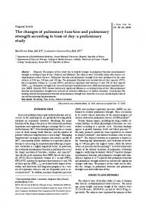
magnification (Maina et al., 1989). The ratio St/τht, multiplied by Krogh’s coefficient of oxygen diffusion, provides the anatomical (tissue) diffusing capacity of the blood–gas barrier. The value for Krogh’s coefficient was assumed to be 4.1×10⁻¹⁰·cm2·s–1·mbar–1, to be comparable to the study of Maina et al. (1989). Statistics Statistics include linear regressions (model 1, least squares) on log10-transformed data, excluding the single adult bird. The units of mass are grams. Regression statistics include coefficients of determination (r2) and 95% confidence intervals of the slope (CI), calculated in Excel with StatistiXL add-in software (statistixl.com), as were tests for relationships among residuals and for outlier points. Differences in regression slopes and elevations were tested with analysis of covariance (ANCOVA) according to Zar (1998). Where slopes were significantly different, the regions of significantly different elevations were identified with the Johnson–Neyman technique (White, 2003). Means of other values are given with 95% confidence intervals. Results Respiration Four newly hatched pelicans (body mass 88–121·g), had metabolic rates of 1.5±0.3·ml·min–1 (mean ± 95% CI) at 36°C, near the mean of 1.18·ml·min–1 for 18 hatchlings in a previous study of Australian pelicans (Pearson et al., 2002). It was not possible to increase metabolic rate by exposure to cold, because the hatchlings were naked and had not developed any thermoregulatory ability. They were helpless and uncoordinated and thus incapable of exercise. Therefore, metabolic rates measured on the first day of hatching were used for both resting and maximal values (Fig.·1). Resting and maximal metabolic rates diverged in birds aged 11 days or older. The increases in resting metabolic rate were not linear on a double log plot, because 11- and 21-dayold birds had relatively high values. Maximal rate, however, was linear over two orders of magnitude body mass, with a slope of 1.28±0.07 CI (Fig.·1). At the maximum body mass of 8834·g, the regression equations indicate a resting metabolic rate of 144·ml·min–1 and a maximum metabolic rate of 436·ml·min–1. This indicates a total metabolic scope of 292·ml·min–1 or a factorial scope of 3.0. There was no apparent difference in maximal rate of the adult and the juveniles of similar mass. Pulmonary morphometry Lung volume data were available for 13 post-hatch pelicans (Fig.·2). There was a strong, linear relationship between logtransformed lung volume and body mass, and the slope of the relationship (1.05±0.06 CI) was not significantly greater than 1.0. The value from the adult bird was not exceptional. The surface area of the blood–gas barrier increased allometrically with body size with a slope of 1.25±0.15 CI
2666 R. S. Seymour and others 1000
Oxygen consumption (ml min–1)
VO2max VO2rest 100
Adult birds Flight
Pelicans
Cold
10
1 0.01
0.1
1 Body mass (kg)
(Fig.·3). There appeared to be further increase in surface area in the single adult, and the difference was significant (Outlier test: Studentised residual 2.66 > critical value 2.23 at α=0.05). Harmonic mean thickness of the blood–gas tissue barrier did not change significantly in the juvenile pelicans during development, the slope being 0.02±0.08 CI (Fig.·4). However, the value from the adult was significantly lower than the other juveniles of similar body mass (Outlier test: Studentised residual 4.58 > critical value 2.23 at α=0.05). The anatomical diffusing capacity of the blood–gas tissue barrier was significantly allometric, with a slope of 1.23±0.20 CI. Because of the dramatic decrease in barrier thickness, diffusing capacity in the adult was significantly higher
10
Fig.·1. Allometry of resting (VO∑rest) and maximum (VO∑max) rate of oxygen consumption of 12 Australian pelicans during post-hatching growth (filled circles), excluding one adult bird (open circle). Regression equations: VO∑rest=0.0149M1.01 (r2=0.98); VO∑max=0.0040M1.28 (r2=0.99). Data from 33 species of adult birds during flight (open triangles) are from Norberg (1996), converting W to ml·min–1 assuming 20·J·ml–1, and averaging multiple data from the same species; VO∑max=0.641M0.80 (r2=0.86). Data from nine species of adult birds exposed to cold (filled triangles) are from Hinds et al. (1993); VO∑max=0.787M0.62 (r2=0.99). Dotted lines are extrapolations.
than values for the other juveniles of similar mass (Outlier test: Studentised residual 3.78 > critical value 2.23 at α=0.05).
Lung volume (ml)
Discussion This study demonstrates that maximum metabolic rate and diffusing capacity of the blood–gas barrier of highly altricial pelicans increase greatly during post-hatching development, as the animals progress from helpless neonate to a state capable of flight. The scaling exponents (slopes) are greater than 1 (Figs·1 and 5). To illustrate these increases in terms of massspecific values for an 88·g hatchling and an 8.8·kg juvenile, maximum metabolic rate increases by 357% and diffusing capacity by 291%. 1000 The maximum metabolic rates we recorded during exercise in the 8.8·kg juvenile (402·ml·min–1) and the 7.2·kg adult Pelicans (357·ml·min–1) were apparently sufficient to 100 power flight. The cost of flight for such a large bird has not been measured, but it is estimated that the largest flying bird would weigh about 12·kg when the power required for flight is equal 10 to the available power (Norberg, 1996). This point is 115·W, or 345·ml·O2 min–1, assuming Adult birds 20·J·ml–1. Although the cardio-pulmonary system 1 may have the capacity to deliver enough oxygen for powered flight, it is unlikely that the oldest juveniles could sustain flight. Indeed, older juveniles (partial primary feather development) 0.1 that were observed at the colony were only 0.01 0.1 1 10 capable of short gliding flight, and then only after Body mass (kg) long run-ups on open ground. We judge that the juveniles we sampled had never flown Fig.·2. Allometry of lung volume (VL) of 13 Australian pelicans during postsustainably. It appears that the capacity of the hatching growth (filled circles), excluding one adult (open circle). Data from 26 oxygen delivery system is developed in advance species of adult birds (triangles) are from Maina et al. (1989). Regression equations: pelicans VL=0.018M1.07 (r2=0.99); adult birds VL=0.022M1.02 (r2=0.98). of their need to use it.
Pelican pulmonary structure and function 2667 the adult. This bird is the only one in this study with a barrier thickness in the range for other adult birds (Fig.·4). Although only one adult was available to us, it was a statistically significant outlier that leads to the conclusion that the gasexchange barrier decreases in thickness and increases in area during maturation of sedentary juveniles to volant adults, although body mass and 106 lung volume do not change. It is instructive to compare the allometries of growing pelicans and adults of other species to distinguish the 105 effects of development and inherent scaling relationships of the products of development. Fig.·1 compares maximum metabolic rate in pelicans with 33 species 4 10 of flying birds (Norberg, 1996) and nine species exposed to cold (Hinds et al., Adult birds 1993). Analysis of covariance reveals 3 significant differences in slope between Pelicans 10 growing pelicans and both studies of adult birds (P