bioRxiv preprint first posted online Jul. 23, 2018; doi: http://dx.doi.org/10.1101/374538. The copyright holder for this preprint (which was not peer-reviewed) is the author/funder, who has granted bioRxiv a license to display the preprint in perpetuity. It is made available under a CC-BY 4.0 International license.
Dynamic functional connectivity changes in dementia with Lewy bodies and Alzheimer’s disease Authors: Julia Schumachera, Luis R. Perazaa,b, Michael Firbanka, Alan J. Thomasa, Marcus Kaiserb,c, Peter Gallagherc, John T. O’Briend, Andrew M. Blamiree, John-Paul Taylora Affiliations: a
Institute of Neuroscience, Newcastle University, Campus for Ageing and Vitality, Newcastle upon
Tyne, NE4 5PL, United Kingdom b
Interdisciplinary Computing and Complex BioSystems (ICOS) research group, School of
Computing, Newcastle University, Newcastle upon Tyne, NE4 5TG, United Kingdom c
Institute of Neuroscience, Newcastle University, The Henry Wellcome Building, Newcastle upon
Tyne, NE2 4HH, United Kingdom d
Department of Psychiatry, University of Cambridge School of Medicine, Cambridge, CB2 0SP,
United Kingdom e
Institute of Cellular Medicine & Newcastle Magnetic Resonance Centre, Campus for Ageing and
Vitality, Newcastle upon Tyne, NE4 5PL, United Kingdom Corresponding Author: Julia Schumacher, Biomedical Research Building 3rd floor, Campus for Ageing and Vitality, Institute of Neuroscience, Newcastle University, NE4 5PL Newcastle upon Tyne,
[email protected]
bioRxiv preprint first posted online Jul. 23, 2018; doi: http://dx.doi.org/10.1101/374538. The copyright holder for this preprint (which was not peer-reviewed) is the author/funder, who has granted bioRxiv a license to display the preprint in perpetuity. It is made available under a CC-BY 4.0 International license.
Abstract We studied the dynamic functional connectivity profile of dementia with Lewy bodies (DLB) and Alzheimer’s disease (AD) and the relationship between dynamic connectivity and the temporally transient symptoms of cognitive fluctuations and visual hallucinations in DLB. Resting state fMRI data from 31 DLB, 29 AD, and 31 healthy control participants were analysed using dual regression to determine between-network functional connectivity. We used a sliding window approach followed by k-means clustering and dynamic network analyses to study dynamic functional connectivity changes associated with AD and DLB. Network measures that showed significant group differences were tested for correlations with clinical symptom severity. AD and DLB patients spent more time than controls in sparse connectivity configurations with absence of strong positive and negative connections and a relative isolation of motor networks from other networks. Additionally, DLB patients spent less time in a more strongly connected state and the variability of global brain network efficiency was reduced in DLB compared to controls. However, there were no significant correlations between dynamic connectivity measures and clinical scores. The loss of global efficiency variability in DLB might indicate the presence of an abnormally rigid brain network and the lack of economical dynamics, factors which could contribute to an inability to respond appropriately to situational demands. However, the absence of significant clinical correlations indicates that the severity of transient cognitive symptoms such as cognitive fluctuations and visual hallucinations might not be directly related to these dynamic connectivity changes observed during a short resting state scan. Keywords: Resting state fMRI, dual regression, sliding window, dynamic network analysis, neurodegeneration, cognitive fluctuations
bioRxiv preprint first posted online Jul. 23, 2018; doi: http://dx.doi.org/10.1101/374538. The copyright holder for this preprint (which was not peer-reviewed) is the author/funder, who has granted bioRxiv a license to display the preprint in perpetuity. It is made available under a CC-BY 4.0 International license.
1. Introduction Resting state functional MRI has been used to study changes in functional connectivity associated with different forms of dementia such as dementia with Lewy bodies (DLB) and Alzheimer’s disease (AD) (Kenny et al., 2012; Lowther et al., 2014; Peraza et al., 2014; Schumacher et al., 2018). To date, most functional connectivity studies have focused on mean connectivity over the duration of a scan of several minutes, thereby implicitly assuming that functional connectivity remains stationary during that time. However, it has recently been shown that functional connectivity can vary substantially in both strength and directionality on a timescale of seconds to minutes (Chang and Glover, 2010; Hutchison et al., 2013b) and that studying these dynamics can provide important complementary information to the traditional analysis of stationary functional connectivity (Calhoun et al., 2014; Hutchison et al., 2013a). A large number of DLB patients experience fluctuations in cognition and attention/arousal, mostly spontaneously occurring without any situational explanation, which results in pronounced variation in cognitive ability over time (Bradshaw et al., 2004; McKeith et al., 2005). In addition, the majority of DLB patients present with visual hallucinations that recur over time (Aarsland et al., 2001). The transient nature of these DLB symptoms suggests that changes in functional connectivity dynamics might play a role in their aetiology. We therefore studied dynamic functional connectivity in DLB compared to healthy controls and patients with AD to (1) identify the differential dynamic connectivity profile of the two dementia subtypes and (2) investigate how abnormal dynamics might relate to the clinical DLB symptoms, especially with respect to cognitive fluctuations and visual hallucinations. 2. Materials and methods 2.1 Participants The study involved 102 participants over 60 years of age: 33 were diagnosed with probable DLB, 36 with probable AD, and 33 were age-matched healthy controls (HCs) with no history of psychiatric or neurological illness. Participants from two contemporary and independent dementia studies conducted at one research center (Newcastle) were combined for this analysis. Both studies recruited patients from the local community-dwelling population who had been referred to old age psychiatry and
bioRxiv preprint first posted online Jul. 23, 2018; doi: http://dx.doi.org/10.1101/374538. The copyright holder for this preprint (which was not peer-reviewed) is the author/funder, who has granted bioRxiv a license to display the preprint in perpetuity. It is made available under a CC-BY 4.0 International license.
neurology services. DLB and AD diagnoses were performed independently by two experienced old age psychiatrists using consensus criteria for probable DLB (McKeith et al., 2005) and probable AD (McKhann et al., 1984, 2011). Written informed consent was obtained from all participants and both studies were approved by the local ethics committee. 2.2 Data acquisition Imaging for both studies was performed on the same 3T Philips Intera Achieva scanner. The imaging protocol was the same in both studies except for a different resolution of the structural scans. Structural images were acquired with a magnetization prepared rapid gradient echo (MPRAGE) sequence, sagittal acquisition, echo time 4.6ms, repetition time 8.3ms, inversion time 1250ms, flip angle = 8°, SENSE factor = 2, and in-plane field of view 256 x 256 mm2 with slice thickness 1.2 mm, yielding a voxel size of 0.93 x 0.93 x 1.2 mm3 (study 1) and in-plane field of view 240 x 240 mm2 with slice thickness 1.0 mm, yielding a voxel size of 1.0 x 1.0 x 1.0 mm3 (study 2). Resting state scans were obtained with a gradient echo echo-planar imaging sequence with 25 contiguous axial slices, 128 volumes, anterior-posterior acquisition, in plane resolution = 2.0 x 2.0 mm, slice thickness = 6 mm, repetition time (TR) = 3000ms, echo time = 40ms, and field of view = 260 x 260 mm2. DLB patients who were taking dopaminergic medication were scanned in the ON state. 2.3 Preprocessing FEAT (FMRI Expert Analysis Tool, version 6.0) which is part of the FMRIB's software library (FSL, www.fmrib.ox.ac.uk/fsl) was used to perform motion correction with MCFLIRT, slice-timing correction, and spatial smoothing with a 6.0mm full width at half maximum Gaussian kernel. Participants were excluded if the MCFLIRT motion parameters exceeded 2 mm translation and/or 2° rotation. To ensure that there were no group differences in motion due to the presence of Parkinsonian symptoms in the DLB patients, motion was compared between the groups using the formula introduced in (Liao et al., 2010). A denoising procedure was performed with ICA-AROMA in FSL which performs single-subject independent component analysis (ICA) to remove motion components from each participant’s functional data (Pruim et al., 2015). Subsequently, eroded CSF and white matter masks were
bioRxiv preprint first posted online Jul. 23, 2018; doi: http://dx.doi.org/10.1101/374538. The copyright holder for this preprint (which was not peer-reviewed) is the author/funder, who has granted bioRxiv a license to display the preprint in perpetuity. It is made available under a CC-BY 4.0 International license.
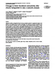
estimated using FAST in FSL and the mean signal inside the mask was regressed out of each participant’s cleaned functional data. Functional and structural images were co-registered using boundary based registration in FSL, and normalized to standard MNI space using Advanced Normalization Tools (Avants et al., 2011). As a final step, functional data were temporally high-pass filtered with a cut-off of 150 s and resampled to a resolution of 4 x 4 x 4 mm3. 2.4 Analysis of resting state data Resting state networks (RSNs) were estimated with an independent set of 42 healthy control participants from two previous studies that were conducted on the same MR scanner using a similar imaging protocol (see Supplementary Table S1). Data from all 42 HCs were temporally concatenated and subjected to a group-ICA using FSL’s MELODIC. A meta ICA approach was adopted to obtain more reliable components (Biswal et al., 2010) using a model order of 70 independent components which has been shown to be optimal for assessing disease-related group differences (Abou Elseoud et al., 2011) (see Schumacher et al. (2018) for a more detailed description). Meta ICA components were visually inspected with respect to their spatial maps (Kelly et al., 2010) and 27 were identified as being of biological interest according to previous literature (Agosta et al., 2012; Beckmann et al., 2005) (Figure 1 and Supplementary Table S2). Subsequently, FSL-dual regression was run with all 27 identified RSNs concatenated in a single 4D image, to obtain subject-specific representations of the RSN spatial maps and associated subjectspecific time courses (Figure 2A). Results from a static connectivity analysis have been published previously using the same data (Schumacher et al., 2018). The subject-specific time courses resulting from dual regression were further processed in Matlab (R2016b) using functions from the Group ICA of fMRI toolbox (GIFT, http://mialab.mrn.org/software/gift/index.html) to remove remaining noise sources. Postprocessing included (1) detrending to remove linear, quadratic, and cubic trends, (2) outlier detection based on AFNI’s 3dDespike function (http://afni.nimh.nih.gov/afni) and interpolation of outliers using a thirdorder spline fit to the clean parts of the time courses, and (3) low-pass filtering using a fifth-order Butterworth filter with a cutoff frequency of 0.15 Hz.
bioRxiv preprint first posted online Jul. 23, 2018; doi: http://dx.doi.org/10.1101/374538. The copyright holder for this preprint (which was not peer-reviewed) is the author/funder, who has granted bioRxiv a license to display the preprint in perpetuity. It is made available under a CC-BY 4.0 International license.
Figure 1: Resting state networks. Spatial maps of the 27 (RSNs) obtained from the independent healthy control group. RSN maps are thresholded at 3