NIH Public Access Author Manuscript Cardiovasc Res. Author manuscript; available in PMC 2010 March 17.
NIH-PA Author Manuscript
Published in final edited form as: Cardiovasc Res. 2008 May 1; 78(2): 324–332. doi:10.1093/cvr/cvn055.
Effect of Mechanical Boundary Conditions on Orientation of Angiogenic Microvessels Laxminarayanan Krishnan1, Clayton J. Underwood1, Steve Maas1, Benjamin J. Ellis1, Tejas C. Kode1, James B. Hoying2, and Jeffrey A. Weiss1 1 Department of Bioengineering, University of Utah 2 Division of Cardiovascular Therapeutics Cardiovascular Innovation Institute University of Louisville
Abstract
NIH-PA Author Manuscript
Aim—Mechanical forces are important regulators of cell and tissue phenotype. We hypothesized that mechanical loading and boundary conditions would influence neovessel activity during angiogenesis. Methods—Using an in vitro model of angiogenesis sprouting and a mechanical loading system, we evaluated the effects of boundary conditions and applied loading. The model consisted of rat microvessel fragments cultured in a 3D collagen gel, previously shown to recapitulate angiogenic sprouting observed in vivo. We examined changes in neovascular growth in response to four different mechanical conditions. Neovessel density, diameter, length and orientation were measured from volumetric confocal images of cultures exposed to no external load (free-floating shape control), intrinsic loads (fixed ends, no stretch), static external load (static stretch) or cyclic external load (cyclic stretch). Results—Neovessels sprouted and grew by the 3rd day of culture and continued to do so during the next 3 days of loading. The numbers of neovessels and branch points were significantly increased in the static stretch group when compared to the free-floating shape control, no stretch or cyclic stretch groups. In all mechanically loaded cultures, neovessel diameter and length distributions were heterogeneous, while they were homogeneous in shape control cultures. Neovessels were significantly more oriented along the direction of mechanical loading than those in the shape controls. Interestingly, collagen fibrils were organized parallel and adjacent to growing neovessels.
NIH-PA Author Manuscript
Conclusion—Externally applied boundary conditions regulate neovessel sprouting and elongation during angiogenesis, affecting both neovessel growth characteristics and network morphometry. Furthermore, neovessels align parallel to the direction of stress/strain or internally generated traction, and this may be due to collagen fibril alignment induced by the growing neovessels themselves. Keywords boundary conditions; angiogenesis; strain; orientation; morphometry; image analysis
Corresponding Author: Jeffrey A. Weiss, Department of Bioengineering, University of Utah, 50 South Central Campus Drive, Room 2480, Salt Lake City, UT 84112, Phone: 801-587-7834, Fax: 801-535-6801,
[email protected]. CONFLICT OF INTERESTS None.
Krishnan et al.
Page 2
INTRODUCTION NIH-PA Author Manuscript
Physiological organ morphology is achieved by migration and orientation of cells under the influence of chemotaxis, haptotaxis, and mechanotaxis. Both externally applied and internally generated mechanical forces influence cell migratory, proliferative, and secretory activities and matrix orientation 1–3. The current understanding of the morphogenesis of natural 3D tissue structure is limited. Given its importance in development, repair, tumorigenesis, and design of artificial constructs, significant resources have been directed at understanding the 3D morphology of the microvasculature, especially the vascularity of soft connective tissues 4, 5, where vasculature is often oriented along the direction of strain experienced in vivo and/or structural features of the extracellular matrix. Cellular orientation in engineered tissue is often achieved by mechanical loading or contact guidance 6–9.
NIH-PA Author Manuscript
Mechanical forces affect the behavior of nearly all cell types (e.g., 7, 10–12). The mechanical environment of endothelial cells influences migration, proliferation and tube formation 11, 13, protease and other secretory activities 14, cellular and cytoskeletal organization 15, expression of genes and surface molecules 16, and cell signaling 12. A large body of literature has focused on the responses of endothelial cells as monolayers, spheroids or cords, cultured in natural or engineered matrices and surfaces, to biochemical and biomechanical stimuli 11– 18. These studies have shown that cells align both along 19, 20 and transverse 15, 17, 18, 21 to the direction of principal stretch in response to mechanical loading. The causes of this discrepancy are elusive and have been attributed to cell seeding density, matrix density, genetic framework, cell age, 2D vs. 3D substrates, cell subpopulation and the nature of loading. The angiogenic response of intact preformed microvessels to mechanical perturbations is relatively unexplored. The objective of the present study was to determine the morphological changes in sprouting microvessels in a 3D in vitro model of angiogenesis 22 due to alterations in construct boundary conditions and external loading, in previously unaligned collagen matrices. We quantified the orientation of microvessel segments in free-floating cultures, in cultures under traction against fixed anchors, and in cultures under externally applied strain. This is the first study to examine the orientation of multicellular microvessel structures rather than individual cells or cell clusters. The results of this study demonstrate that neovessel orientation and during angiogenesis depends more on culture boundary conditions than externally applied strains.
MATERIALS AND METHODS In vitro culture model
NIH-PA Author Manuscript
The 3D in vitro angiogenesis model has been characterized in our prior publications 22,23–25. Unlike angiogenesis models that use isolated endothelial cells, this model uses isolated microvessel fragments. “Parent” vessels contain associated perivascular cells and retain their basement membrane after initial harvest and seeding in 3D collagen gels. Sprouting begins predictably at Day 2–3. Pericytes disassociate from parent vessels and endothelial cells, consistent with observations that perivascular cell withdrawal relaxes the parent endothelial cell tube and permits sprouting and vessel elongation during angiogenesis. Sprouts elongate as patent tubes, branching and anastamosing with other vessels, and forming a new vascular network that ultimately fills the construct. Neovessel constructs form a functional vascular tree when implanted 24, rapidly inosculating with the recipient host circulation after implantation and carrying blood. Epididymal fat pads from male retired breeder Sprague-Dawley rats (>500g) were aseptically harvested conforming to the Guide for the Care and Use of Laboratory Animals (NIH Publication No. 85-23, revised 1996), and washed with Leibowitz (L-15) media containing 2%
Cardiovasc Res. Author manuscript; available in PMC 2010 March 17.
Krishnan et al.
Page 3
NIH-PA Author Manuscript
fetal bovine serum. Fat pads were minced into ~1 mm3 pieces and subjected to limited digestion with 2 mg/ml Clostridium collagenase (Worthington Biochemicals, Lakewood, NJ) and 2 mg/ ml bovine serum albumin (BSA, Sigma-Aldrich, St. Louis, MO) in Dulbecco’s cation free phosphate buffered saline (DCF-PBS, pH 7.4) at 37°C for 4 minutes. The solution was immediately diluted with L-15 media to halt digestion and centrifuged. The pellet was washed twice and resuspended in L-15 media with 2% serum and sequentially filtered through a 350 μm and then a 30 μm sterile nylon filter. The larger filter eliminates undigested particulate debris while the fine filter retains larger vessel fragments, allowing single cells and smaller fragments to pass through. The 30 μm filter was transferred to a sterile Petri dish and washed with serum containing L-15 media to suspend the fragments. The solution was analyzed for number of microvessels and centrifuged to obtain a pellet of microvessel fragments. Reagents were obtained from GIBCO (Grand Island, NY) unless mentioned otherwise.
NIH-PA Author Manuscript
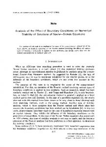
Rat tail collagen type I (BD Biosciences, Bedford, MA) was mixed with concentrated Dulbecco’s modified Eagle medium (DMEM, GIBCO-Invitrogen, Carlsbad, CA) to yield a final concentration of 3 mg/ml in 1X DMEM. Microvessels were suspended in collagen solution at 15,000 fragments/ml, and 1.2–1.5 ml of this suspension was pipetted into custom culture chambers and polymerized at 37°C and 95% humidity for 30 min (Figure 1). Labtek II chambers (Nunc-Nalge, Rochester, NY) were modified to accommodate a 5×20 mm specimen between fixed and mobile anchor posts, based on our earlier design 26. A removable Teflon mold was used to cast the collagen constructs and was then removed after polymerization. The fixed post was attached to the chamber base near one end (Loctite® 3301, Henkel Corp., Rocky Hill, CT). Stainless steel pins were inserted through holes in the sides of the chamber for temporary anchorage of the mobile post. The acrylic posts had holes (1 mm dia) to allow the collagen solution to pass through. Gels were overlaid with serum free culture media composed of 1:1 mixture of 1X DMEM (low glucose) and F12 nutrient mixture with additional components (Insulin 5 μg/ml, Transferrin 100 μg/ml, Progesterone 20 nM, Selenium 30 nM, and Putrescine 100 μM) 27, rhVEGF (10 ng/ml, PeproTech, Rocky Hill, NJ), and Gentamycin (50 μg/ml). Three chambers and one chamber without the acrylic anchors were prepared simultaneously and allowed to grow undisturbed for 3 days. The interval between preparation of microvessel seeded collagen constructs and the beginning of mechanical intervention was based on the time required for initial sprout formation as we previously reported 22, 28. On the third day, culture dimensions were measured with digital calipers, photographed, and given a 70% media change. Mechanical conditioning and experimental groups
NIH-PA Author Manuscript
The mechanical conditioning device used two motorized linear actuators for simultaneous testing of two constructs within the incubator (Aerotech, Pittsburgh, PA, range = ±25 mm, resolution = ±5 nm) (Figure 1A). Chambers were mounted on raised platforms to minimize any influence of heat, magnetic or electric fields and vibration. Two conditioning regimens and two boundary conditions were evaluated (Figure 1E). For each experiment, one construct was subjected to 6% “Static Stretch” (SS group) for 3 days and the second to 6% “Cyclic Stretch” (CS group) at 1 Hz for 12 hrs every 24 hours for 3 days. This cyclic routine was chosen since vasculature in tissues that are subjected to cyclic stretch such as muscle and tendon experience a recovery period during the sleep cycle. The free floating constructs without acrylic anchors, referred to as “Shape Control” (SC), were placed on the conditioning platform along with the SS construct. Similarly, the anchored but unstretched construct, referred to herein as “No Stretch” (NS), was placed on the cyclic conditioning platform along with CS construct. To maintain comparability across different groups, four constructs, one for each group, were cast using the same starting material. Some samples
Cardiovasc Res. Author manuscript; available in PMC 2010 March 17.
Krishnan et al.
Page 4
became contaminated due to the open culture system and hence the final number of samples in each group over 9 experiments were SC – 9, NS – 8, SS - 6, and CS – 6.
NIH-PA Author Manuscript
Confocal microscopy On the 6th day of culture constructs were cut free from the anchor posts with a scalpel, gels were measured and then fixed in 4% formaldehyde. Note that fixation caused a reduction in construct volume (approx. 20% or 7.5% along each dimension, nearly uniform). Microvessels within the gel were permeabilized with 0.1% Triton-X-100 in PBS. Endothelial cells were stained with 2 μg/ml Isolectin IB4-Alexa 488 conjugate (Invitrogen, Carlsbad, CA). Confocal microscopy of 6 fields adjacent to the centers of the top and bottom surfaces of the constructs was performed on an Olympus FV1000 with a 10× objective. Individual z-axis images were acquired to a depth of 300 μm at 2.5 μm intervals and 512×512 resolution. Volumetric image reconstruction and skeletonization
NIH-PA Author Manuscript
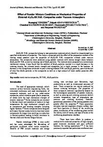
Image stacks from the six adjacent fields (1.27×1.27 mm each) forming a 2×3 grid were stitched together to generate a single mosaic (3.8×2.5×300 μm) (Figure 2). The Amira™ software (Mercury Computer Systems, Carlsbad, CA) was used for image analysis and skeletonization. Intensity artifacts in images with slice depth 29 were reduced using an algorithm that fit an exponential curve to average intensities. Mosaic stacks were further deconvolved using a point spread function generated from the numerical aperture (NA = 0.4, 10X – air), wavelength of light (520 nm) and the refractive index of the gel 30. An automated thresholding algorithm based on a Gaussian distribution fit to the image histogram was used to binarize each mosaic stack (ImageJ, NIH). A size filter of 600 μm3 was applied to remove debris and small cell clusters. Images were then skeletonized 31,32. A Chamfer distance map was generated as a part of this process, and these data were used to determine average diameter of each segment 31. Data analysis
NIH-PA Author Manuscript
Coordinates of points from the skeletonization, forming line equivalents of the vasculature, were processed by a custom C++ program (available at http://mrl.sci.utah.edu) to calculate morphometric parameters, including branch points, end points, individual segments, and vessels (all connected segments). Total vascular volume was determined from lengths and diameters. Additional parameters were orientation with respect to stretch axis, segment and total network length, vessel diameters, number of branches, junctions, segments, segment lengths, and vascular volume fractions. A minimum vessel size of 150 μm length was used to avoid smaller segments and debris. This cut off was based on microvessel dimensions at the time of seeding, which were about 150 μm long. A segment length cutoff of 25 μm was used to eliminate branches that were not much larger than the average vessel diameters. Segment orientation was examined along the direction of stretch (X-axis, θ) and through the culture depth (Z- axis, δ). Additionally, each segment was projected onto the XY-plane and the angle of the projected segment with respect to the X-axis was determined (Projected-X, φ). Segments were sorted into 10° bins from 0–90°. Results were expressed as percentage of total fragments per bin and as percentage of length-weighted segments per bin. The angle distribution of segments was compared between groups and within groups using 2-way ANOVAs and Tukey tests (or Kruskal-Wallis ANOVA and Dunn’s post hoc tests for nonparametric distributions) with significance at α=0.05. Confocal reflectance microscopy Confocal reflection microscopy 33 was performed to visualize collagen fibril orientation in the presence and absence of anchorage and external loading, and modulation by growing microvessels. Samples were illuminated with a 633 nm laser and reflected light was collected between 632–633 nm. Several image stacks (1 micron step size × 50) were collected for each
Cardiovasc Res. Author manuscript; available in PMC 2010 March 17.
Krishnan et al.
Page 5
NIH-PA Author Manuscript
condition. To see whether fibril orientation was induced by the boundary/loading conditions, gels lacking microvessels were prepared and subjected to the same mechanical regimens. After 6 days these gels were fixed while still attached to the anchors.
RESULTS In vitro culture model All constructs showed well formed sprouts by Day 3 and well established vascular networks by Day 6 (Figure 3A). Total volume reduction between the 3rd and 6th day of culture was 60% for free-floating SC constructs and 40% for the other three groups (p=0.001, ANOVA). The SC constructs reduced to about 32% of their Day 3 length, while the length of constructs in the other groups was maintained by the anchoring posts. The average height reduction in anchored construct groups was 17.6% and not significantly different from the SC constructs. Similarly, there was 27.6% reduction in width of the anchored constructs, which was not significantly different from SC constructs. SC constructs showed a near uniform percent contraction along the two directions. Construct vascularity and complexity
NIH-PA Author Manuscript
Microvessel network morphometry at Day 6 was determined from the 3D image reconstructions (Figure 2). Qualitatively, the SC constructs showed random microvessel orientation, while the NS, SS and CS constructs showed various degrees of orientation along the construct long axis. The number of vessels per construct was highest in the SS group and lowest in the SC group (Figure 3B). All treatment groups had significantly more vessels per construct than the SC group (Kruskal-Wallis ANOVA, p