Satoru Nogami Ð Fumi Sano-Kumagai Ð Ayaka Saka. Masashi Yukawa Ð Taro L. Saito Ð Jun Sese. Dai Hirata Ð Shinichi Morishita Ð Yoshikazu Ohya.
Curr Genet (2006) 49: 237–247 DOI 10.1007/s00294-005-0051-0
R ES E AR C H A RT I C L E
Genjiro Suzuki Æ Hiroshi Sawai Æ Miwaka Ohtani Satoru Nogami Æ Fumi Sano-Kumagai Æ Ayaka Saka Masashi Yukawa Æ Taro L. Saito Æ Jun Sese Dai Hirata Æ Shinichi Morishita Æ Yoshikazu Ohya
Evaluation of image processing programs for accurate measurement of budding and fission yeast morphology Received: 1 September 2005 / Revised: 27 October 2005 / Accepted: 28 October 2005 / Published online: 6 January 2006 Ó Springer-Verlag 2006
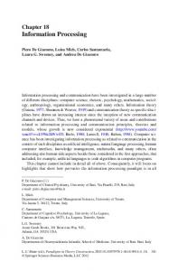
Abstract To study the cellular functions of gene products, various yeast morphological mutants have been investigated. To describe yeast morphology objectively, we have developed image processing programs for budding and fission yeast. The programs, named CalMorph for budding yeast and F-CalMorph for fission yeast, directly process microscopic images and generate quantitative data about yeast cell shape, nuclear shape and location, and actin distribution. Using CalMorph, we can easily and quickly obtain various quantitative data reproducibly. To study the utility and reliability of CalMorph, we evaluated its data in three ways: (1) The programs extracted three-dimensional bud information from two-dimensional digital images with a low error rate (200) and the 6.0-lm beads (lane 3, n>200) and these data as provided by the manufacturer (lanes 2 and 4, respectively) are shown. e–g The percentages of budding yeast cells at the indicated morphological stages counted by hand and by
CalMorph. Wild-type cells at exponential phase were sampled and stained with FITC-ConA, DAPI, and rhodamine-phalloidin. The same person classified cells by bud index (e), nuclear stage (f), and actin stage (g) by hand three times (lanes 1–3). The same images were processed by CalMorph (e–g lane 4). h–j Cell length and cell width of fission yeast cells were measured by hand (h) and by F-CalMorph (i, j). The same person measured three times (h square, triangle, and circle), and the averages of the data are shown (h, i x mark). The lengths of the same cells were also measured by F-CalMorph (i diamond). An example of processed image is shown (j). Cell length and width of an asterisked cell are colored with purple and blue, respectively. k, l The percentages of fission yeast cells at the indicated stages counted by hand and by F-CalMorph. Wild-type cells at exponential phase were sampled and triple-stained. The same person classified cells by nuclear stage (k) and actin stage (l) by hand three times (k, l lanes 1–3). The same images were processed by F-CalMorph (k, l lane 4)
photographed cells were observed from the extension of their short axis and that the data processed with CalMorph from 2D yeast images reflects the 3D morphology of yeast cells well.
CalMorph were within the ranges specified by the manufacturer (Fig. 6d, lanes 1 and 3). These data indicate that CalMorph accurately recognizes bead shapes and calculates bead sizes, suggesting that CalMorph can accurately recognize cellular morphology and calculate cell size. Moreover, data processing by CalMorph required less time and labor than performing the same procedures by hand. We also determined the bud index, nuclear stage, and actin stage by hand and with CalMorph (Fig. 6e–g). These parameters have been used for describing yeast cell morphology and cell cycle progression (Pruyne and Bretscher 2000; Hayashi et al. 2004; Martin and Chang 2005; Castagnetti et al. 2005). The data calculated by hand showed slight variation (lanes 1–3). On the other hand, the data calculated by CalMorph were always the same and were close to the data collected by hand (lane 4). These results suggest that CalMorph can accurately recognize nuclear shape and the distribution of actin patches and can accurately classify nuclear and actin states in the yeast cell cycle. Similarly, we compared the fission yeast morphological data obtained by hand and that obtained with F-CalMorph. The cell length and cell width of fission yeast cells were measured three times by hand (Fig. 6h), and these parameters of the same cells were determined with F-CalMorph (Fig. 6i, j). As was the case for budding yeast, the data obtained with FCalMorph were close to the average of the data collected by hand. For example, a cell asterisked in Fig. 6j was approximated to a circumscribed rectangle and the cell length and width were calculated from this rectangle. The cell length and width were calculated by CalMorph as 10.67 lm (purple line) and 4.79 lm (blue line) while by hand three times as 11.25, 11.05, and 11.36 lm; 4.88, 4.68, and 4.86 lm, respectively. Next, we determined the nuclear stage and actin stage by hand and using F-CalMorph (Fig. 6k, l). The fission yeast data for nuclear stage and actin stage calculated by F-CalMorph (lane 4) were close to the data counted by hand (lanes 1–3). These results indicate that, in the study of budding and fission yeast cell morphology, the collection of data using CalMorph
CalMorph processes images easily and generates reproducible results We have previously had to count images by hand to investigate cell morphology, bud index, nuclear position, or actin distribution. This process was laborious and time consuming, and the data often varied from experiment to experiment owing to differences among observers and in experimental conditions. To compare the measurements made with CalMorph and those made by hand in terms of labor and reproducibility, we measured the long and short axes of budding yeast cells. First, the same person measured the cells three times by hand (Fig. 6a). As expected, these results differed slightly from each other, and we therefore could not obtain strictly reproducible results. Next, the same cells were processed by CalMorph and these parameters were calculated (Fig. 6b, c). CalMorph gave completely reproducible data, and the data were close to the average of the data measured by hand (Fig. 6b). For example, a part of processed images is shown (Fig. 6c). CalMorph calculates long and short axes of cells from approximated ellipse. Long and short axes of an asterisked cell were colored by purple and blue, respectively. Long axes of this cell measured by hand three times were 4.50, 4.48, and 4.48 lm. Short axis were 3.76, 3.86, and 3.86 lm. But long and short axes of this cell calculated by CalMorph were always 4.57 and 3.78 lm. Moreover, it took few minutes to measure by hand the long and short axes of ten budding yeast cells; with CalMorph, the measuring took few seconds. Next, to evaluate the absolute values calculated by CalMorph, we measured the diameters of manufactured fluorescent beads using the program (Fig. 6d). Compared with the average values listed on the product label for beads of different sizes (Fig. 6d, lanes 2 and 4), the values calculated by
246
could generate reproducible data more quickly and easily than can be done by hand.
We previously reported that, for budding yeast, CalMorph could detect cellular morphological differences between haploid cells and diploid cells, and between logphase cells and stationary-phase cells (Ohtani et al. 2004). However, these differences were judged only from asynchronous cell cultures. Therefore, we determined whether changes in morphology, nuclear shape, and actin distribution resulting from the progression of the cell cycle could be recognized using CalMorph (Pruyne and Bretscher 2000). We collected budding yeast G1 cells (cells without buds) by elutriation and sampled and fixed them at the indicated time points after release into the medium (Walker 1999). The samples were stained and photographed, and the images were processed with CalMorph. The formation of a bud coinciding with the progression of the cell cycle is shown by the bud index data (Fig. 7a). The transitions of nuclear and actin
stages are also shown (Fig. 7b, c). The data calculated by CalMorph indicate the bud growth, nuclear division, and the changes of actin patch distribution during the cell cycle. These data indicate that nuclei were divided when mature buds were formed (simultaneous increasing in ‘‘nuclear C’’ and ‘‘large bud’’ after 140 min). The data also show that distribution of actin patches were changed through the cell cycle. In G1 cells, actin patches were distributed thoroughout cells in most of the cells (0 min, ‘‘actin A’’). In apical growth phase, actin patches were concentrated at the bud tip (120–140 min, increasing in ‘‘actin C’’). In G2/M phase, apical-isotropic growth switch was occurred and actin patches were distributed throughout bud (160–180 min, increasing in ‘‘actins D and E’’). After nuclear division, actin patches were concentrated at bud neck for cytokinesis (180 min, increasing in ‘‘actin F’’). These morphological changes of budding yeast cells are consistent with previous report (Pruyne and Bretscher 2000). These results indicate that CalMorph could correctly chase the morphological changes of budding yeast during the cell cycle. Next, we performed the same experiments to examine the use of F-CalMorph in recognizing morphological
Fig. 7 Cell cycle progression monitored by CalMorph. a–c Morphological transition of budding yeast cells resulting from cell cycle progression. Budding yeast cells at G1 phase were collected by centrifugal elutriation, released into fresh medium, and sampled at the indicated time points. The samples were triple-stained and photographed. The images were processed and the cells were classified by CalMorph. The percentages of cells classified by bud index (a), nuclear stage (b), and actin stage (c) are shown. d–f Morphological transition of fission yeast cells resulting from cell cycle progression. Small fission yeast cells were collected by
centrifugal elutriation, released into fresh medium, and sampled at the indicated time points. The fission yeast images were processed and cells were classified by F-CalMorph. The percentages of cells classified by cell length (d), nuclear stage (e), and actin stage (f) are shown. Actin stages were classified into three stages. Actin 1 indicates monopolar actin distribution and consists of actin A stage. Actin 2 indicates bipolar or delocalized actin distribution and consists of actin B, actin C, and actin F stages. Actin 3 indicates center localization and consists of actin D and actin E stages
Cell cycle progression monitored with CalMorph
247
changes of fission yeast cells. Fission yeast cells initially grow in a monopolar manner. At a point during G2 phase, the cells change their growth to a bipolar manner. During mitosis, the cells stop growing, and after nuclear division, the cells undergo cell division (Martin and Chang 2005). To confirm that F-CalMorph can correctly recognize these morphological transitions, we collected small fission yeast cells by elutriation, sampled the cells at the indicated time points, and triple-stained the cells. The images were processed using F-CalMorph. These data indicate that the fission yeast cells grew and underwent nuclear division about 140 min after release (Fig. 7d, e). Then, the cells underwent cell division (increasing in small cells at 180 min). To describe the actin state in fission yeast, we newly defined three actin stages: actin 1 (monopolar distribution of actin), actin 2 (bipolar distribution and delocalization of actin), and actin 3 (center localization of actin). Actin 1 consisted of an actin A stage; actin 2 comprised actin B, actin C, and actin F stages; and actin 3 comprised actin D and actin E stages (Figs. 3e, 7f). The data indicate that many cells showed monopolar actin distribution in the small cells (0 min, ‘‘actin 1’’). As cells grew, most of the cells showed bipolar actin distribution (100–120 min, ‘‘actin 2’’). After nuclear division, actin patches concentrated at division plane for cytokinesis (after 140 min, ‘‘actin 3’’). These changes of actin distribution are consistent with previous report (Martin and Chang 2005). These results indicate that F-CalMorph could correctly recognize the morphological changes of the fission yeast cells during the cell cycle. These data indicate that CalMorph and F-CalMorph can recognize budding and fission yeast cell morphology at every cell cycle stage and that the data calculated using CalMorph are consistent with those of previous reports (Pruyne and Bretscher 2000; Martin and Chang 2005). We can easily obtain data on bud index, nuclear stage, and actin stage using CalMorph, in contrast to the excessive time and labor needed to collect these data by hand. Using CalMorph, we can readily detect irregular cell cycle progression, such as a delay of a particular cell cycle stage or the uncoupling of cell cycle progression with bud formation. Moreover, CalMorph may enable the collection of in silico synchronized cell cycle data from bulk cell images by selecting cells based on cell size, nuclear stage, or actin stage. For example, we could collect nuclear C-stage cells as budded cells with two nuclei and investigate their morphology instead of using synchronized M phase cells. It is possible to use CalMorph to detect novel and modest phenotypic changes resulting from defective cell cycle progression. Acknowledgements We thank Tamao Goto, Yuka Kitamura, and Emi Shimoi for technical assistance; and the members of the Ohya and Morishita groups at the University of Tokyo for stimulating discussions. This work was supported by the Institute for Bioinformatics and Research and Development of the Japan Science and Technology Corporation.
References Beach D, Durkacz B, Nurse P (1982) Functionally homologous cell cycle control genes in budding and fission yeast. Nature 300:706–709 Brachmann CB, Davies A, Cost GJ, Caputo E, Li J, Hieter P, Boeke JD (1998) Designer deletion strains derived from Saccharomyces cerevisiae S288C: a useful set of strains and plasmids for PCR-mediated gene disruption and other applications. Yeast 14:115–132 Castagnetti S, Behrens R, Nurse P (2005) End4/Sla2 is involved in establishment of a new growth zone in Schizosaccharomyces pombe. J Cell Sci 118:1843–1850 Chang F (2001) Establishment of a cellular axis in fission yeast. Trends Genet 17:273–278 Chang F, Peter M (2003) Yeasts make their mark. Nat Cell Biol. 5:294–299 Coelho MA, Belo I, Pinheiro R, Amaral AL, Mota M, Coutinho JAP, Ferreira EC (2004) Effect of hyperbaric stress on yeast morphology: study by automated image analysis. Appl Microbiol Biotechnol 66:318–324 Drubin DG, Nelson WJ (1996) Origins of cell polarity. Cell 84:335–344 Hartwell LH, Unger MW (1977) Unequal division in Saccharomyces cerevisiae and its implications for the control of cell division. J Cell Biol 75:422–435 Hayashi T, Fujita Y, Iwasaki O, Adachi Y, Takahashi K, Yanagida M (2004) Mis16 and Mis18 Are required for CENPA loading and histone deacetylation at centromeres. Cell 118:715–729 Hayles J, Nurse P (2001) A journey into space. Nat Rev Mol Cell Biol 2:647–656 Hirata D, Kishimoto N, Suda M, Sogabe Y, Nakagawa S, Yoshida Y, Sakai K, Mizunuma M, Miyakawa T, Ishiguro J, Toda T (2002) Fission yeast Mor2/Cps12, a protein similar to Drosophila Furry, is essential for cell morphogenesis and its mutation induces Wee1-dependent G(2) delay. EMBO J 21:4863–4874 Johnson DI (1999) Cdc42: an essential Rho-type GTPase controlling eukaryotic cell polarity. Microbiol Mol Biol Rev 63:54–105 Madden K, Costigan C, Snyder M (1992) Cell polarity and morphogenesis in Saccharomyces cerevisiae. Trends Cell Biol 2:22–29 Martin SG, Chang F (2005) New end take off: regulating cell polarity during the fission yeast cell cycle. Cell Cycle 4:1046–1049 Nurse P, Thuriaux P, Nasmyth K (1976) Genetic control of the cell division cycle in the fission yeast Schizosaccharomyces pombe. Mol Gen Gene 146:167–178 Ohtani M, Saka A, Sano F, Ohya Y, Morishita S (2004) Development of image processing program for yeast cell morphology. J Bioinform Comput Biol 4:695–709 Pruyne D, Bretscher A (2000) Polarization of cell growth in yeast. I. Establishment and maintenance of polarity states. J Cell Sci 113:365–75 Reid BJ, Hartwell LH (1977) Regulation of mating in the cell cycle of Saccharomyces cerevisiae. J Cell Biol 75:355–365 Saito TL, Ohtani M, Sawai H, Sano F, Saka A, Watanabe D, Yukawa M, Ohya Y, Morishita S (2004) SCMD: Saccharomyces cerevisiae morphological database. Nucleic Acids Res 32:D319–D322 Saito TL, Sese J, Nakatani Y, Sano F, Yukawa M, Ohya Y, Morishita S (2005) Data mining tools for the Saccharomyces cerevisiae morphological database. Nucleic Acids Res 33:W753–W757 Verde F, Mata J, Nurse P (1995) Fission yeast cell morphogenesis: identification of new genes and analysis of their role during the cell cycle. J Cell Biol 131:1529–1538 Walker GM (1999) Synchronization of yeast cell populations. Methods Cell Sci 21:87–93