Sep 4, 2012 - Laura Hokkanen. References. 1. Itti, L., & Koch, C. (2001). ... 194â203. doi:10.1038/35058500. 2. Bush, G., Valera, E.M., Seidman, L.J. (2005).
Experimental Investigation of Saliency Processing in ADHD
C O G N I T I V E S C I E NC E U N I V E R S I T Y OF H E L S I N K I
Benjamin Cowley, Cognitive Science Unit, Department of Behavioural Sciences
4th SEPTEMBER 2012
UNIVERSITY OF HELSINKI, FINLAND
STUDY DESIGN
We investigate the role of visual salience processing in attention deficit disorders (ADHD) using a novel Event-Related Potential (ERP) protocol. Saliency refers to the order of importance attached to visual features by the attention system, making certain features ‘stand out’, with processing time 25-50ms. Task-related or top-down attention can modulate saliency but is much slower, 200ms or more [1]. Findings of brain structural alteration in ADHD include significantly smaller volumes in the dorsolateral prefrontal cortex (dPFC) and associated regions: caudate, pallidum, anterior cingulate, and cerebellum [2]. This motivates the study of attentional neural processing in a task requiring top-down redirection of attention. To simulate this, our protocol manipulates the saliency of targets in a choice-response task, testing interference inhibition at the task-response level, and saliency-processing at the pre-attentive level. Here we present initial findings.
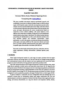
Figure 1: Protocol of the experiment conditions: Congruent vs Incongruent and contour vs no contour. The fixation cross 100ms is used for baseline subtraction in ERPs. Time P marks the primer presentation. Time T marks the presentation of targets and is set as time zero in ERPs shown.
Project Leader: Prof. Christina Krause, Lead Researcher: Benjamin Cowley, PhD, Manager: Markus Kivikangas
52 adult ADHD patients (27 female, mean age 36 years, SD 10) and 17 normal controls (11 female, mean age 34, SD 11) were recruited. All subjects were measured using the Biosemi active electrode amplifier with 128 electrodes. After briefing and a practice session, subjects were tested in a two-Choice Response Task (CRT), with five blocks of 110 trials. A trial, as shown in Figure 1, presented acute-angle line primers followed by Kanizsa stimuli targets which depict Pacman-like shapes: these may induce a subjective contour illusion (SCI) if angled coherently. The task was to respond with left or right hand to the presence or absence of an SCI in Kanizsa stimuli. In each block, trials were evenly randomised between 2x2 conditions as shown in Figure 4: either congruent or incongruent primer-target pairs showing either SCI or no SCI stimuli. The congruency of primers was designed to modulate the saliency of edges in the Kanizsa. Primers were flashed for 150ms; targets were held until response. Thus the task is not difficult by design, because the aim is to study the cortical global processing associated with the attention paid to more or less salient stimuli; not to act as an attention test per se.
References
Research Assistants: Mona Moisala, Kristiina Juurmaa, Svetlana Kirjanen, Marko Repo, Jari Torniainen Psychiatric Consultant: Dr. Levas Kovarskis
1. Itti, L., & Koch, C. (2001). Computational modelling of visual attention. Nature reviews. Neuroscience, 2(3), 194–203. doi:10.1038/35058500.
Psychological Consultant: Prof. Laura Hokkanen
2. Bush, G., Valera, E.M., Seidman, L.J. (2005). Functional neuroimaging of attention-deficit/hyperactivity disorder: a review and suggested future directions, Biological Psychiatry, 57, 1273- 1284.
RESULTS On behavioural measures, Patients generally performed worse than Controls. In all hit trials, Patients had higher response time means and variability; in all conditions they had lower hit rates and higher false alarms; their d’prime index of sensitivity was lower overall: though it was higher for incongruent trials. This exception might be explained by the EEG results. Of all these comparisons, statistically significant ones are shown in Figure 1. ERPs from four conditions show that Incongruent stimuli had a strong effect on processing, as expected. In general, amplitudes are higher for Incongruent trials, reflecting that an additional neuronal recruitment is required.
Figure 1: Behavioural measure group differences, left: response time variability shows that patients had poorer self-regulation of attention; right: proportionate error rate difference in Incongruent no-SCI condition shows patients had more difficulty when earlyto-late attentional incongruity was highest.
Conclusions Diminished late frontal incongruent-condition N-waves (highlighted with no gradient) of ADHD patients shows their reduced response when resolving competing (rather than reinforcing) stimuli. This provides support for the hypothesis that reduced fronto-cortical volume may result in diminished capacity for selfregulation of attention.
Figure 2: ERPs from healthy controls and ADHD adults, for Congruent SCI vs no SCI conditions and four bilateral electrodes (left hemisphere on the left, odd numbers; right hemisphere on the right, even numbers). Bootstrapped t-test shows that these ERPs are statistically significantly different between groups, primarily at N1 and P3 waves, at the p