tion in the adult central nervous system (CNS; Ramón y. Cajal, 1928). ...... Bovolenta, P., Wandosell, F., and Nieto-Sampedro, M. (1993). Charac- terization of a ...
Molecular and Cellular Neuroscience 13, 143–166 (1999) Article ID mcne.1999.0738, available online at http://www.idealibrary.com on
MCN
Expression of the Gene Encoding the Chemorepellent Semaphorin III Is Induced in the Fibroblast Component of Neural Scar Tissue Formed Following Injuries of Adult But Not Neonatal CNS R. J. Pasterkamp, R. J. Giger, M-J. Ruitenberg, A. J. G. D. Holtmaat, J. De Wit, F. De Winter, and J. Verhaagen Graduate School for Neurosciences Amsterdam, Netherlands Institute for Brain Research, Meibergdreef 33, Amsterdam ZO 1105 AZ, The Netherlands
This study evaluates the expression of the chemorepellent semaphorin III (D)/collapsin-1 (sema III) following lesions to the rat CNS. Scar tissue, formed after penetrating injuries to the lateral olfactory tract (LOT), cortex, perforant pathway, and spinal cord, contained numerous spindle-shaped cells expressing high levels of sema III mRNA. The properties of these cells were investigated in detail in the lesioned LOT. Most sema III mRNA-positive cells were located in the core of the scar and expressed proteins characteristic for fibroblast-like cells. Neuropilin-1, a sema III receptor, was expressed in injured neurons with projections to the lesion site, in a subpopulation of scar-associated cells and in blood vessels around the scar. In contrast to lesions made in the mature CNS, LOT transection in neonates did not induce sema III mRNA expression within cells in the lesion and was followed by vigorous axonal regeneration. The concomitant expression of sema III and its receptor neuropilin-1 in the scar suggests that sema III/neuropilin-1-mediated mechanisms are involved in CNS scar formation. The expression of the secreted chemorepellent sema III following CNS injury provides the first evidence that chemorepulsive semaphorins may contribute to the inhibitory effects exerted by scars on the outgrowth of injured CNS neurites. The vigorous regrowth of injured axons in the absence of sema III following early neonatal lesions is consistent with this notion. The inactivation of sema III in scar tissue by either antibody perturbation or by genetic or pharmacological intervention could be a powerful means to promote long-distance regeneration in the adult CNS.
INTRODUCTION The vigorous regrowth of injured axons in the adult mammalian peripheral and neonatal central nervous 1044-7431/99 $30.00 Copyright r 1999 by Academic Press All rights of reproduction in any form reserved.
systems is in contrast with the failure of axonal regeneration in the adult central nervous system (CNS; Ramo´n y Cajal, 1928). Several studies have shown that, although many adult CNS neurons are intrinsically capable of regrowth following injury, the environment of the damaged CNS does not have the capacity to support extensive axonal growth (e.g., Richardson et al., 1980; David and Aguayo, 1981; Reier et al., 1992; Cheng et al., 1996; Li et al., 1997; Ramo´n-Cueto et al., 1998). The factors thought to play important roles in this regenerative failure include the lack of trophic factors and the presence of growth inhibitory molecules in the neural scar (Richardson, 1991; Carbonetti and David, 1993; Hagg et al., 1993; Schwab et al., 1993). Inhibition of regenerative neurite outgrowth has been attributed to enhanced expression of extracellular matrix proteins, including tenascin and several proteoglycans (Rudge and Silver, 1990; Smith et al., 1990; Snow et al., 1990; McKeon et al., 1991; Laywell et al., 1992; Bovolenta et al., 1993; Pindzola et al., 1993; Levine, 1994; Mukhopadhyay et al., 1994; Gates et al., 1996; Davies et al., 1997; Zhang et al., 1997), and to neurite growth inhibitory proteins (NI-35 and NI-250) produced by mature oligodendrocytes (Caroni and Schwab, 1988; Schwab et al., 1993). Recently, several additional chemorepulsive molecules have been identified, including members of the ephrin, netrin, and semaphorin gene families. Distinct and dynamic expression patterns during development, together with observations in genetically manipulated mice, demonstrate that these proteins are involved in constraining axons to specific regions of the embryo (for
143
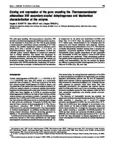
FIG. 1. Expression of the chemorepellent semaphorin III following lesions to distinct parts of the adult CNS. (A) Schematic representation of a horizontal section through the adult rat CNS showing the approximate location of stab wound lesions (gray boxes) performed in the present study. Letters refer to corresponding panels. At different postoperative survival times, in situ hybridization for sema III was performed on horizontal
144
145
Semaphorin III in CNS Regeneration
reviews see, Luo and Raper, 1994; Kolodkin, 1996; Tessier-Lavigne and Goodman, 1996). Recent observations suggest that, in addition to their role during development, recapitulation of these chemorepulsive guidance mechanisms following injury to the adult primary olfactory system is involved in the regeneration of newly formed primary olfactory axons (Pasterkamp et al., 1998b). To further examine a possible role for chemorepulsive signals in CNS regeneration, we evaluated the expression of semaphorin III (D)/collapsin-1 (sema III) following stab wound injuries to the CNS of neonatal and adult rats. Sema III is a secreted member of the semaphorin gene family (Kolodkin et al., 1992, 1993; Luo et al., 1993, 1995; Pu¨schel et al., 1995; Pu¨schel, 1996) and acts as a chemorepellent for subsets of neurons in vitro (Luo et al., 1993; Messersmith et al., 1995; Pu¨schel et al., 1995; Sheperd et al., 1996, 1997; Kobayashi et al., 1997; ValeraEchavarrı´a et al., 1997; Bagnard et al., 1998; Che´dotal et al., 1998; Polleux et al., 1998; Rochlin and Farbman, 1998). Targeted mutation of the sema III gene leads to defects in the trajectories of developing nerve tracts and malformation of several nonneuronal tissues (Behar et al., 1996; Taniguchi et al., 1997). Neuropilin-1 is a high affinity receptor for sema III (He and Tessier-Lavigne, 1997; Feiner et al., 1997; Kolodkin et al., 1997) and is present on sema III-responsive neurons in the developing nervous system (Kawakami et al., 1996). Sema III and neuropilin-1 persist in distinct sets of adult CNS neurons (Luo et al., 1993; Giger et al., 1996, 1998a; Kawakami et al., 1996; Pasterkamp et al., 1998a,b) and ectopic expression of sema III in the cornea of adult rabbits causes repulsion of existing mature sensory fibers (Tanelian et al., 1997). This indicates that sema III is likely to contribute to morphological plasticity of neuritic arbors during adulthood.
Here we report that scar tissue formed after traumatic injury to the adult rodent CNS contains fibroblast-like cells expressing high levels of sema III mRNA. These sema III-expressing cells are observed in scars formed following stab lesions of the lateral olfactory tract (LOT), cortex, and spinal cord and are present in close proximity to severed axon stumps. Neuropilin-1 is expressed in injured neurons that have projections to the lesion site, in a subpopulation of scar-associated cells and in blood vessels within the lesion zone. Interestingly, LOT transection in neonates is followed by successful axonal regeneration (Devor, 1975, 1976; Grafe, 1983; Small and Leonard, 1983; observations in this study) and does not induce sema III mRNA expression at the lesion site. The concomitant expression of sema III and its receptor neuropilin-1 in scar tissue argues that sema III/neuropilin-1-mediated signaling may be involved in adult CNS scar formation and provides the first evidence for a contribution of chemorepellents, like sema III, to the inhibitory effects exerted by glial scars on axonal regeneration in the adult CNS.
RESULTS Semaphorin III mRNA in CNS Scar Tissue The expression of the gene encoding the chemorepellent sema III was studied by in situ hybridization following stab wound lesions to the adult CNS (Fig. 1A). Since different areas of the CNS display a differential cellular response to penetrating injuries (Alonso and Privat, 1993; Hill et al., 1996; Schnell et al., 1997), we first examined the distribution of sema III transcripts following lesions to distinct regions of the CNS, including the lateral olfactory tract (LOT), cerebral cortex, perforant
(B, C; rostral is toward the top) or transverse (D–I; dorsal is toward the top) cryosections through the area of the lateral olfactory tract (LOT; B), the main olfactory bulb (C), the cerebral cortex (D, E), the area including entorhinal cortex, parasubiculum and presubiculum (F, G), and the thoracic spinal cord (H, I). Arrows in B, D, and F point to the lesion site. (B) At 10 days after transection of the LOT, a solid mass of sema III-positive cells occupies the lesioned portion of the LOT. (C) Heavy lesions of the LOT result in a large area of secondary degeneration expanding into the ipsilateral olfactory bulb. At 60 days after lesioning, the glial scar occupying the bulbar cavity is packed with cells expressing high levels of sema III mRNA, presumably fibroblast-like cells (see Fig. 4). Scar tissue in the lesion is encapsulated by sema III mRNA-expressing cells (asterisks). The olfactory nerve layer (onl) of the contralateral, control, bulb is clearly devoid of sema III signals. (D) Stab lesions of the cerebral cortex result in strong expression of sema III transcripts in and around the lesion site at 10 days postlesion. (E) Higher magnification of D showing the presence of sema III-labeled cells within the myelinated external capsule. (F, G) Prominent sema III mRNA expression is associated with scar tissue at the surface of the stab lesion after transection of the angular bundle. (B, F, G) Interestingly, sema III mRNA-positive cells are often found in close association to blood vessels (stars) within the lesion zone. (H) After transection of the dorsal spinal cord, numerous sema III-positive cells are present in and immediately around the injured dorsal roots (asterisk). Strings of sema III mRNA-expressing cells (arrowheads) originating from the injured roots appear to be invading the lesion site in the dorsal portion of the upper thoracic spinal cord. (I) In the intact situation, moderate-to-weak sema III signals (arrowheads) are limited to the leptomeninges covering the spinal cord and dorsal roots (asterisk). dh, dorsal horn; ec, external capsule; lot, lateral olfactory tract; mb, midbrain; onl, olfactory nerve layer. Scale bar, 160 µm (B, E, G, H, I), 625 µm (C), and 390 µm (D, F).
FIG. 2. Time course of semaphorin III mRNA expression following transection of the adult LOT. In situ hybridization for sema III in horizontal sections of the lesion site at 3 days (A), 4 days (B), 10 days (C, D, E, G, H), and 60 days (F) after LOT transection. Rostral is toward the top. (A) At 3 days postlesion, strong sema III hybridization signals are associated with scar tissue at the surface of the stab lesion. The star indicates the LOT.
146
Semaphorin III in CNS Regeneration
pathway and spinal cord. Lesions at all 4 locations induced robust expression of sema III mRNA in spindleshaped non-neuronal cells in the lesion zone. Sema III-positive cells were organized in rounded masses and elongated cords in the lesion, often in close association with the meningeal sheet lining the site of the lesion. In addition, the sema III-positive cells contained a characteristically elongated to ovoid nucleus and were present predominantly in the inner core of the glial scar, especially after long postoperative survival times (Figs. 1B–1H). Stab lesions of the LOT result in damage to CNS parenchyme and in transection of mitral and tufted cell axons that run through the LOT. At 10 days postlesion, a solid mass of sema III-positive cells occupied the lesioned portion of the LOT (Fig. 1B). In addition to relatively small LOT lesions, heavy lesions were performed by extending the knife cut more medially, completely disconnecting the ipsilateral olfactory bulb from the frontal pole of the cortex. This procedure resulted in a large area of secondary degeneration in the ipsilateral bulb. At 60 days postlesion, sema III mRNAcontaining nonneuronal cells had formed a cellular network occupying the bulbar cavity (Fig. 1C). Stab lesions to the cerebral cortex resulted in abundant expression of sema III mRNA around the lesion cavity (Fig. 1D). In situ hybridization revealed a halo of intensely labeled cells in the cortical gray matter immediately adjacent to the lesion site. In addition, a number of sema III-labeled cells was present in the fiber tracts in animals in which the lesion infringed upon the subcortical white matter (Fig. 1E). These sema III-positive cells had an appearance similar to that observed in the injured LOT. Moderate-to-weak sema III mRNA expression was also observed in cortical neurons adjacent to the lesion. Lesions of the perforant pathway, i.e., transection of the angular bundle, disrupt fibers originating from the medial and lateral entorhinal cortex and induce glial scar formation in an area which includes the entorhinal area, parasubiculum and presubiculum (Schauwecker et
147 al., 1995). Scar tissue displayed prominent expression of sema III mRNA at 7 days postaxotomy, especially confined to cells in close association with the pia mater at the border of the lesion site (Figs. 1F and 1G). Following transection of the thoracic spinal cord, restricting damage to the dorsolateral aspect, numerous sema III-positive cells were detected in and around the lesion area (Fig. 1H). Although a faint sema III hybridization signal was present in the leptomeninges covering the dorsal horn and dorsal roots of unlesioned spinal cord (Fig. 1I), a striking induction of sema III mRNA expression was observed in cells in and around dorsal roots injured as a result of the lesion (Fig. 1H). Moreover, strings of sema III-positive cells, formed in association with the injured roots, penetrated the lesioned dorsal horn (Fig. 1H). In all types of lesions, sema III signal was often found in the vicinity of blood vessels in the lesion area (Figs. 1C, 1F, and 1G; see also Figs. 7C and 7E).
Injury-Induced Expression of Semaphorin III mRNA Persists for up to 2 Months after Injury To study the temporal expression profile of sema III mRNA in the scar, sema III mRNA expression was studied following unilateral transection of the LOT at 1, 3, 4, 7, 10, 25, 30, and 60 days postlesion (Figs. 2 and 4). As early as 1 day after lesioning, a significant increase in the intensity of sema III hybridization signals in meningeal cells at the margin of the lesion relative to cells lying within undamaged areas of the meningeal sheet was observed (data not shown). By 3 days postlesion, the intensity of sema III labeling in nonneuronal cells at the surface of the stab lesion had further increased (Fig. 2A). Numerous sema III-positive cells accumulated in the meningeal sheet and started to migrate into the lesion site between 3 and 7 days postlesion. In many instances, sema III mRNA-expressing cells had already extended into the lesion site at 4 days (Fig. 2B). Sections processed with sense probe failed to label any scarrelated cells (Fig. 2I). By 10 days, numerous strings and
(B) By 4 days, sema III-labeled cells appear in the lesion site. The open arrows indicate the lesion. (C, D) At 10 days postlesion, strings and clusters of sema III-positive cells, occasionally continuous with the meningeal sheet (arrowheads in C), occupy the lesion site. Elevated expression of sema III messenger extends distantly into the meningeal sheet adjacent to the site of injury (arrows in D). (E) Higher magnification of boxed area in D showing sema III-labeled cells in the lesion site in close proximity to the leptomeninges (asterisk). (F) Subsets of moderately and strongly labeled cells persist in the inner core of the lesion until at least 60 days after lesioning. (G) Heavy lesions of the LOT damage the connections between the olfactory bulb and the CNS and induce extensive scar formation in the ipsilateral (right) bulb. At 10 days postlesion, sema III transcripts are confined to rounded masses and elongated cords of cells filling up the injured bulb. (H) Higher magnification of the lesion core in G (indicated by an asterisk) showing strings of intensely labeled sema III-positive cells containing a characteristic elongated to ovoid nucleus. (I) Consecutive section hybridized with sense probe. ml, mitral cell layer. Scale bar, 390 µm (A, F), 625 µm (B, D), 160 µm (C), 62.5 µm (E, H, I), and 1600 µm (G).
148 patches of sema III-labeled cells, occasionally continuous with the pia mater, were present in the lesion (Fig. 2C). Elevated expression of sema III messenger was not restricted to the lesion area but extended for several hundreds of microns into the leptomeninges adjacent to the lesion (Fig. 2D). Sema III cells were also observed in the CNS parenchyme in close association with the meningeal sheet, even at some distance from the lesion site (Fig. 2E). Clusters of positive cells were occasionally grouped in the tissue immediately adjacent to the lesion (Figs. 4A–4C). The intensity of the staining in individual cells and the density of the sema III-positive cells appeared similar until 30 days postlesion (Fig. 4). By 60 days postlesion, the longest survival time examined, the number of sema III mRNA-positive cells had somewhat decreased as compared with 30 days. However, at 60 days postlesion weakly and strongly labeled cells were still clearly detectable and intermixed in the scar (Fig. 2F). As described above, heavy lesions of the LOT induced robust expression of sema III mRNA in the ipsilateral olfactory bulb. At 10 days postlesion, a large number of sema III-positive cells appeared in the degenerating bulb, while at 30 and 60 days a cellular network of sema III cells fully occupied the bulbar cavity (Figs. 1C, 2G, 2H, 7C, and 7E).
Semaphorin III mRNA Is Expressed by Fibroblast-like Cells in the Intact and Injured CNS To determine the identity of the sema III-positive cells in intact leptomeninges and during CNS scar formation, sema III mRNA-positive cells were characterized with a series of immunohistochemical markers. The results are illustrated in Figs. 3 and 4 and summarized in Table 1. In the intact CNS, relatively strong mRNA expression for sema III was found in meningeal cells lining the adult olfactory bulb, whereas meningeal expression of sema III decreased to moderate-to-weak levels in more caudal regions of the nervous system (Figs. 3A and 3B; Giger et al., 1998a). To characterize the sema III-positive nonneuronal cells in undamaged nervous tissue we therefore used the olfactory bulb. Consecutive sections through the bulb were either subjected to in situ hybridization for sema III or immunostained with antibodies for GFAP, vimentin, laminin, fibronectin, or ED-14 (Fig. 3). In the intact bulb, immunoreactivity for glial fibrillary acidic protein (GFAP), an intermediate filament protein found in astrocytes, was present in astrocytes scattered throughout the olfactory nerve and glomerular layers and in the glia limitans, leaving the meningeal sheet unstained (Figs. 3C and 3D). Antibod-
Pasterkamp et al.
ies to vimentin, another intermediate filament protein, stained the leptomeninges and astrocytic processes of the glia limitans. Consistent with previous observations, the walls of blood vessels and primary olfactory axons were also vimentin-positive (Figs. 3E and 3F; Schwob et al., 1986). Laminin immunoreactivity was associated with the basal lamina in between the glia limitans and the leptomeninges and around blood vessels. In addition, patchy laminin-like staining was observed in sema III mRNA-positive meningeal cells (Figs. 3G and 3H). The expression pattern of fibronectin was very similar to that of laminin, with the exception of a subset of cells in the meningeal sheet, which was strongly fibronectin immunoreactive (Figs. 3I and 3J). ED-14 and ED-15, antibodies raised against the fibrotic component of peripheral lymphoid organs (Van den Berg et al., 1989), strongly labeled meningeal cells, basal lamina, and subsets of blood vessels (Figs. 3K and 3L). Staining of the pia mater was also found with antibodies against type IV collagen and p75 (Table 1 and data not shown). Taken together, the results indicate that cells expressing sema III mRNA in the intact leptomeninges correspond to fibroblast-like meningeal cells. To identify the cellular source of sema III transcripts in CNS scar tissue and to determine its relationship to other cellular components of the scar, a series of established immunohistochemical markers for neurons, astrocytes, oligodendrocytes, fibroblasts, and extracellular matrix proteins was used in combination with in situ hybridization for sema III (Fig. 4 and summarized in Table 1). Unfortunately, a number of antigens, i.e., ED-15, Gal-C, type IV collagen, OX-6, OX-8, OX-42, were destroyed by the harsh conditions of the in situ hybridization procedure. Therefore, staining with antibodies recognizing these particular antigens was performed on sections adjacent to ones subjected to in situ hybridization. Stab lesions to the LOT induced a vigorous astrocytic reaction. Strong GFAP immunoreactivity was present in a halo of astrocytes around the lesion and in the astrocytes of the LOT. At 30 days postlesion, a rim of highly reactive astrocytes and their interdigitating processes (which form the new glia limitans) delineated the area of tissue damage. Sema III-positive cells lacked GFAP immunoreactivity and most sema III cells were located directly adjacent to the newly formed glia limitans in the core of the scar (Fig. 4A). Clusters of sema III-positive cells in the tissue directly adjacent to the lesion were always encapsulated by reactive astrocytes (Fig. 4B). Consistent with previous reports, laminin immunoreactivity in the newly formed basal lamina demarcated the lesion interface (Fig. 4E; Maxwell et al.,
Semaphorin III in CNS Regeneration
149
FIG. 3. Characterization of semaphorin III mRNA expression in the leptomeninges of adult rat. In situ hybridization for sema III (A, B) and immunohistochemistry for GFAP (C, D), vimentin (E, F), laminin (G, H), fibronectin (I, J), and ED-14 (K, L) in consecutive horizontal sections of the intact rat olfactory bulb. B, D, F, H, J, L are higher magnifications of A, C, E, G, I, K, respectively. Rostral is toward the top. (A, B) Sema III hybridization signals are present in the leptomeninges in between the olfactory bulbs (arrowheads in A). (C–L) Note that whereas GFAP is absent from the meningeal sheet, meningeal cells display varying degrees of vimentin-, laminin-, fibronectin-, and ED-14-immunoreactivity. gl, glomerular layer; onl, olfactory nerve layer. Scale bar, 290 µm (A, C, E, G, I, K) and 115 µm (B, D, F, H, J, L).
FIG. 4. Identification of the cellular source of semaphorin III mRNA in CNS scar tissue. Double labeling combining nonradioactive in situ hybridization for sema III (in purple) and immunohistochemistry (in brown) for GFAP (A, B), laminin (E), fibronectin (H), ED-1 (I), and S-100 (L) in consecutive horizontal cryosections of a lesion site 30 days after a stab wound lesion to the LOT. Double labeling for sema III mRNA and vimentin was performed in a horizontal section at 25 days after LOT transection (F, G). In addition, in case of type IV collagen, in situ for sema III and immunolabeling for collagen type IV were performed in adjacent sections (J, K). Inserts in E, H, I, and K show labeled cells at a high magnification.
150
151
Semaphorin III in CNS Regeneration
TABLE 1 Immunohistochemical Identification of Semaphorin III and Neuropilin-1 mRNA-Expressing Cells in CNS Scar Tissue Formed Following Lesions of the LOT Antibody Neurons RT 97 NF 200 Glial cells GFAP S-100 OX-42 OX-6 Gal-C MBP Blood-derived cells ED-1 OX-8 R-73 ECM and basal lamina components IV Collagen ED-14 ED-15 Fibronectin Laminin Miscellaneous p75 Vimentin
Marker for
Sema III
NP-1
Figure
Reference
Neurons Neurons
— —
— —
6A, B
Anderton et al., 1983 Shaw et al., 1986
Astrocytes Astrocytes, Schwann cells Macrophages, microglia Microglia a Oligodendrocytes, myelin Myelin
— — — — — —
— — nd — — —
4A, B 4L
Bignami and Dahl, 1973 Malhotra et al., 1990 Robinson et al., 1986 Fukomoto et al., 1982 Ranscht et al., 1982 Bartholdi and Schwab, 1998
Macrophages, monocytes a Cytotoxic T-cells, ␥␦T-Cells, NK cells ␣T-Cells
— — —
nd — —
4I
Dijkstra et al., 1985 Brideau et al., 1980 Hu¨nig et al., 1989
Meningeal cells, fibroblasts Fibroblasts Fibroblasts Meningeal cells, fibroblasts a Meningeal cells, astrocytes
⫹ ⫹ ⫹ ⫹ ⫹
⫹ ⫹ ⫹ ⫹ ⫹
4K
4H 4E
Sanes et al., 1990 Van den Berg et al., 1989 Van den Berg et al., 1989 Ruoslahti et al., 1982 Sanes et al., 1990
Meningeal cells, neurons a Meningeal cells, fibroblasts a
⫹ ⫹
⫹ ⫹
4F, G
Junier et al., 1994 Osborn et al., 1984
Note. Sema III, semaphorin III; NP-1, neuropilin-1; ⫹ colocalization/overlap with sema III or neuropilin-1 hybridization signals; — no colocalization/overlap; nd, not determined. a Predominantly labeling the indicated cell types but may occasionally label other cell types as well (see Materials and Methods).
1990). In the lesion, laminin was deposited on a large subset of sema III-labeled cells (Fig. 4E). Whereas a number of cells were strongly labeled, most doublelabeled cells displayed a patchy laminin-like immunoreactivity. Laminin immunoreactivity was also associated with numerous blood vessels in the highly vascularized zone around the lesion (Fig. 4E). Strong vimentin immu-
noreactivity was observed in the lesion and in the adjacent zone of reactive gliosis (Fig. 4F). All sema III-positive cells in the lesion contained vimentin intermediate filaments (Figs. 4F and 4G). Fibronectin and type IV collagen immunoreactivity was found throughout the lesion area and in surrounding blood vessels, as well as more superficially at the pial surface (Figs. 4H
Note that whereas double labeled cells are dark brown (e.g., G), single labeled profiles are either purple (e.g., B) or light brown (e.g., L). (A, B) Numerous GFAP-positive astrocytes delineate the lesion and are situated in close proximity to sema III-positive cells in the lesion core. (B) Note that clusters of sema III-labeled cells (asterisk) are tightly encapsulated by astrocytic processes. Double labeling for sema III mRNA (C) and BrdU (D; same field as C, fluorescein isothiocyanate immunofluorescence) shows that several sema III-positive cells have incorporated BrdU (arrowheads in C and D point to double-labeled profiles), indicating that these cells display mitotic activity. (E) Laminin immunoreactivity is confined to the basal lamina demarcating the lesion interface and to blood vessels in the area adjacent to the lesion. In the lesion, laminin is deposited on a large subset of sema III-labeled cells (arrowheads). (F) Asterisk indicates the lesion cavity. Sema III-positive cells throughout the lesion colocalize with the intermediate filament protein vimentin. (G) Higher magnification showing cells double-labeled for sema III mRNA and vimentin. (H) Fibronectin colocalizes with sema III mRNA in cells associated with the meningeal sheet and lesion site. (I) Although numerous ED-1-positive macrophages have invaded the lesion, no colocalization with sema III transcripts is observed. Insert shows ED-1-positive macrophages (arrowhead) adjacent to a cluster of sema III mRNA-expressing cells. (J, K) Arrowheads indicate that sema III-labeled cells in the lesion (J) clearly correspond to cells expressing type IV collagen in an adjacent section (K). The relatively dark staining of the white matter in J is a nonspecific artifact of the in situ technique. (L) S-100, a marker for astrocytes and Schwann cells, does not colocalize with sema III transcripts. Arrowhead points to a S-100-positive glial cell. lot, lateral olfactory tract. Scale bar, 155 µm (A, E, F, H, I, J, K), 60 µm (C, D), and 50 µm (B, G, L, and inserts in E, H, I, and K).
152 and 4K). Fibronectin antibodies labeled the surface of sema III-positive cells in the lesion and of sema III cells in the intact meningeal sheet (Fig. 4H), whereas type IV collagen immunoreactivity clearly corresponded to the expression of sema III transcripts in the lesion (Figs. 4J and 4K). Immunolabeling with ED-14 and ED-15, two markers for fibrotic tissue (Van den Berg et al., 1989), yielded laminin-like expression patterns, although expression of ED-14 and ED-15 in sema III-positive cells was more abundant as compared to laminin (Table 1 and data not shown). Myelin loss had occurred in a large zone adjacent to the lesion as visualized by the lack of myelin basic protein (MBP) immunoreactivity. In addition, Gal-C-positive oligodendrocytes were absent from the area of sema III expression and were only detected in the degenerated distal portion of the LOT (Table 1 and data not shown). The low-affinity neurotrophin receptor, p75, was coexpressed by most sema III-positive cells (Table 1). Elevated expression of S-100, a molecule expressed by astrocytes and Schwann cells, was restricted to astrocytes constituting the de novo glia limitans. Labeled astrocytes scattered in the area directly adjacent to the lesion displayed control levels of S-100, while S-100 was absent from sema III-positive cells (Fig. 4L). The stab lesions resulted in recruitment of a large number of inflammatory cells into the lesion and surrounding parenchyme. At 30 days postlesion, a dense infiltrate of ED-1- and OX42-positive macrophages and activated microglial cells occupied the lesion site. Although sema III-positive cells were intermingled with numerous ED-1-positive cells, no double-labeled cell profiles were observed (Fig. 4I and Table 1). In adjacent sections immunolabeling with OX-6, OX-8, and R73 antibodies was performed (see Table 1 and data not shown). At 30 days postlesion, abundant MHC class II expression (labeled with antibody OX6) was present in the lesion zone but did not correspond to the expression of sema III messenger. An appreciable number of T-lymphocytes, predominantly ␣ T-cells (labeled with antibody R73), had infiltrated the lesion site and surrounding tissue. Their size, distribution, and number did not correspond to that of sema III-positive cells. The progressive increase in the number of cells expressing sema III mRNA after injury suggests that this population of cells may expand by cell division. To examine this possibility, animals were injected with BrdU at 2 days after lesioning and were analyzed 8 days later. At 10 days postlesion, numerous BrdU-positive profiles were present in and around the lesion, whereas only a few BrdU-positive cells were present in contralat-
Pasterkamp et al.
eral undamaged CNS parenchyme. Double labeling revealed that the nuclei of a number of sema III mRNA-containing cells in the lesion area had incorporated BrdU (Figs. 4C and 4D), indicating that these cells indeed display mitotic activity.
Neuronal Expression of Neuropilin-1 mRNA Is Unchanged Following Transection of the LOT Mitral and tufted cells in the intact mature olfactory bulb express the sema III receptor neuropilin-1. To examine whether lesion-induced changes occur in the expression of neuropilin-1 following axotomy of mitral and tufted cell axons, neuropilin-1 mRNA expression was studied after transection of the LOT (Fig. 5). Expression of B-50/GAP-43 mRNA was employed as a marker for injured mitral cells, since induction of this growthassociated protein in mitral cells has been reported following LOT transection (Verhaagen et al., 1993). Four and 10 days after LOT transection, B-50/GAP-43 mRNA signals were clearly elevated in ipsilateral axotomized mitral cells as compared with the contralateral control bulb (Figs. 5A and 5B). Messenger expression for neuropilin-1 was unchanged in mitral and tufted cells (Figs. 5C and 5D). At 30 and 60 days postaxotomy, B-50/GAP-43 mRNA expression had returned to preaxotomy levels in line with previous observations (Verhaagen et al., 1993). Although a number of mitral and tufted cells had degenerated at these longer postoperative time intervals (Verhaagen et al., 1993), neuropilin-1 mRNA expression in the remaining mitral and tufted cells resembled control (data not shown).
Relationship between Injured Axon Endings and Semaphorin III mRNA Expression Following transection of the LOT, injured adult mitral and tufted cell axons are unable to penetrate the glial scar (Devor, 1975, 1976; Grafe, 1983; Sijbesma and Leonard, 1986). To visualize regenerating LOT fibers in relation to the scar, we initially performed immunohistochemistry using both affinity-purified neuropilin-1 antibodies (AN-1; Pasterkamp et al., 1998b) and a previously described neuropilin-1 antiserum (Kolodkin et al., 1997). Although mitral and tufted cells constitutively expressed neuropilin-1 mRNA following axotomy (Figs. 5C and 5D), both antibodies failed to label LOT axons. Faint neuropilin-1-immunoreactivity was restricted to layer Ia of the piriform cortex immediately adjacent to the LOT (data not shown), probably reflecting terminal labeling of mitral cell axons as has been observed
Semaphorin III in CNS Regeneration
153
FIG. 5. Neuropilin-1 mRNA expression is unaffected by transection of the lateral olfactory tract. In situ hybridization for B-50/GAP-43 (A, B) and neuropilin-1 (C, D) in horizontal sections through the contralateral (A, C) or ipsilateral (B, D) main olfactory bulb at 10 days after unilateral LOT transection. Note the increased mRNA expression for the growth-associated protein B-50/GAP-43 in axotomized mitral cells as compared with contralateral control neurons (A, B). In contrast, hybridization signals for the sema III receptor neuropilin-1 are unaffected by axotomy (C, D). con, contralateral control side; epl, external plexiform layer; gl, glomerular layer; les, ipsilateral lesioned side; ml, mitral cell layer. Scale bar, 160 µm.
previously for synaptic proteins (Ramakers et al., 1992). In subsequent experiments, we therefore combined immunohistochemistry for the high molecular weight neurofilament subunit protein (NF), which labels mitral and tufted cell axons (Ramakers et al., 1992), with in situ hybridization for sema III. Three and 10 days after injury, the lesion and the distal portion of the LOT contained a considerable amount of NF-immunoreactive debris. At 30 days, most debris had been cleared and NF-positive axons in the proximal portion of the LOT were located close to (30 to 60 µm) the sema III-labeled cells in the lesion (Figs. 6A and 6B). Occasionally, axons grew parallel to the zone of sema III mRNA expression towards the midline, however, these neurites never penetrated the area of the scar occupied by sema III-positive cells (Fig. 6B). No axons were seen in the degenerated distal portion of the LOT (Fig. 6A). The leptomeninges laterally from the NFpositive axons in the LOT also displayed strong sema III signals (Fig. 6A). At 60 days postlesion, the density of NF-positive axons had somewhat decreased, probably due to mitral and tufted cell degeneration, but an appreciable number of axons remained present in close association with the lesion and were never found to cross the sema III-positive core of the lesion.
Developmental Changes in Semaphorin III Expression in Response to Injury In contrast to abortive regenerative sprouting of mature LOT axons (Figs. 6A and 6B; Devor, 1975, 1976),
transection of the LOT within the first week after birth is followed by a substantial regrowth of injured axons (Devor, 1975, 1976; Grafe, 1983; Small and Leonard, 1986). Several studies have provided evidence that developmental changes in the cellular response to injury largely account for the differential capacity of neonatal and mature olfactory axons to regenerate through an injury site (Grafe, 1983; Friedman and Aguayo, 1985; Sijbesma and Leonard, 1986). To examine the expression of sema III in the context of these developmental changes, 3-day-old pups (P3) were subjected to LOT lesions and the regeneration of mitral and tufted cell axons was studied in relation to sema III mRNA expression in the scar. At 1, 2, and 4 days after axotomy, an increased number of GFAP-positive astrocytes was present in the lesion site and surrounding tissue (Figs. 6C and 6D). The extent of the astrocytic reaction, however, was considerably smaller as compared with adult lesions (compare Figs. 4A and 4B to 6C and 6D). Following neonatal lesions, elevated sema III mRNA expression was restricted to the leptomeninges laterally from the lesion site (Figs. 6C and 6D). In contrast to adult lesions, no sema III messenger expression was observed in cells in the lesion at any of the postlesion time intervals examined (Figs. 6C–6I). By 10 days postlesion, a few astrocytes demarcated the lesion. Sema III mRNA expression in the meningeal sheet had decreased to control levels (Fig. 6E). A small number of NF-positive axons penetrated the lesion site and extended into the undamaged
FIG. 6. Relationship between regenerating olfactory axons and semaphorin III mRNA expression following LOT lesions in neonatal and adult rats. Double labeling combining in situ hybridization for sema III (in purple) and immunohistochemistry (in brown) for neurofilament (A, B, F, H, I) or GFAP (C, D, E, G) in horizontal sections through the lesion site following adult (A, B) or neonatal (C–I) transection of the LOT. Rostral is towards the top. Open arrows in A, C, D, E, and G point to the lesion. (A) At 30 days after transection of the LOT during adulthood (A30),
154
Semaphorin III in CNS Regeneration
CNS parenchyme distal to the lesion (Fig. 6F). At 30 and 60 days postlesion, the lesion site had largely disappeared and GFAP immunoreactivity in the lesion zone had further decreased (Fig. 6G). In addition, numerous NF-positive fibers had passed the lesion and were present in the region caudal to the lesion, although these regenerating fibers were not organized in a discrete bundle as observed in the intact situation (Figs. 6H and 6I).
The Semaphorin III Receptor Neuropilin-1 Is Expressed in CNS Scar Tissue When we examined the expression of neuropilin-1 messenger in axotomized mitral cells (Figure 5), we also observed nonneuronal mRNA expression for this sema III receptor at the site of the lesion. Neuropilin-1 transcripts were present in cells in the lesion and in cells associated with blood vessels surrounding the lesion site, both in close proximity to sema III-positive fibroblast-like cells (Figs. 7A–7F). In situ hybridization revealed that a small subset of scar-associated cells displayed moderate-to-weak neuropilin-1 signals. At 25 days after lesioning, the meningeal sheet contained a small number of neuropilin-1-positive cells, whereas most cells were grouped in small strings and clusters just beneath the pial surface at the border of the lesion (Fig. 7B). The intensity and density of neuropilin-1 labeling was highest between 1 and 3 weeks postlesion, while at 60 days postlesion, few neuropilin-1positive cells were detected in the lesion. As observed for sema III mRNA (Figs. 7C and 7E), heavy lesions of the LOT induced robust expression of neuropilin-1 transcripts in a large number of cells in the injury site and the ipsilateral olfactory bulb (Figs. 7D and 7F). To visualize neuropilin-1 protein in these scar-related cells, immunohistochemistry was performed using affinity-
155 purified anti-neuropilin-1 antibodies (AN-1) (Figs. 7G and 7H). Strings and clusters of nonneuronal cells throughout the lesion site displayed moderate-to-weak neuropilin-1 immunoreactivity, consistent with the distribution of neuropilin-1 messenger (Figs. 7G and 7H). Interestingly, neuropilin-1-labeled cells in the lesion were present in the same area as the cells that expressed the neuropilin-1 ligand, sema III (Figs. 7A and 7B). Double labeling combining in situ hybridization for sema III and immunohistochemistry for neuropilin-1 showed colocalization of sema III transcripts and neuropilin-1 protein in a small subset of cells (data not shown). In addition, both sema III-positive/neuropilin1-negative and sema III-negative/neuropilin-1-positive cell profiles could be detected in the lesion site (data not shown). To identify the cellular source of neuropilin-1 in CNS scar tissue, a series of established immunohistochemical markers was used in combination with in situ hybridization for neuropilin-1. No neuropilin-1 mRNA was found in association with astrocytes, oligodendrocytes, and lymphocytes (Table 1). However, neuropilin-1 messenger was present in a subset of cells exhibiting the same immunohistochemical profile observed for cells expressing sema III mRNA (Table 1). Thus, both sema III and neuropilin-1 mRNA appear to be expressed by fibroblast-like cells in the scar. One of the cellular responses associated with penetrating injuries is the neovascularization of the lesion zone (see for example Fig. 4E). Following LOT transection, we observed a large number of blood vessels, predominantly at the interface of the undamaged CNS parenchyme. As mentioned above, blood vessels at the lesion interface were often surrounded by sema III mRNApositive cells (Figs. 1C, 1F, 1G, 7C, and 7E). In addition, neuropilin-1 mRNA-expressing cells were observed as well around blood vessels (Figs. 7D and 7F). Whereas no sema III hybridization signals were found associated
neurofilament (NF)-positive axons in the proximal portion of the LOT are located in close proximity to the sema III mRNA-expressing cells in the core of the lesion. Strong sema III hybridization signals are also present in the leptomeninges laterally from the LOT (arrowheads). Axons fail to grow across the lesion site and are not found in the degenerated distal portion of the LOT (asterisk). (B) Higher magnification of A showing that NF-positive axons are located at some distance from the sema III-positive lesion core. Occasionally, axons grow parallel to sema III-labeled cells in the lesion site (arrow). (C, D) At 2 and 4 days after transection of the LOT at postnatal day 3 (N2 and N4), a moderate astrocytic reaction can be observed in the lesion site. Sema III messenger is absent from cells in the lesion site. However, elevated sema III mRNA expression is observed in the leptomeninges lining the lesion (arrowheads). (E) By 10 days (N10), GFAP-immunoreactivity in the lesion has decreased and sema III mRNA expression in the meningeal sheet has returned to moderate-to-weak control levels (arrowheads). The dashed lines in F, H, and I indicate the lesion site. (F) Section adjacent to one shown in E. A number of regenerating NF-positive axons has started to penetrate the lesion zone. (G) At 30 days after neonatal transection (N30), a few remaining GFAP-positive astrocytes mark the lesion site. (H, I) Section adjacent to the one shown in G. Numerous axons have penetrated the lesion and grow into the undamaged CNS parenchyme distal to the lesion (arrow in H). Sema III mRNA expression is confined to the leptomeninges (arrowhead in H) and to neurons in the primary olfactory cortex (arrowheads in I). lot, lateral olfactory tract. Scale bar, 160 µm (A, D, E, F, I), 62.5 µm (B), and 390 µm (C, G, H).
FIG. 7. Expression of the semaphorin III receptor neuropilin-1 in CNS scar tissue. In situ hybridization for sema III (A, C, E) and in situ hybridization (B, D, F) or immunohistochemistry (G–I) for neuropilin-1 in horizontal sections through a lesion site at 30 days after a normal (A, B, I) or heavy (C–H) LOT lesion in adult rat. (A, B) Rostral is toward the top and the asterisk indicates a lesion cavity. The population of cells
156
Semaphorin III in CNS Regeneration
with blood vessels, in situ hybridization for neuropilin-1 clearly labeled cells within the blood vessel wall (Figs. 7D and 7F). In addition, strong neuropilin-1 immunoreactivity was observed on blood vessels surrounding the lesion site (Figs. 7G–7I). In the undamaged CNS parenchyme blood vessels only displayed weak neuropilin-1 immunoreactivity.
DISCUSSION The data presented here demonstrate that following penetrating injuries to the adult CNS the gene encoding the chemorepellent sema III is expressed by fibroblastlike cells in the core of the glial scar. Interestingly, the sema III receptor neuropilin-1 is expressed in a subset of scar-associated cells as well and in blood vessels surrounding the lesion. Following LOT lesions, axons of mitral and tufted cells, which constitutively express neuropilin-1 messenger after axotomy, do not grow across the sema III-positive lesion center. In contrast to adult lesions, however, following early neonatal LOT transection vigorous regrowth of injured olfactory axons occurs in the absence of sema III mRNA expression in the lesion site. These results suggest that following penetrating brain injuries scar-associated expression of the chemorepellent sema III may contribute to the repulsive characteristics of neural scar tissue known to prevent axonal regeneration in the adult CNS. Furthermore, the concomitant expression of sema III and neuropilin-1 in CNS scar tissue suggests a role for semaphorin/neuropilin-mediated signaling in glial scar formation. The glial scar is a complex mixture of astroglial cells, blood-borne cells, mesodermal cells, and other cells. The present data indicate that fibroblast-like cells are the cellular source of sema III transcripts in the scar. Double labeling showed that astroglial and blood-borne cells do not express sema III mRNA. In addition to astrocytes and blood-borne cells, Schwann cells may penetrate the
157 injury site. Previous studies, however, have shown that Schwann cells do not express sema III mRNA during development, adulthood, or following peripheral nerve injury (Wright et al., 1995; Pu¨schel et al., 1996; Giger et al., 1996, 1998a; Pasterkamp et al., 1998a). In addition, following penetrating injuries to the CNS, sema IIIpositive cells did not express the Schwann cell marker S-100. Furthermore, sema mRNA colocalizes with the fibroblast markers ED14 and ED15 while Schwann cells do not express these proteins in the intact or injured situation (R. J. Pasterkamp and J. Verhaagen, unpublished observations). Sema III transcripts, however, colocalized with several proteins expressed by fibroblastlike cells, including vimentin, fibronectin, laminin, type IV collagen, and p75 (Raff et al., 1979; Kru¨ger et al., 1986; Rutka et al., 1986; Maxwell et al., 1990; Matthiessen et al., 1991; Colombo et al., 1994; Li and David, 1996; DeGiorgio et al., 1997; Frise´n et al., 1998). The spindle-shaped appearance and elongated nucleus of sema III mRNApositive cells, as well as their position directly externally to the glia limitans and their close association with blood vessels, corresponds well to previous observations on the properties of fibroblasts of meningeal origin in scar tissue (Matthews et al., 1979; Carbonell and Boya, 1988; Maxwell et al., 1990; Li and David, 1996). Furthermore, the organization of cells expressing sema III messenger in rounded masses or elongated cords is strikingly similar to that observed for fibroblasts and meningeal cells both in in vitro studies and in vivo after stab injury or in meningiomas (Rubinstein, 1972; Matthews et al., 1979; Krikorian et al., 1981; Rutka et al., 1986; Carbonell and Boya, 1988; Kobata et al., 1998). The observation that, like fibroblast-like cells, sema IIIlabeled cells penetrate the lesion within the first days after lesioning to become a permanent constituent of the inner core of the scar provides further evidence for a fibroblast-like phenotype (Berry et al., 1983; Kru¨ger et al., 1986; Carbonell and Boya, 1988; Maxwell et al., 1990; Ajemian et al., 1994). Although fibroblasts can be derived from different sources following injury, the lepto-
expressing messenger encoding the sema III receptor neuropilin-1 (B) clearly overlaps with sema III-positive fibroblast-like cells in the lesion site (A). Inset in A and B shows a blow up of sema III and neuropilin-1 mRNA-expressing cells, respectively. (C, D) Rostral is to the right. At 30 days after a heavy stab lesion to the LOT, the ipsilateral bulb is filled with numerous sema III (C) and neuropilin-1-positive (D) cells. Large-diameter blood vessels are formed in response to the injury and are located predominantly at the interface of the olfactory nerve layer of the undamaged contralateral bulb (onl). Blood vessels are surrounded by strings and patches of sema III and neuropilin-labeled cells. (E, F) Higher magnification of C and D, respectively. Note that whereas blood vessels are devoid of sema III hybridization signals, in situ for neuropilin-1 clearly labels cells in the blood vessel wall (open arrows). (G, H) Immunohistochemistry for neuropilin-1 at two distinct sites in a section adjacent to the one shown in F. Numerous cells in the lesion display neuropilin-1 immunoreactivity (arrowheads). In addition, blood vessels throughout the lesion zone are neuropilin-1-positive (asterisks). Arrows in H point to small-sized cells associated with the wall of a large diameter blood vessel (asterisk) at the lesion interface. (I) Section adjacent to the one shown in B. Numerous neuropilin-1 blood vessels (arrowheads) are present in the area adjacent to the lesion (star in B). Scale bar, 160 µm (A–D) and 62.5 µm (Inset A and B, E–I).
158 meninges, i.e., pia and arachnoid mater, is a major source for these nonneuronal cells (Ross et al., 1968; Tobin et al., 1980; Krikorian et al., 1981; Kru¨ger et al., 1986; Carbonell and Boya, 1988; Maxwell et al., 1990). The progressive development of sema III-labeled scar tissue from the pial surface of the lesion into deeper regions suggests that sema III-positive cells have a meningeal origin and is consistent with the presence of sema III transcripts in fibroblast-like cells in both the intact and injured leptomeninges. Noticeably, no sema III-positive cells were found in the lesion site following early neonatal lesions. This is consistent with the inability of fibroblasts to penetrate neonatal lesions (Berry et al., 1983). Fibroblasts and associated connective tissue are thought to be essential for appropriate wound healing (Ross et al., 1968; Tobin et al., 1980; Ehrlich, 1988). Following CNS injury, mesodermal elements (e.g., fibroblasts, meningeal cells) and astrocytes cooperate in the formation of a new glia limitans and concomitant basal lamina thereby preventing further spread of damage (Abnet et al., 1991; Sievers et al., 1994; Li and David, 1996; Ness and David, 1997). Soluble factors released by meningeal cells cause astrocytic alterations rendering them less permissive for axon growth, whereas astrocyteconditioned medium can induce process formation in meningeal cells (Colombo et al., 1994; Ness and David, 1997). The precise mechanisms underlying these molecular interactions are unknown, but are likely to include factors released by scar-associated cells, like sema III. Although most studies on the function of sema III have focused on its chemorepulsive effects on developing neurites, data obtained from sema III null mutants suggests a role for this glycoprotein in the regulation of the differentiation and growth of several nonneuronal tissues (Behar et al., 1996). Sema III binds to T cell, B cell, macrophage, and mast cell lines, suggesting sema III to be involved in immune function as well (Xu et al., 1998). In addition, chemorepellents have been shown to participate in the regulation of cellular migration (Hu and Rutishauser, 1996). These data suggest that sema III released from the fibrotic core of the scar may, in addition to its putative chemorepulsive effects, contribute to the regulation of migration, proliferation, or differentiation of sema III-responsive cells in the lesion zone. In view of this it is interesting that a subset of scar-related cells expresses a receptor for sema III, neuropilin-1, and is therefore potentially responsive to sema III. In addition, astrocytes and oligodendrocytes express CRMP-2, a cytoplasmic protein essential for sema III functioning (Goshima et al., 1995), in the intact situation (Kamata et al., 1998), while CRMP-2 is persis-
Pasterkamp et al.
tently expressed following CNS stab injuries (R. J. Pasterkamp and J. Verhaagen, unpublished observations). It is conceivable that these CRMP-2-positive cells express additional semaphorin receptors, such as neuropilin-2 or VESPR/plexin (Chen et al., 1997, 1998; Kolodkin et al., 1997; Comeau et al., 1998; Giger et al., 1998b; Winberg et al., 1998), rendering them responsive to semaphorin-mediated signaling. After penetrating injuries to the CNS, neovascularization occurs in the tissue surrounding the lesion (Beggs and Waggener, 1979; Imperato-Kalmar et al., 1997). This angiogenic response involves complex interactions between factors present in and around blood vessels (Clark, 1993). The abnormal development of the cardiovascular system of neuropilin-1 null mutant mice suggests a role for neuropilin-1 in angiogenesis (Kitsukawa et al., 1997). In the present study, neuropilin-1 mRNA and protein were detected in blood vessels in and around the lesion. Recently, neuropilin-1 has been identified as a (co-) receptor for vascular endothelial growth factor (VEGF; Soker et al., 1998) and placenta growth factor-2 (PlGF-2; Migdal et al., 1998), while tumor necrosis factor-␣ (TNF-␣) was shown to regulate endothelial neuropilin-1 expression (Giraudo et al., 1998). The concomitant expression of these angiogenic factors and neuropilin-1 in CNS scar tissue (Taupin et al., 1993; Bartholdi et al., 1997; and present study) argues that neuropilin-1 is a crucial component of the injuryinduced angiogenic response. Neuropilin-1-immunoreactive blood vessels at the lesion interface were often surrounded by sema III-positive fibroblasts. It is therefore tempting to speculate that sema III secreted from these cells contributes, in concert with other angiogenic factors, to the neovascularization of the lesion zone by interacting with neuropilin-1 on developing blood vessels. Although injured mitral and tufted cells are intrinsically capable of regenerating their injured axons, inhibitory influences associated with the glial scar prevent them from elongating axons beyond the lesion site (Devor, 1975, 1976; Grafe, 1983; Friedman and Aguayo, 1985; Sijbesma and Leonard, 1986). Several studies have demonstrated putative neurite outgrowth inhibitory molecules in association with the scar, including myelinassociated proteins and ECM proteins (Caroni and Schwab, 1988; Rudge and Silver, 1990; Smith et al., 1990; Snow et al., 1990; McKeon et al., 1991; Laywell et al., 1992; Bovolenta et al., 1993; Pindzola et al., 1993; Schwab et al., 1993; Levine, 1994; Mukhopadhyay et al., 1994; Gates et al., 1996; Davies et al., 1997; Zhang et al., 1997). Additional inhibitory molecules may include recently identified repellents, such as members of the netrin, ephrin
159
Semaphorin III in CNS Regeneration
and semaphorin gene families. During development these molecules are involved in constraining axons to their correct pathways (Tessier-Lavigne and Goodman, 1996) and thus retention or injury-induced recapitulation of their expression may constitute repulsive barriers for regenerative axonal growth as well. Here we show that penetrating injuries to the adult CNS induce robust mRNA expression of the developmentally important chemorepellent sema III in the scar. Since there are currently no antibodies available that can detect sema III in tissue sections, the distribution of sema III protein is unknown. However, it has been suggested that following secretion, sema III protein is able to bind to the extracellular matrix (Luo et al., 1993). It is conceivable that following CNS injury sema III forms a chemorepulsive gradient in the newly deposited extracellular matrix of the lesion. Axotomized mitral and tufted cells continue to express neuropilin-1 mRNA, suggesting a sustained sensitivity to sema III, while their severed axons do never regrow across the sema III-positive lesion site. Although early embryonic olfactory bulb neurons are not repelled by sema III in vitro (Sheperd et al., 1996), it is likely that the role of sema III in guidance of olfactory bulb axons is restricted to later stages of development. At early embryonic days no sema III is expressed in the vicinity of the developing LOT, while at later stages target cells throughout the olfactory cortex express sema III (Giger et al., 1996, 1998a). Stagedependent effects of sema III have already been reported in several other neuronal systems (Messersmith et al., 1995; Pu¨schel et al., 1996; Sheperd et al., 1997; Rochlin and Farbman, 1998). Failure of regeneration in the adult CNS contrasts with the vigorous regrowth of axons after injuries to the PNS or neonatal CNS (Ramo´n y Cajal, 1928; Devor, 1975; Kalil and Reh, 1979). This differential regenerative capacity is thought to be largely due to the absence of significant glial scar formation in the injured PNS and neonatal CNS. Stab wound lesions to the neonatal CNS only induce a moderate and transient astrocytic response, while other scar-associated cells such as fibroblasts fail to invade the lesion (Bignami and Dahl, 1973; Berry et al., 1983; Sijbesma and Leonard, 1986; Moore et al., 1987; Trimmer and Wunderlich, 1990). In line with this, no sema III-positive fibroblast-like cells were seen in the lesion site following early neonatal LOT lesions. Interestingly, recent observations show that sema III is also absent from injured peripheral nerve (Pasterkamp et al., 1998a). Together this shows that both peripheral and central neonatal projections regenerate vigorously in the absence of sema III expression at the site of the lesion. The only site of enhanced sema III expression
after neonatal LOT lesion is the meningeal sheet laterally from the lesion. Several studies have shown that the leptomeninges can serve as a conduit for regenerating axons (Bohn et al., 1982; Fawcett, 1985; Risling et al., 1991). Therefore, meningeal sema III expression during the first weeks after neonatal injury may serve to guide sema III-responsive axons through the lesion and may prevent them from leaving the CNS parenchyme. The concomitant expression of sema III and its receptor neuropilin-1 following penetrating injuries to the CNS suggests that sema III/neuropilin-1-mediated mechanisms are involved in scar formation. The expression of the secreted chemorepellent sema III following CNS injury provides evidence that chemorepulsive semaphorins may contribute to the inhibitory effects exerted by scars on the outgrowth of injured CNS neurites. Therefore, the inactivation of chemorepulsive factors, like sema III, following CNS injury may become an essential component of future strategies to stimulate CNS regeneration.
EXPERIMENTAL METHODS Materials The following materials were obtained from the indicated sources: Vectashield and Vectastain ABC kit from Brunschwig (Amsterdam, The Netherlands); Biotinylated anti-mouse IgG (H ⫹ L) and biotinylated antirabbit IgG (H ⫹ L) from Vector Laboratories (Burlingame, CA); Dormicum from Roche Nederland B.V. (Mijdrecht, The Netherlands); Bromodeoxyuridine, 5-bromo-4-chloro-3-indolylphosphate, and 3,38-diaminobenzidine tetraHCl from Sigma (Deisenhofen, Germany); Graefe microknife from Tiemann & Co. (Hauppauge, NY); 2-methylbutane from Merck (Darmstadt, Germany); Hypnorm from Janssen Pharmaceutical Ltd. (Oxford, England); Nembutal from Sanofi Sante (Maassluis, The Netherlands); Superfrost plus slides from Menzel Gla¨ser (Darmstadt, Germany); pQE30 vector and Ni-NTA-agarose column from Qiagen (Hilden, Germany); Nitro-bluetetrazolium chloride, proteinase K, anti-DIG Fab fragment conjugated to alkaline phosphatase, glycerin, monoclonal anti-bromodeoxyuridine antibody, and T3, T7, SP6 polymerases from BoehringerMannheim (Mannheim, Germany); Temgesic from Schering-Plough (Amstelveen, The Netherlands); TissueTek (OCT compound) from Miles Inc. (Elkhart, IN); Isoflurane (Forene; Abbott Laboratories Ltd., Queenborough, UK); Donkey anti-mouse fluorescein isothiocyanate (FITC) from Jackson Immunoresearch Laboratories Inc. (West Grove).
160
Animals In total 120 adult male Wistar rats (225–450 g; Harlan CPB-Zeist, The Netherlands) and 46 Wistar rat pups were used in the present study. The pups were obtained from random-bred females in our colony. Adult female Wistar rats were originally purchased from Harlan CPB-Zeist. Pups remained with their mothers until sacrifice or were weaned at the age of 21 days. Adult animals were housed in group cages, maintained on a 12-h light/12-h dark cycle and food and water were available ad libitum.
Surgical Procedures Lesions in adult rats. All surgical and animal care procedures were carried out according to local guidelines of the experimental animal care committee. Rats were anesthetized for aseptic surgery with Hypnorm (0.04 ml/100 g, i.m.) and Dormicum (0.08 ml/100 g, s.c.). Buprenorphine hydrochloride (Temgesic; 0.03 ml/100 g, s.c.) was given postoperatively. Four areas were lesioned encompassing both brain and spinal cord: (1) transection of the lateral olfactory tract, (2) stab wound lesion of the cerebral cortex, (3) transection of the perforant pathway, and (4) transection of the thoracic spinal cord (Fig. 1A); (1) Transection of the lateral olfactory tract (LOT) was performed as described previously (Verhaagen et al., 1993). Briefly, a small groove was made in the orbital surface of the left frontal bone and a knife was lowered into the right side of the groove until it touched the base of the skull. Subsequently, the knife was moved from right to left transecting the LOT. Care was taken to avoid damage to the frontal pole of the cortex. Animals were sacrificed at 1 (n ⫽ 4), 2 (n ⫽ 5), 3 (n ⫽ 7), 4 (n ⫽ 13), 7 (n ⫽ 3), 10 (n ⫽ 15), 25 (n ⫽ 12), 30 (n ⫽ 13), and 60 days (n ⫽ 8) postlesion. Postmortem inspection of the lesion revealed that this procedure resulted in complete transection of the LOT. In a number of animals more extensive lesions were produced by extending the knife cut more medially towards the midline. Animals were sacrificed at 4 (n ⫽ 3), 10 (n ⫽ 4), 30 (n ⫽ 3), and 60 days (n ⫽ 4). (2) Unilateral stab lesions of the cerebral cortex were made by inserting a Graefe microknife along the mediolateral axis of the frontal cerebral cortex. Animals (n ⫽ 4) were sacrificed following a survival time of 10 days. (3) Stereotaxic lesions of the perforant pathway were performed according to Schauwecker et al. (1995). Animals (n ⫽ 3) were allowed to recover and were sacrificed at 7 days postlesion. (4) Transection of the dorsolateral spinal cord was performed at thoracic 11. After the dura
Pasterkamp et al.
was opened, the dorsal funiculus of the spinal cord was transected with microsurgical scissors. The animals were allowed to recover and were sacrificed at 14 days postlesion (n ⫽ 3). Lesions in neonatal rats. A unilateral transection of the LOT was performed at postnatal day 3 (P3). Pups were anesthetized with isoflurane and a mixture of oxygen and nitrous oxide (1:2). A small opening was made in the orbital surface of the left frontal bone and the LOT was cut using a microknife, taking care not to cause unnecessary additional damage. After surgery pups were returned to their mother and were sacrificed at 1 (n ⫽ 2), 2 (n ⫽ 3), 4 (n ⫽ 9), 10 (n ⫽ 9), 30 (n ⫽ 9), and 60 days (n ⫽ 2) postlesion. The location of the lesion was identified by postmortem visualization of the scar using immunohistochemistry for GFAP (visualizing the astrocytic reaction) or Nissl counterstaining in adjacent sections. The data from four pups were discarded because the lesions were at the wrong location. In all lesion experiments the unoperated side served as a control. In addition, age-matched unoperated animals were used to analyze the normal expression patterns of the molecules studied here [adult rats (n ⫽ 15) and neonates (n ⫽ 2 at each postlesion time interval)].
Tissue Preparation At the appropriate postoperative survival time, animals were deeply anesthetized with Nembutal (0.125 ml/100 g (adult rats) and 0.30 ml/100 g (pups); i.p.), exsanguinated by transcardial perfusion with ice-cold 0.1 M phosphate-buffered saline (pH 7.4, PBS), and fixed by perfusion with 4% paraformaldehyde (PFA) in icecold 0.1 M PBS. Perfusion pressure was adjusted to the age of the animal. The brains were dissected and postfixed overnight in 4% PFA at 4°C. The tissue was then immersed in a solution containing 0.01 to 0.15 M EDTA (depending on the age of the animal) and 0.1 M phosphate buffer (pH 7.4, PB) for 8 h at 4°C to enhance tissue penetration, cryoprotected in 10% and 25% sucrose in 0.1 M PB (both overnight at 4°C), covered with Tissue Tek (OCT compound), and rapidly frozen in dry-ice-cooled 2-methylbutane. For neuropilin-1 immunohistochemistry rats were perfused with ice-cold PBS followed by PLP fixative (2% PFA, 0.075 M L-lysine, 0.214% sodium metaperiodate) in PB. Following overnight postfixation in the same fixative, brains were cryoprotected and frozen. A number of animals was sacrificed by an intraperitoneal injection of a lethal dose of Nembutal (0.25 ml/100 g; i.p.) and decapitation. Brains were rapidly dissected,
161
Semaphorin III in CNS Regeneration
embedded in Tissue-Tek (OCT compound), and immediately frozen in dry-ice-cooled 2-methylbutane. Consecutive horizontal or transverse cryosections (5, 10, or 20 µm) were cut at ⫺15°C (unfixed tissue) or ⫺20°C (fixed tissue), collected onto microscope slides, air dried, and used for nonradioactive in situ hybridization, immunohistochemistry, or double labeling combining in situ hybridization and immunohistochemistry.
In Situ Hybridization Digoxigenin (DIG)-labeled cRNA probes, message complementary (antisense), and noncomplementary (sense), were generated from completely linearized cDNA template using the appropriate RNA polymerases (SP6, T3, or T7). The following cDNA templates were used: rat semaphorin(D)III/collapsin-1 (entire coding region (Giger et al., 1996)), rat B-50/GAP-43 (entire coding region, a gift from Dr. L. H. Schrama, Rudolf Magnus Institute for Neurosciences, Utrecht, The Netherlands) and the extracellular domain of rat neuropilin-1 (nucleotides 181–2593, a gift from Dr. A. L. Kolodkin, Johns Hopkins School of Medicine). To enhance tissue penetration and avoid nonspecific background, the cRNA probes were adjusted to an average length of 100–200 bases by limited alkaline hydrolysis (SchaerenWiemers and Gerfin-Moser, 1993). Nonradioactive in situ hybridization was performed as described previously (Giger et al., 1996) with minor modifications. Briefly, cryostat sections obtained from unfixed tissue were fixed in 4% PFA in 0.1 M PBS before acetylation with 0.25% acetic anhydride in 0.1 M triethanolamine. To perform in situ hybridization on perfused tissue, sections were pretreated at room temperature (RT) with proteinase K for 10 min (10 µg/ml and 0.1% Triton X-100), subsequently fixed in freshly prepared 4% PFA and acetylated. One hundred-and-fifty microliters of hybridization mixture (containing 50% formamide, 5⫻ Denhardt’s solution, 5⫻ SSC, 250 µg/ml bakers yeast tRNA, 500 µg/ml sheared, heat-denatured herring sperm DNA, and 200 ng/ml of the appropriate cRNA probe) were applied per slide, slides were sealed and incubated overnight in a humidified chamber at 55°C (sema III and B-50/GAP-43) or 60°C (neuropilin-1). Binding of DIG-labeled RNA hybrids with an anti-DIG Fab fragment conjugated to alkaline phosphatase was followed by the color reaction with nitro-bluetetrazolium chloride and 5-bromo-4-chloro-3-indolylphosphate as substrates. Controls, consisting of sections subjected to the complete in situ hybridization procedure, but with no probe added or hybridized with corresponding sense probe, exhibited no specific hybrid-
ization signal. Following photodocumentation, some sections were also processed for immunohistochemistry with different antibodies (see below).
Neuropilin-1 Antibody Production Anti-neuropilin-1 antibodies (AN-1) were produced as described by Kolodkin et al. (1997). A fragment of rat neuropilin-1, corresponding to amino acids C583-I856 was cloned in the BamHI and HindIII sites of the pQE30 vector, which was subsequently used to produce 6-histidine-tagged neuropilin-1 fragments in Escherichia coli. Rabbits were immunized with approximately 0.5 mg of protein in complete Freunds adjuvant and two times boosted in incomplete Freunds adjuvant. Anti-neuropilin-1 antibodies were affinity purified on a neuropilin-1 protein immunosorbent column according to a method described by Oestreicher et al. (1983). In an attempt to visualize regenerating LOT fibers we used affinity-purified neuropilin-1 antibodies (AN-1; Pasterkamp et al., 1998b) and a previously described IgG-enriched neuropilin-1 antiserum (Kolodkin et al., 1997).
Antibodies Sections were immunolabeled with the following mouse monoclonal antibodies: ED-1 (specific for macrophages, monocytes and activated microglia; 1:1,000; Serotec, Oxford, UK), OX-42 (specific for macrophages, microglia and granulocytes; 1:300; a gift from Dr. I. Huitinga, Netherlands Institute for Brain Research, Amsterdam, The Netherlands), ED-14 and ED-15 (specific for connective tissue; both 1:250; a gift from Dr. T. Van den Berg, Vrije Universiteit, Amsterdam, The Netherlands), OX-6 (specific for MHC class II expression; 1:500; a gift from Dr. C. D. Dijkstra, Vrije Universiteit, Amsterdam, The Netherlands), OX-8 (specific for cytotoxic T-cells, ␥␦T-cells, and NK cells; 1:1,000; Cedarlane Laboratories, Hornby, Canada), R73 (specific for ␣Tcells; 1:50; Serotec), RT97 (specific for the 200-kDa phosphorylated neurofilament subunit; 1:100; Sigma, Deisenhofen, Germany), and antibodies to vimentin (vimentin is an intermediate filament present in a variety of cells, e.g., reactive glial cells, meningeal cells and fibroblasts; 1:20; Boehringer-Mannheim, Mannheim, Germany), galactocerebroside (Gal-C; specific for oligodendrocytes; 1:50; a gift from Dr. G. Wolswijk, The Netherlands Institute for Brain Research, Amsterdam, The Netherlands), p75 (the low-affinity neurotrophin receptor; 1:200; Boehringer-Mannheim, Mannheim, Germany), and type IV collagen (type IV collagen is a major
162 constituent of the basal lamina associated with fibroblasts and meningeal cells; 1:250; Sigma). In addition, we used the following polyclonal antisera: rabbit anti-rat fibronectin (a marker for meningeal cells and fibroblasts; 1:40; Boehringer-Ingelheim, Heidelberg, Germany), rabbit anti-mouse laminin (laminin is a noncollagenous connective tissue glycoprotein and a major constituent of basement membranes; 1:40; Sigma), rabbit anti-rat glial fibrillary acidic protein (GFAP; a marker of mature astrocytes; 1:800; Dako, Glostrup, Denmark), rabbit anti-myelin basic protein (MBP; a major constituent of CNS myelin; 1:200; a gift from Dr. H. van Noort, TNO, Leiden, The Netherlands), affinity-purified rabbit anti-rat neuropilin-1 (AN-1; 1:100 (Pasterkamp et al., 1998b)), IgG-enriched rabbit anti-rat neuropilin-1 (1:2,000; a gift from Dr. A. L. Kolodkin, The Johns Hopkins School of Medicine), rabbit anti-NF-200 (200kDa nonphosphorylated neurofilament subunit protein, a marker for neurons and their processes; 1:100; Sigma), and rabbit anti S-100 (marker for astrocytes and Schwann cells; 1:100; Sigma).
Immunohistochemistry Initially, nonradioactive in situ hybridization was combined with indirect immunofluorescence. However, the dark blue/purple in situ precipitate was found to interfere with the immunofluorescent signals. Therefore, immunocytochemistry was conducted following standard immunohistochemical procedures incorporating the avidin–biotin–peroxidase complex using 3,38diaminobenzidine tetrachloride (DAB) as a chromophore. To confirm colocalization, sema III mRNA-expressing cells were monitored before and after immunohistochemical labeling. In addition, the possibility of colocalization of sema III mRNA and all proteins used here was further examined on consecutive sections. Following in situ hybridization, sections were rinsed in Tris-buffered saline (TBS; pH 7.4), blocked for 1 h in TBS containing 2% heat-inactivated horse serum and then incubated overnight at 4°C with optimal concentrations of primary antibodies in THZT (0.05 M Tris, pH 7.6, 0.5 M NaCl, 0.5% Triton X-100) containing 0.1% bovine serum albumine (BSA). The following day sections were washed three times in TBS and incubated with the appropriate biotinylated secondary antibodies (1:200) in TBS containing 0.1% BSA for 1 h at RT. Subsequent sections were washed three times in TBS and incubated with avidin–biotin–peroxidase complex (ABC; 1:400) in TBS containing 0.1% BSA for 1.5 h at RT. Following incubation with ABC, sections were washed
Pasterkamp et al.
twice in TBS, rinsed in 0.05 M Tris–HCl, pH 7.6, and treated with 0.05% DAB and 0.003% H2O2 in 0.05 M Tris (pH 7.6), which gives a brown peroxidase reaction product. The reaction was halted in 0.05 M Tris–HCl (pH 7.6) and the sections were mounted in glycerin. In some instances, in situ hybridization completely destroyed labeling by particular antibodies (type IV collagen, ED-15, Gal-C, OX-6, OX-8, OX-42). In these cases, in situ hybridization for sema III and immunocytochemistry were conducted on consecutive cryosections (20 µm) obtained from unfixed tissue or fixed tissue. In case of unfixed tissue, sections were air dried overnight, lightly fixed for 10 min in ice-cold acetone, and again air dried. Subsequent incubation with optimal concentrations of primary antibodies for 1 h at RT (OX-8, OX-42) or overnight at 4°C (ED-15, OX-6) was followed by the immunocytochemical procedures described above. Immunolabeling with antibodies against Gal-C and type IV collagen was conducted on fixed cryosections. Control sections were subjected to the complete immunocytochemical procedure with omission of the primary antibody, which resulted in no specific immunoreactivity in any of the experiments.
Bromodeoxyuridine (BrdU) Labeling Bromodeoxyuridine (BrdU) was used to label cells in the S-phase of the cell cycle. LOT transected rats were lightly anesthetized with Hypnorm (0.03 ml/100 g, i.m.) and the femoral vein exposed. Rats were injected intravenously with BrdU at a dose of 50 mg/kg bodyweight in normal saline at 2 days (n ⫽ 4) postlesion and were sacrificed at 10 days after unilateral transection of the LOT. Animals were perfused and brains were treated as described above. Cryostat sections (20 µm) were cut and were subjected to in situ hybridization for sema III, as described above. When the colorimetric reaction was completed, sections were washed twice in PBS, incubated in 0.2 M HCl for 1 h at 37°C, washed twice in 0.1 M Tris–HCl (pH 7.5), twice in PBS containing 0.1% Triton X-100 (PBS-T), and incubated overnight at 4°C with mouse monoclonal anti-BrdU antibody (1:20 in PBS-T containing 0.1% BSA). Sections were then incubated with donkey anti-mouse FITC (1:100 in PBS containing 0.1% BSA), washed twice in Tris–HCl and mounted in Vectashield. Here, in situ hybridization could be combined with immunofluorescence since the in situ precipitate was located in the cytoplasm whereas BrdU labeling was confined to the nucleus.
Semaphorin III in CNS Regeneration
ACKNOWLEDGMENTS This study was supported by a NWO-GMW Pioneer Grant 030-94142 and by grants of the Van den Houten Fonds and the KNAW Vernieuwingsfonds. We thank Dr. T. Van Den Berg, Dr. C. D. Dijkstra, Dr. I. Huitinga, Dr. H. van Noort, and Dr. G. Wolswijk for their gift of antibodies; Dr. A. L. Kolodkin for his gift of the neuropilin-1 cDNA and neuropilin-1 antibodies; Dr. L. H. Schrama for her gift of the B-50/GAP-43 cDNA; Chris Pool and Joke Wortel are thanked for assistance with preparation of neuropilin-1 antibodies; Gerben van der Meulen is thanked for assistance with preparation of the figures. Thanks also to Guus Wolswijk and Bob Baker for critical reading of this manuscript.
REFERENCES Abnet, K., Fawcett, J. W., and Dunnett, S. B. (1991). Interactions between meningeal cells and astrocytes in vivo and in vitro. Dev. Brain. Res. 59: 187–196. Ajemian, A., Ness, R., and David, S. (1994). Tenascin in the injured rat optic nerve and in nonneuronal cells in vitro: Potential role in neural repair. J. Comp. Neurol. 340: 233–242. Alonso, G., and Privat, A. (1993). Reactive astrocytes involved in the formation of lesional scars differ in the mediobasal hypothalamus and in other forebrain regions. J. Neurosci. Res. 34: 523–538. Anderton, B., Coakham, H. B., Garson, J. A., Harper, A. A., Harper, E. I., and Lawson, S. N. (1983). A monoclonal antibody against neurofilament protein specifically labels the large light cell population in rat dorsal root ganglia. J. Physiol. (London) 334: 97–98P. Bagnard, D., Lohrum, M., Uziel, D., Pu¨schel, A. W., and Bolz, J. (1998). Semaphorins act as attractive and repulsive guidance signals during the development of cortical projections. Development 125: 5043–5053. Bartholdi, D., Rubin, B. P., and Schwab, M. E. (1997). VEGF mRNA induction correlates with changes in the vascular architecture upon spinal cord damage in the rat. Eur. J. Neurosci. 9: 2549–2560. Bartholdi, D., and Schwab, M. E. (1998). Oligodendroglial reaction following spinal cord injury in rat: Transient upregulation of MBP mRNA. Glia 23: 278–284. Beggs, J. L., and Waggener, J. D. (1979). The acute microvascular responses to spinal cord injury. Adv. Neurol. 22: 179–189. Behar, O., Golden, J. A., Mashimo, H., Schoen, F. J., and Fishman, M. C. (1996). Semaphorin III is needed for normal patterning and growth of nerves, bones and heart. Nature 383: 525–528. Berry, M., Maxwell, W. L., Logan, A., Mathewson, A., McConnell, P., Ashhurst, D. E., and Thomas, G. H. (1983). Deposition of scar tissue in the central nervous system. Acta. Neurochir. suppl. 32: 31–53. Bignami, A., and Dahl, D. (1973). Astrocyte-specific protein and radial glia in the cerebral cortex of newborn rat. Nature 252: 55–56. Bohn, R. C., Reier, P. J., and Sourbeer, E. B. (1982). Axonal interactions with connective tissue and glial substrata during optic nerve regeneration in Xenopus larvae and adults. Am. J. Anat. 165: 317–419. Bovolenta, P., Wandosell, F., and Nieto-Sampedro, M. (1993). Characterization of a neurite outgrowth inhibitor expressed after CNS injury. J. Neurosci. 5: 454–465. Brideau, R. J., Carter, P. B., McMaster, W. R., Mason, D. W., and Williams, A. F. (1980). Two subsets of rat T lymphocytes defined with monoclonal antibodies. Eur. J. Immunol. 10: 609–615. Carbonell, A. L., and Boya, J. (1988). Ultrastructural study on meningeal and meningo-glial relationships after cerebral stab wound in the adult rat. Brain Res. 439: 337–344.
163 Carbonetto, S., and David, S. (1993). Adhesive molecules of the cell surface and extracellular matrix in neural regeneration. In Neuroregeneration (A. Gorio, Ed.), pp. 77–100. Raven Press, New York. Caroni, P., and Schwab, M. E. (1988). Two membrane protein fractions from rat central myelin with inhibitory properties for neurite outgrowth and fibroblast spreading. J. Cell. Biol. 106: 1281–1288. Che´dotal, A., Del Rio, J. A., Ruiz, M., He, Z., Borell, V., de Castro, F., Ezan, F., Goodman, C. S., Tessier-Lavigne, M., Sotelo, C., and Soriano, E. (1998). Semaphorins III and IV repel hippocampal axons via two distinct receptors. Development 125: 4313–4323. Chen, H., Che´dotal, A., He, Z., Goodman, C. S., and Tessier-Lavigne, M. (1997). Neuropilin-2, a novel member of the neuropilin family, is a high affinity receptor for the semaphorins sema E and sema IV but not sema III. Neuron 19: 547–559. Chen, H., He, Z., Bagri, A., and Tessier-Lavigne, M. (1998). Semaphorinneuropilin interactions underlying sympathetic axon responses to class III semaphorins. Neuron 21: 1283–1290. Cheng, H., Cao, Y., and Olson, L. (1996). Spinal cord repair in adult paraplegic rats: Partial restoration of hind limb function. Science 273: 510–513. Clark, R. A. F. (1993). Biology of dermal wound repair. Dermatol. Clin. 11: 647–666. Colombo, J. A., Napp, M. I., and Puissant, V. (1994). Leptomeningeal and skin fibroblasts: Two different cell types? Int. J. Dev. Neurosci. 12: 57–61. Comeau, M. R., Johnson, R., DuBose, R. F., Petersen, M., Gearing, P., Van den Bos, T., Park, L., Farrah, T., Buller, R. M., Cohen, J. I., Strockbine, L. D., Rauch, C., and Spriggs, M. K. (1998). A poxvirusencoded semaphorin induces cytokine production from monocytes and binds to a novel cellular semaphorin receptor, VESPR. Immunity 8: 473–482. David, S., and Aguayo, A. J. (1981). Axonal elongation into peripheral nervous system bridges after central nervous system injury in adult rats. Science 214: 931–933. Davies, S. J. A., Fitch, M. T., Memberg, S. P., Hall, A. K., Raisman, G., and Silver, J. (1997). Regeneration of adult axons in white matter tracts of the central nervous system. Nature 390: 680–683. DeGiorgio, L. A., Bernstein, J. J., and Blass, J. P. (1997). Implantation of cultured human leptomeningeal cells into rat brain. Int. J. Dev. Neurosci. 15: 231–238. Devor, M. (1975). Neuroplasticity in the sparing or deterioration of function after early olfactory tract lesions. Science 190: 998–1000. Devor, M. (1976). Neuroplasticity in the rearrangement of olfactory tract fibers after neonatal transection in hamsters. J. Comp. Neurol. 166: 49–72. Dijkstra, C. D., Do¨pp, E. A., Joling, P., and Kraal, G. (1985). The heterogeneity of mononuclear phagocytes in lymphoid organs: Distinct macrophage subpopulations in the rat recognized by monoclonal antibodies ED1, ED2 and ED3. Immunology 54: 589–599. Ehrlich, H. P. (1988). Wound closure: Evidence of cooperation between fibroblasts and collagen matrix. Eye 2: 149–157. Fawcett, J. W. (1985). Factors guiding regenerating retinotectal fibres in the frog Xenopus laevis. J. Embryol. exp. Morph. 90: 233–250. Feiner, L., Koppel, A. M., Kobayashi, H., and Raper, J. A. (1997). Secreted chick semaphorins bind recombinant neuropilin with similar affinities but bind different subsets of neurons in situ. Neuron 19: 539–545. Friedman, B., and Aguayo, A. J. (1985). Injured neurons in the olfactory bulb of the adult rat grow axons along grafts of peripheral nerve. J. Neurosci. 5: 1616–1625. Frisen, J., Risling, M., Korhonen, L., Zirrgiebel, U., Johansson, C. B., Cullheim, S., and Lindholm, D. (1998). Nerve growth factor induces
164 process formation in meningeal cells: Implications for scar formation in the injured CNS. J. Neurosci. 18: 5714–5722. Fukomoto, T., McMaster, W. R., and Williams, A. F. (1982). Mouse monoclonal antibodies against rat major histocompatibility antigens. Two Ia antigens and expression of Ia and class I antigens in rat thymus. Eur. J. Immunol. 12: 237–243. Gates, M. A., Fillmore, H., and Steindler, D. A. (1996). Chondroitin sulfate proteoglycan and tenascin in the wounded adult mouse neostriatum in vitro: Dopamine neuron attachment and process outgrowth. J. Neurosci. 16: 8005–8018. Giger, R. J., Wolfer, D. P., De Wit, G. M. J., and Verhaagen, J. (1996). Anatomy of rat semaphorinIII/collapsin mRNA expression and relationship to developing nerve tracts during neuroembryogenesis. J. Comp. Neurol. 375: 378–392. Giger, R. J., Pasterkamp, R. J., Heijnen, S., Holtmaat, A. J. G. D., and Verhaagen, J. (1998a). Anatomical distribution of the chemorepellent semaphorin III/collapsin-1 in the adult rat and human brain: Predominant expression in structures of the olfactory-hippocampal pathway and motor system. J. Neurosci. Res. 52: 27–42. Giger, R. J., Rowe Urquhart, E., Gillespie, S. K. H., Levengood, D. V., Ginty, D. D., and Kolodkin, A. L. (1998b). Neuropilin-2 is a receptor for semaphorin IV: Insight into the structural basis of receptor function and specificity. Neuron 21: 1079–1092. Giraudo, E., Primo, L., Audero, E., Gerber, H-P., Koolwijk, P., Soker, S., Klagsburn, M., Ferrara, N., and Bussolino, F. (1998). Tumor necrosis factor-␣ regulates expression of vascular endothelial growth factor receptor-2 and of its co-receptor neuropilin-1 in human vascular endothelial cells. J. Biol. Chem. 273: 22128–22135. Goshima, Y., Nakamura, F., Strittmatter, P., and Strittmatter, S. M. (1995). Collapsin-induced growth cone collapse mediated by an intracellular protein related to UNC-33. Nature 376: 509–514. Grafe, R. G. (1983). Developmental factors affecting regeneration in the central nervous system: Early but not late formed mitral cells reinnervate olfactory cortex after neonatal tract transection. J. Neurosci. 3: 617–630. Hagg, T., Louis, J-C., and Varon, S. (1993). Neurotrophic factors and CNS regeneration. In Neuroregeneration (A. Gorio, Ed.), pp. 265–288. Raven Press, New York. He, Z., and Tessier-Lavigne, M. (1997). Neuropilin is a receptor for the axonal chemorepellent semaphorin III. Cell 90: 739–751. Hill, S. J., Barbarese, E., and McIntosh, T. K. (1996). Regional heterogeneity in the response of astrocytes following traumatic brain injury in the adult rat. J. Neuropath. Exp. Neurol. 55: 1221–1229. Hu, H., and Rutishauser, U. (1996). A septum-derived chemorepulsive factor for migrating olfactory interneuron precursors. Neuron 16: 933–940. Hu¨nig, T., Wallny, H. J., Hartley, J. K., Lawerzky, A., and Tiefenthaler, G. (1989). A monoclonal antibody to a constant determinant of the rat T cell antigen receptor that induces T cell activation. Differential reactivity with subsets of immature and mature T lymphocytes. J. Exp. Med. 169: 73–86. Imperato-Kalmar, E. L., McKinney, R. A., Schnell, L., Rubin, B. P., and Schwab, M. E. (1997). Local changes in vasculature architecture following partial spinal cord lesions in the rat. Exp. Neurol. 145: 322–328. Junier, M. P., Suzuki, F., Onteniente, B., and Peschanski, M. (1994). Target-deprived CNS neurons express the NGF gene while reactive glia around their terminals contain low and high affinity NGF receptors. Mol. Brain Res. 24: 247–260. Kalil, K., and Reh, T. (1979). Regrowth of severed axons in the neonatal central nervous system. Establishment of normal connections. Science 205: 1158–1161.
Pasterkamp et al.
Kamata, T., Subleski, M., Hara, Y., Yuhki, N., Kung, H-F., Copeland, N. G., Jenkins, N. A., Yoshimura, T., Modi, W., and Copeland, T. D. (1998). Isolation and characterization of a bovine neural specific protein (CRMP-2) cDNA homologous to unc-33, a C. elegans gene implicated in axonal outgrowth and guidance. Mol. Brain Res. 54: 219–236. Kawakami, A., Kitsukawa, T., Takagi, S., and Fujisawa, H. (1996). Developmentally regulated expression of a cell surface protein, neuropilin, in mouse nervous system. J. Neurobiol. 29: 1–17. Kitsukawa, T., Shimizu, M., Sanbo, M., Hirata, T., Taniguchi, M., Bekku, Y., Yagi, T., and Fujisawa, H. (1997). Neuropilin-semaphorin III/D-mediated chemorepulsive signals play a crucial role in peripheral nerve projection in mice. Neuron 19: 995–1005. Kobata, H., Kondo, A., Iwasaki, K., Kusaka, H., Ito, H., and Sawada, S. (1998). Chordoid meningioma in a child. J. Neurosurg. 88: 319–323. Kobayashi, H., Koppel, A. M., Luo, Y., and Raper, J. A. (1997). A role for collapsin-1 in olfactory and cranial sensory axon guidance. J. Neurosci. 17: 8339–8352. Kolodkin, A. L. (1996). Semaphorins: Mediators of repulsive growth cone guidance. Trends Cell. Biol. 6: 15–22. Kolodkin, A. L., Matthes, D. J., O’Conner, T. P., Patel, N. H., Admon, A., Bentley, D., and Goodman, C. S. (1992). Fasciclin IV: Sequence, expression, and function during growth cone guidance in the grasshopper embryo. Neuron 9: 831–845. Kolodkin, A. L., Matthes, D. J., and Goodman, C. S. (1993). The semaphorin genes encode a family of transmembrane and secreted growth cone guidance molecules. Cell 75: 1389–1399. Kolodkin, A. L., Levengood, D. V., Rowe, E. G., Tai, Y-T., Giger, R. J., and Ginty, D. D. (1997). Neuropilin is a semaphorin III receptor. Cell 90: 753–762. Krikorian, J. G., Guth, L., and Donati, E. J. (1981). Origin of the connective tissue scar in the transected rat spinal cord. Exp. Neurol. 72: 698–707. Kru¨ger, S., Sievers, J., Hansen, C., Sadler, M., and Berry, M. (1986). Three morphologically distinct types of interface develop between adult host and fetal brain transplants: Implications for scar formation in the adult central nervous system. J. Comp. Neurol. 249: 103–116. Laywell, E. D., Do¨rries, U., Bartsch, U., Faissner, A., Schachner, M., and Steindler, D. A. (1993). Enhanced expression of the developmentally regulated extracellular matrix molecule tenascin following adult brain injury. Proc. Natl. Acad. Sci. USA 89: 2634–2638. Levine, J. M. (1994). Increased expression of NG2 chondroitin-sulfate proteoglycan after brain injury. J. Neurosci. 14: 4716–4730. Li, M. S., and David, S. (1996). Topical glucocorticoids modulate the lesion interface after cerebral cortical stab wounds in adult rats. Glia 18: 306–318. Li, Y., Field, P. M., and Raisman, G. (1997). Repair of adult rat corticospinal tract by transplants of olfactory ensheating cells. Science 277: 2000–2002. Luo, Y., and Raper, J. A. (1994). Inhibitory factors controlling growth cone motility and guidance. Curr. Opin. Neurobiol. 4: 648–654. Luo, Y., Raible, D., and Raper, J. A. (1993). Collapsin: A protein in brain that induces the collapse and paralysis of neuronal growth cones. Cell 75: 217–227. Luo, Y., Sheperd, I., Li, J., Renzi, M. J., Chang, S., and Raper, J. A. (1995). A family of molecules related to collapsin in the embryonic chick nervous system. Neuron 14: 1–20. Malhotra, S., Shintka, T., and Albrink, J. (1990). Reactive astrocytes—a review. Cryobios. 61: 133–160. Matthews, M. A., Onge, M. F. St., Faciane, C. L., and Gelderd, J. B. (1979). Spinal cord transection: a quantitative analysis of elements of
Semaphorin III in CNS Regeneration
the connective tissue matrix formed within the site of lesion following administration of piromen, cytoxan or trypsin. Neuropathol. Apll. Neurobiol. 5: 161–180. Matthiesen, H. P., Schmalenbach, C., and Muller, H. W. (1991). Identification of meningeal cell released neurite promoting activities for embryonic hippocampal neurons. J. Neurochem. 56: 759–768. Maxwell, W. L., Follows, R., Ashhurst, D. E., and Berry, M. (1990). The response of the cerebral hemisphere of the rat to injury. I. The mature rat. Phil. Trans. R. Soc. Lond. 328: 479–500. McKeon, R. J., Schreiber, R. C., Rudge, J. S., and Silver, J. (1991). Reduction of neurite outgrowth in a model of glial scarring following CNS injury is correlated with the expression of inhibitory molecules on reactive astrocytes. J. Neurosci. 11: 3398–3411. Messersmith, E. K., Leonardo, E. D., Shatz, C. J., Tessier-Lavigne, M., Goodman, C. S., and Kolodkin, A. L. (1995). Semaphorin III can function as a selective chemorepellent to pattern sensory projections in the spinal cord. Neuron 14: 949–959. Migdal, M., Huppertz, B., Tessler, S., Comforti, A., Shibuya, M., Reich, R., Baumann, H., and Neufeld, G. (1998). Neuropilin-1 is a placenta growth factor-2 receptor. J. Biol. Chem. 273: 22272–22278. Moore, I. E., Buontempo, J. M., and Weller, R. O. (1987). Response of fetal and neonatal rat brain to injury. Neuropathol. Appl. Neurobiol. 13: 219–228. Mukhopadhyay, G., Doherty, P., Walsh, F. S., Crocker, P. R., and Filbin, M. Y. (1994). A novel role of myelin-associated glycoprotein, MAG, as an inhibitor of axonal regeneration. Neuron 13: 757–767. Ness, R., and David, S. (1997). Leptomeningeal cells modulate the neurite growth promoting properties of astrocytes in vitro. Glia 19: 47–57. Oestreicher, A. B., Van Dongen, C. J., Zwiers, H., and Gispen, W. H. (1983). Affinity purified anti-B50 protein antibody: Interference with the function of the phosphoprotein B-50 in synaptic plasma membranes. J. Neurochem. 41: 331–340. Osborn, M., Debus, E., and Weber, K. (1984). Monoclonal antibodies specific for vimentin. Eur. J. Cell Biol. 34: 137–143. Pasterkamp, R. J., Giger, R. J., and Verhaagen, J. (1998a). Regulation of semaphorin III/collapsin-1 gene expression during peripheral nerve regeneration. Exp. Neurol. 153: 313–327. Pasterkamp, R. J., De Winter, F., Holtmaat, A. J. G. D., and Verhaagen, J. (1998b). Evidence for a role of the chemorepellent semaphorin III and its receptor neuropilin-1 in the regeneration of primary olfactory axons. J. Neurosci. 18: 9962–9976. Pindzola, R. R., Doller, C., and Silver, J. (1993). Putative inhibitory extracellular matrix molecules at the dorsal root entry zone of the spinal cord during development and after root and sciatic nerve lesions. Dev. Biol. 156: 34–48. Polleux, F., Giger, R. J., Ginty, D. D., Kolodkin, A. L., and Gosh, A. (1998). Patterning of cortical efferent projections by semaphorinneuropilin interactions. Science 282: 1904–1906. Pu¨schel, A. W. (1996). The semaphorins: A family of axonal guidance molecules? Eur. J. Neurosci. 8: 1317–1321. Pu¨schel, A. W., Adams, R. H., and Betz, H. (1995). Murine semaphorin D/collapsin is a member of a diverse gene family and creates domains inhibitory for axonal extension. Neuron 14: 941–948. Pu¨schel, A. W., Adams, R. H., and Betz, H. (1996). The sensory innervation of the mouse spinal cord may be patterned by differential expression of and differential responsiveness to semaphorins. Mol. Cell. Neurosci. 7: 419–431. Raff, M. C., Fields, K. L., Hakomori, S-I., Mirsky, R., Pruss, R. M., and Winter, J. (1979). Cell-type-specific markers for distinguishing and studying neurons and the major classes of glial cells in culture. Brain Res. 174: 283–308.
165 Ramakers, G. J. A., Verhaagen, J., Oestreicher, A. B., Margolis, F. L., Van Bergen en Henegouwen, P. M. P., and Gispen, W. H. (1992). Immunolocalization of B-50 (GAP-43) in the mouse olfactory bulb: Predominant presence in preterminal axons. J. Neurocytol. 21: 853–869. Ramo´n y Cajal, S. (1928). Degeneration and Regeneration of the Nervous System. Hafner, New York. Ramo´n-Cueto, A., Plant, G. W., Avila, J., and Bunge, M. B. (1998). Long-distance axonal regeneration in the transected adult rat spinal cord is promoted by olfactory ensheating glia transplants. J. Neurosci. 18: 3803–3815. Ranscht, B., Clapshaw, P. A., Price, J., Noble, M., and Seifert, W. (1982). Development of oligodendrocytes and Schwann cells studied with a monoclonal antibody against galactocerebroside. Proc. Natl. Acad. Sci. USA 79: 2709–2713. Reier, P. J., Stokes, B. T., Thompson, F. J., and Anderson, D. K. (1992). Fetal cell grafts into resection and contusion/compression injuries of rat and cat spinal cord. Exp. Neurol. 115: 177–188. Richardson, P. M. (1991). Neurotrophic factors in regeneration. Curr. Opin. Neurobiol. 1: 401–406. Richardson, P. M., McGuiness, U. M., and Aguayo, A. J. (1980). Axons from the CNS neurones regenerate into PNS grafts. Nature 284: 264–265. Risling, M., Carlstedt, T., Lind, H., and Cullheim, S. (1991). The pia mater-a conduit for regenerating axons after ventral root replantation. Res. Neurol. Neurosci. 3: 157–160. Robinson, A. P., White, T. M., and Mason, D. W. (1986). Macrophage heterogeneity in the rat as delineated by two monoclonal antibodies MRC OX-41 and MRC OX-42, the latter recognizing complement receptor type 3. Immunology 57: 239–247. Rochlin, M. W., and Farbman, A. I. (1998). Trigeminal ganglion axons are repelled by their presumptive targets. J. Neurosci. 18: 6840–6852. Ross, R. (1968). The fibroblast and wound repair. Biol. Rev. 43: 51–96. Rubinstein, L. J. (1972). Tumors of the central nervous system. In Atlas of Tumor Pathology, 2nd series, Fascicle 6 (H. I. Firminger, Ed.) Armed Forces Institute of Pathology, Washington. Rudge, J., and Silver, J. (1990). Inhibition of neurite outgrowth on astroglial scars in vitro. J. Neurosci. 10: 3594–3603. Ruoslahti, E., Hayman, E. G., Pierschbacher, M., and Engvall, E. (1982). Fibronectin: Purification, immunological properties and biological activities. Methods Enzymol. 82 Pt: 803–831. Rutka, J. T., Giblin, J., Dougherty, D. V., McCulloch, J. R., DeArmond, S. J., and Rosenblum, M. L. (1986). An ultrastructural and immunocytochemical analysis of leptomeningeal and meningioma cultures. J. Neuropathol. Exp. Neurol. 45: 285–303. Sanes, J. R., Engvall, E., Butkowski, R., and Hunter, D. D. (1990). Molecular heterogeneity of basal laminae: Isoforms of laminin and collagen type IV at the neuromuscular junction and elsewhere. J. Cell. Biol. 111: 1685–1699. Schaeren-Wiemers, N., and Gerfin-Moser, A. (1993). A single protocol to detect transcripts of various types and expression levels in neural tissue and cultured cells: In situ hybridization using digoxigeninlabeled cRNA probes. Histochemistry 100: 431–440. Schauwecker, P. E., Cheng, H-W., Serquinia, R. M. P., Mori, N., and McNeill, T. (1995). Lesion-induced sprouting of commissural/ associational axons and induction of GAP-43 mRNA in hilar and CA3 pyramidal neurons in the hippocampus are diminished in aged rats. J. Neurosci. 15: 2462–2470. Schnell, L., Klassen, H. J., Schwab, M. E., and Perry, V. H. (1997). Inflammatory responses in brain and spinal cord injury: Influence of recruited cells on secondary lesion area abd BBB-damage. Soc. Neurosci. Abstract 23: 899.
166 Schwab, M. E., Kapfhammer, J. P., and Bandtlow, C. E. (1993). Inhibitors of neurite growth. Annu. Rev. Neurosci. 16: 565–595. Schwob, J. E., Farber, N. B., and Gottlieb, D. I. (1986). Neurons of the olfactory epithelium in adult rats contain vimentin. J. Neurosci. 6: 208–217. Shaw, G., Osborn, M., and Weber, K. (1986). Reactivity of a panel of neurofilament antibodies on phosphorylated and dephosphorylated neurofilaments. Eur. J. Cell. Biol. 42: 1–9. Sheperd, I., Luo, Y., Raper, J. A., and Chang, S. (1996). The distribution of collapsin-1 mRNA in the developing chick nervous system. Dev. Biol. 173: 185–199. Sheperd, I. T., Luo, Y., Lefcort, F., Reichardt, L. F., and Raper, J. A. (1997). A sensory axon repellent secreted from ventral spinal cord explants is neutralized by antibodies raised against collapsin-1. Development 124: 1377–1385. Sievers, J., Pehlemann, F. W., Gude, S., and Berry, M. (1994). Meningeal cells organize the superficial glia limitans of the cerebellum and produce components of both the interstitial matrix and the basement membrane. J. Neurocytol. 23: 135–149. Small, R., and Leonard, C. M. (1983). Rapid fiber reorganization after early olfactory tract section and bulbectomy in the hamster. J. Comp. Neurol. 214: 353–369. Smith, G. M., Rutishauser, U., Silver, J., and Miller, R. H. (1990). Maturation of astrocytes in vitro alters the extent and molecular basis of neurite outgrowth. Dev. Biol. 138: 377–390. Snow, D. M., Lemmon, V., Carrino, D. A., Caplan, A. I., and Silver, J. (1990). Sulfated proteoglycans in astroglial barriers inhibit neurite outgrowth in vitro. Exp. Neurol. 109: 111–130. Soker, S., Takashima, S., Miao, H. Q., Neufeld, G., Klagsburn, M. (1998). Neuropilin-1 is expressed by endothelial and tumor cells as an isoform-specific receptor for vascular endothelial growth factor. Cell 292: 735–745. Sijbesma, H., and Leonard, C. M. (1986). Developmental changes in the astrocytic response to lateral olfactory tract section. Anat. Rec. 215: 374–382. Tanelian, D. L., Barry, M. A., Johnston, S. A., Le, T., and Smith, G. M. (1997). Semaphorin III can repulse and inhibit adult sensory afferents in vivo. Nature Med. 3: 1398–1401. Taniguchi, M., Yuasa, S., Fujisawa, H., Naruse, I., Saga, S., Mishina, M., and Yagi, T. (1997). Disruption of semaphorin III/D gene causes
Pasterkamp et al.
severe abnormality in peripheral nerve projection. Neuron 19: 519–530. Taupin, V., Toulmond, S., Serrano, A., Benavides, J., and Zavala, F. (1993). Increase in IL-6, IL-1, and TNF levels in rat brain following traumatic lesion. J. Neuroimmunol. 42: 177–186. Tessier-Lavigne, M., and Goodman, C. S. (1996). The molecular biology of axon guidance. Science 274: 1123–1133. Tobin, G. R., Chvapil, M., and Gildenberg, P. L. (1980). Collagen biosynthesis in healing wounds of the spinal cord and surrounding membranes. Surgery 88: 231–238. Trimmer, P. A., and Wunderlich, R. A. (1990). Changes in astroglial scar formation in rat optic nerve as a function of development. J. Comp. Neurol. 296: 359–378. Valera-Echavarrı´a, A., Tucker, A., Pu¨schel, A. W., and Guthrie, S. (1997). Motor axon subpopulations respond differentially to the chemorepellents netrin-1 and semaphorin D. Neuron 18: 193–207. Van den Berg, T. K., Do¨p, E. A., Breve´, J. J. P., Kraal, G., and Dijkstra, C. D. (1989). The heterogeneity of the reticulum of rat peripheral lymphoid tissues identified by monoclonal antibodies. Eur. J. Immunol. 19: 1747–1756. Verhaagen, J., Zhang, Y., Hamers, F. P. T., and Gispen, W. H. (1993). Elevated expression of B-50 (GAP-43)-mRNA in a subpopulation of olfactory bulb mitral cells following axotomy. J. Neurosci. Res. 35: 162–169. Winberg, M. L., Noordermeer, J. N., Tamagnone, L., Comoglio, P. M., Spriggs, M. K., Tessier-Lavigne, M., and Goodman, C. S. (1998). Plexin A is a neuronal semaphorin receptor that controls axon guidance. Cell 95: 903–916. Wright, D. E., White, F. A., Gerfen, R. W., Silos-Santagio, I., and Snider, W. D. (1995). The guidance molecule semaphorin III is expressed in regions of spinal cord and periphery avoided by growing sensory axons. J. Comp. Neurol. 361: 321–333. Xu, X., Ng, S., Wu, Z-L., Nguyen, D., Homburger, S., Seidel-Dugan, C., Ebens, A., and Luo, Y. (1998). Human semaphorin K1 is glycosylphosphatidylinositol-linked and defines a new subfamily of viral-related semaphorins. J. Biol. Chem. 273: 22428–22434. Zhang, Y., Winterbottom, J. K., Schachner, M., Lieberman, A. R., and Anderson, P. N. (1997). Tenascin-C expression and axonal sprouting following injury to the spinal dorsal columns in the adult rat. J. Neurosci. Res. 49: 433–450. Received November 5, 1998 Revised January 26, 1999 Accepted February 3, 1999