Expression Patterns of Non-Coding Spliced Transcripts from Human Endogenous Retrovirus HERV-H Elements in Colon Cancer Qiaoyi Liang1,2,3, Zefeng Xu1,3, Rongzhen Xu1,3, Lijun Wu1,3, Shu Zheng1,3* 1 Cancer Institute, The Second Affiliated Hospital, Zhejiang University School of Medicine, Hangzhou, Zhejiang, China, 2 Department of Medicine and Therapeutics, Li Ka Shing Institute of Health Sciences, The Chinese University of Hong Kong, Hong Kong, China, 3 Key Laboratory of Cancer Prevention and Intervention, China National Ministry of Education, Key Laboratory of Molecular Biology in Medical Sciences, Zhejiang Province, China
Abstract Background: Up-regulation of the most abundant H family human endogenous retrovirus (HERV-H), especially env-related transcripts, correlates with colon cancer. However, expression pattern of spliced non-coding transcripts of HERV-H is not clear. Methodology/Principal Findings: In this study, expression of HERV-H spliced transcripts in colon cancer was investigated by a RT-PCR strategy using primers targeting the tRNAHis primer-binding site and the R region in the 39 long terminal repeat (LTR), followed by cloning and sequencing of the amplicons. Sequences were then assigned to individual HERV-H loci by employing private nucleotide differences between loci. Different expression patterns of HERV-H spliced transcripts from distinct active elements were found in colon cancer cell lines HT29, LS 174T, RKO, SW480 and SW620. Furthermore, the expression patterns in SW480 and RKO were significantly changed by demethylation treatment. Interestingly, more HERV-H elements were found to be transcriptionally active in colon tumor tissues than in adjacent normal tissues (14 vs. 7). Conclusions/Significance: This is the first research to study the character of expression of non-coding spliced transcripts of HERV-H elements in colon cancer. Expression patterns of HERV-H spliced transcripts differed among colon cancer cell lines and could be affected by genomic DNA methylation levels. More importantly, besides the commonly accepted view of upregulation of HERV-H expression in colon tumor tissues, we found more active HERV-H loci in colon tumor as compared with adjacent normal tissues. Citation: Liang Q, Xu Z, Xu R, Wu L, Zheng S (2012) Expression Patterns of Non-Coding Spliced Transcripts from Human Endogenous Retrovirus HERV-H Elements in Colon Cancer. PLoS ONE 7(1): e29950. doi:10.1371/journal.pone.0029950 Editor: Klaus Roemer, University of Saarland Medical School, Germany Received September 15, 2011; Accepted December 7, 2011; Published January 6, 2012 Copyright: ß 2012 Liang et al. This is an open-access article distributed under the terms of the Creative Commons Attribution License, which permits unrestricted use, distribution, and reproduction in any medium, provided the original author and source are credited. Funding: This work was supported by the International Cooperation of Science and Technology Key Foundation of Zhejiang Province (2009C14010), Zhejiang Provincial Natural Science Foundation (R2090353), National Natural Science Foundation of China (30973382 and 81101488). The funders had no role in study design, data collection and analysis, decision to publish, or preparation of the manuscript. Competing Interests: The authors have declared that no competing interests exist. * E-mail:
[email protected]
tRNAHis. There are about one thousand HERV-H elements throughout the human genome, and most of them (800–900) lack almost the entire env region [3,4]. It has been reported that only 18 HERV-H elements in the human genome are relatively complete [5], and only three were identified to contain intact env open reading frames (ORFs) [6]. Although most of the HERV-H elements are structurally incomplete due to mutation and deletion during evolution, there are hundreds of them retaining complete 59 and 39 LTRs and a PBS-tRNAHis immediately downstream of the 59 LTR [7]. The expression of HERV elements is mostly under the control of their LTRs [8]. A complete LTR element has a structure of U3R-U5 (59R39), containing both transcription initiating and terminating signals. The transcription of HERV elements usually starts from the R region in the 59 LTR and terminates at the end of the R region in the 39 LTR. It is also proposed that cell type-dependent expression of HERV elements is usually controlled by specific regulatory sequences located mainly in the U3 region [9].
Introduction Human endogenous retroviruses (HERVs) constitute 8% of the human genome [1]. They are inferred to originate from germ-cell infection by exogenous retroviruses during primate evolution [2]. The proviral structure of HERV elements mainly consists of 59 LTR-gag-pro-pol-env-39 LTR, in which the four genes (gag: groupspecific antigen, pro: protease, pol: polymerase, and env: envelope) encode structural/functional proteins essential to a replicationcompetent retrovirus, and the long terminal repeats (LTR) at both ends differ HERVs from other retrotransposons such as long interspersed nuclear elements (LINEs). Most of the remnants of HERVs are simply isolated LTR copies, with the internal sequence having been lost during integration or via homologous recombination. HERV families are defined by different criteria, such as homogeneity to their exogenous counterparts, sequence similarity of the pol genes, and the primer binding site (PBS) immediately downstream of the 59 LTR. H family HERV (HERV-H) contains a PBS with a sequence similar to human PLoS ONE | www.plosone.org
1
January 2012 | Volume 7 | Issue 1 | e29950
HERV-H Spliced Transcription in Colon Cancer
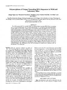
Figure 1. RT-PCR detection of HERV-H spliced transcripts in colon cancer cell lines. A. Schematic demonstration of the primer locations. TSS, transcription start site; TTS, transcription termination site. B. RT-PCR was performed to detect HERV-H spliced transcripts in colon cancer cell lines with/without demethylation/histone acetylation treatment using the DNA demethylation agent DAC and the histone deacetylase inhibitor TSA. H2O and genomic DNA mixture (gDNA mix) were used as controls. Genomic DNA mixture produced bands distinct from cDNA samples, which were reverse transcribed from DNase-treated RNA. doi:10.1371/journal.pone.0029950.g001
referring to the sequence of the HERV-H consensus constructed by Jern et al. [5], with the forward primer targeting the PBStRNAHis downstream of the 59 LTR and the reverse primer targeting the R region of 39 LTR (Fig. 1A). This pair of primers was supposed to generate PCR products from HERV-H transcripts of mixed sizes. Consequently, the total RT-PCR products were cloned into the pGEM-T Easy vectors and transfected into E. coli. More than thirty colonies of each sample were randomly selected and plasmids were then sequenced. Sequencing results were assigned to specific HERV-H loci based on private nucleotide differences between individual loci (one or several nucleotides that are characteristic for an HERV-H locus). Sequencing results of all the randomly selected colonies indicated that no inserts were from non-HERV-H transcripts, demonstrating our strategy worked well.
Due to the immunosuppressive property of the envelope protein of HERV-H, many studies focused on the env-related transcripts [10], and those non-env-related transcripts were rarely paid attention to. Up-regulation of the most abundant H family HERV, especially env-related transcripts, has been reported to be associated with colorectal cancer [5,11,12]. In our previous study, we observed that there were many spliced non-coding RNA transcribed from HERV-H elements, both in normal and cancerous colon tissues, as well as colon cancer cell lines. However, the expression pattern of the spliced non-coding transcripts from HERV-Hs is not clear. During the studies on the three newly identified HERV-H-related genes [13,14,15] in our laboratory, we observed that the overall expression of HERVH elements in colon cancer was complex and different between tumor samples and adjacent normal samples. We also observed that many env-deleted HERV-Hs were transcriptionally active, and many spliced non-coding RNA were transcribed in both tumor and normal tissues, as well as cancer cell lines. In this study, we initiated the first study to find out the exact loci of the most active HERV-H elements in colon cancer.
Expression of HERV-H spliced transcripts in colon cancer cell lines, and altered expression in response to DNA demethylation treatment RT-PCR assays were performed on five colon cancer cell lines, including HT29, LS 174T, RKO, SW480 and SW620. Different band patterns were obtained from these cell lines (Fig. 1B left). A relatively lower level of HERV-H expression was observed in RKO. It has been reported that there is DNA methylation at a global genomic level by targeting repetitive sequences [17], so we further investigated whether demethylation treatment would change the transcription pattern of HERV-H elements. Cancer cells were treated with DNA methylation and histone deacetylase inhibitors (DAC and TSA respectively). RT-PCR results showed that expression patterns of HERV-H were significantly changed in SW480 and RKO cells (Fig. 1B right). These findings suggested that the epigenetic context of a genome affected the expression of some of the HERV-H elements. RT-PCR product of each colon cancer cell line was further analyzed by sequencing after cloning. Sequencing results were assigned to genomic loci by BLAT searches against the human
Results The RT-PCR-sequencing strategy for hooking HERV-H non-coding transcripts Since there are over one thousand members of the HERV-H family throughout the human genome, with sequences of low complexity and homologous to one another, it is difficult to study their transcription profile by taking every member into consideration. In order to profile the expression of HERV-H in colon cancer cell lines, colon tumor and adjacent normal tissues, and to identify the exact loci of the most active HERV-H elements, we developed a RT-PCR-cloning strategy to hook a selected group of HERV-H-related transcripts. The transcription of HERV elements is typically started from the 59 R region and terminated at the end of the 39 R region [16]. We designed the primers by PLoS ONE | www.plosone.org
2
January 2012 | Volume 7 | Issue 1 | e29950
HERV-H Spliced Transcription in Colon Cancer
genome assembly (hg19). Genomic DNA sequences were then retrieved and analyzed with RepeatMasker to determine the HERV-H sequences. Only a limited number of HERV-H elements were found to be transcriptionally active in each of these cell lines, with six loci (seven elements) in HT29 and two or three in each of the other cell lines (Table 1).
More HERV-H elements were transcriptionally active in colon tumor than in adjacent normal tissues Expression of HERV-H transcripts in colon tumor and adjacent normal tissues were analyzed by RT-PCR. A slight difference between the RT-PCR products of colon tumor and adjacent normal samples was observed, with some products of larger sizes present in tumor samples (Fig. 3). After sequencing and sequence analysis of the PCR products, 14 HERV-H elements were found to be transcriptionally active in colon tumor samples. In contrast, only 7 HERV-H elements were found to be active in the adjacent normal colon samples. Details of these HERV-H elements were indicated in Table 2. Sequence analysis by pair-wise alignment against the HERV-H consensus revealed that all the active HERV-H elements were structurally incomplete, with six fragments commonly deleted as indicated in Fig. 2A (Fig. S2). Four HERV-H elements, located at 1q42.2, 16q24.1, 19q13.31 and 5q23.2 respectively, were active in both tumor and adjacent normal tissues. We further analyzed the transcript abundance from each active HERV-H element. Interestingly, three of the commonly active elements (located at 1q42.2, 16q24.1 and 19q13.31) each contributed to more than 10% of the transcripts in both tumor and adjacent normal samples. There was another element, located at 1p31.3, producing more than 10% of the transcripts in tumor. The four most active elements in the tumor sample, i.e. those located at 1q42.2, 16q24.1, 19q13.31 and 1p31.3, in total contributed to 68.89% of the transcripts in the tumor sample. By contrast, the three most active elements in normal samples (located at 1q42.2, 16q24.1 and 19q13.31) contributed to 85.71% of the overall transcripts. These results indicated that there were more HERV-H elements active in colon tumor tissues than in adjacent normal tissues. Furthermore, the
Characterization of the active HERV-H elements and their spliced transcripts in colon cancer cell lines Pair-wise alignments were performed with the 9.0-kb HERV-H consensus constructed by Jern et al. to determine the fragmentdeleting patterns of each HERV-H element. Alignment results showed all the HERV-H elements active in colon cancer cell lines were structurally incomplete, with the longest one being 6,488 bp long (Table 1 and Fig. S1). Six fragments were found to be commonly deleted in these elements, including a short one in the gag region, four in the pol region, and nearly the entire env region (Fig. 2A). There were some exceptions. The element located at 16q24.1 (active in HT29) and the one located at 22q11.1 (active in LS 174T) were extraordinarily short (Fig. 2B). The one at 16q24.1 was only 3,185 bp in length, with the whole gag region and a large fragment encompassing the pol and env deleted. The one at 22q11.1 was 3,842 bp long, with nearly the entire pol and env regions deleted. Most of the amplicons (.90%) from LS 174T contained sequences corresponding to spliced RNAs from the 3.8-kb HERV-H element at 22q11.1, most of which (.60%) were multiply spliced. Interestingly, two HERV-H elements located at 1p32.3 (2.8 kb separated) were found to be active in HT29 cells, making use of 59 LTR of the first HERV-H and 39 LTR of the second one (representative transcript sequence JK017392). Sequence analysis revealed that 59 LTR was missing from the latter 2,298-bp HERV-H element (Fig. 2C).
Table 1. The loci of active HERV-H elements in colon cancer cell lines.
Cell lines
Loci of active HERV-Hs (hg19)
HT29
1p32.3*
LS174T
RKO
SW480
SW620
Chr1+: (53890009–53895701),(53898553–53900850)
Size/bp
Predicted ORFs (Size/nt, homologous protein)***
(5693),(2298)
426, Gag protein
2p14
Chr22: 69670143–69676473
6331
540, Polymerase
3q28
Chr3+: 189862389–189867991
5603
900, partial Gag protein
14q24.3
Chr14+: 74170042–74175916
5875
16q24.1**
Chr16+: 86311701–86314885
3185
19q13.32
Chr192: 47553126–47558666
5541
22q11.1
Chr22+: 17092198–17096039
3842
19q13.31**
Chr192: 43827663–43833536
5874
3q24
Chr3+: 145428282–145433882
5601
6q24.1
Chr6+: 142336803–142342919
6117
19q13.41
Chr19+: 53131048–53136648
5601
1q25.3
Chr1+: 183582345–183588508
6164
6q24.2
Chr6+: 145244311–145249992
5682
20p12.1**
Chr202: 12320915–12326587
5673
1q25.3
Chr1+: 183582345–183588508
6164
5q31.1
Chr52: 135066688–135073175
6488
495, Integrase; 618, conserved domain RT-like
825, Gag protein
525, conserved domain CREB5
528, Gag protein; 747, conserved domain RT-like
*Two adjacent HERV-H elements at 1p32.3 were combinedly active by making use of 59 LTR of the first HERV-H and 39 LTR of the second one (representative transcript sequence JK017392). **The elements located at 16q24.1 and 19q13.31 are also actively transcribed in both tumor and adjacent normal colon tissues, while the one located at 20p12.1 is active in tumor tissue. ***Open reading frames (ORFs) were predicted by the online program ORF Finder and putative peptide sequences were subjected to Blastp search against the Nonredundant protein sequences at http://www.ncbi.nlm.nih.gov/. Only predicted ORFs$303 nt (peptide sequence $100 aa) and with Blastp matches are included. Gag, group-specific antigen; RT, Reverse transcriptase; CREB5, cAMP response element-binding protein 5. doi:10.1371/journal.pone.0029950.t001
PLoS ONE | www.plosone.org
3
January 2012 | Volume 7 | Issue 1 | e29950
HERV-H Spliced Transcription in Colon Cancer
Figure 2. Characterization of the active HERV-H elements in colon cancer cell lines and their spliced transcripts. A. Schematic of the six commonly deleted regions in the active HERV-H elements in colon cancer cell lines. B. Schematics of the two extraordinarily short HERV-H elements and their transcripts. Pair-wise alignments for each HERV-H element were performed with the HERV-H consensus constructed by Jern P, et al. The shortened alignment results were shown to indicate the missing regions precisely. Color density represents the extent of homology with the HERV-H consensus. Gray areas represent deleted regions in the HERV-H elements as compared with the HERV-H consensus. Spliced transcripts are shown above the alignment results accordingly. Thick bars represent exons, and lines represent introns. Regions of LTRs, pre-gag, gag, pro, pol and env are labeled below. C. Schematics of the two combinedly active HERV-H elements located at 1p32.3 in HT29. doi:10.1371/journal.pone.0029950.g002
majority of transcripts in adjacent normal samples were produced from fewer HERV-H elements when compared with tumor samples.
Discussion Due to the abundance of HERV-H elements in the human genome and their high sequence similarity to each other, there seems to be no appropriate approach to investigating the genomewide transcription profile of HERV-H elements while at the same time taking each element into consideration individually. Although findings of this study did not represent the overall actual genomewide expressional profile of HERV-H elements, we took the advantage of the PCR enrichment strategy to hook the most active HERV-H elements that produced transcripts containing the PBStRNAHis and 39 R regions, and successfully identified the exact
Figure 3. Gel electrophoresis of RT-PCR products of HERV-Hrelated transcripts in colon tumor and adjacent normal tissues. M, DL2000 ladder; T, tumor; N, normal. doi:10.1371/journal.pone.0029950.g003
PLoS ONE | www.plosone.org
4
January 2012 | Volume 7 | Issue 1 | e29950
HERV-H Spliced Transcription in Colon Cancer
Table 2. The loci of active HERV-Hs in colon tumor and adjacent normal tissues.
Colon tissues
No.
Loci of active HERV-Hs (hg19)*
Tumor
T1
1q42.2
Chr1+: 230736962–230744940
7979
17.78%
T2
16q24.1
Chr16+: 86311701–86314885
3185
26.67%
618, conserved domain RT-like
T3
19q13.31
Chr192: 43827663–43833536
5874
11.11%
318, CREB5
T4
1p31.3
Chr1+:68851687–68857675
5989
13.33%
T5
2p12
Chr22: 76800751–76806830
6080
T6
3q26
Chr3+: 189862389–189867991
5603
T7
8q22.2
Chr8+: 100955923–100961835
5913
T8
11q24.2
Chr112:127638814–127644790
5977
741, Polymerase
T9
13q33.3
Chr13+: 109917438–109923464
6027
327, Protease
T10
20p12.1
Chr202: 12320915–12326587
5673
T11
5q23.2
Chr52: 121809921–121814541
4621
59LTR incomplete
T12
Xp22.32
ChrX2: 4458515–4464361
5847
HERV-HX**
Adjacent Normal
Size/bp
Abundance (.10%)/Notes 68.89%
Predicted ORFs (Size/nt, homologous protein)**** 399, conserved domain RT-like; 402, Envelope
T13
1q31.3
chr12:195817044–195817727
684
59LTR-39LTR***
T14
12p12.2
Chr122:20970842–20975551
4710
39LTR missing
N1
1q42.2
Chr1+: 230736962–230744940
7979
26.19%
N2
16q24.1
Chr16+: 86311701–86314885
3185
16.67%
(as T2)
N3
19q13.31
Chr192: 43827663–43833536
5874
42.86%
(as T3)
N4
4p15.2
Chr42: 23724496–23729489
4994
N5
19q13.12
Chr192:36750410–36756122
5713
N6
Xq23
ChrX2: 115854285–115860680
6396
564, Gag protein
N7
5q23.2
Chr52: 121809921–121814541
4621
(as T11)
85.71%
327, conserved domain Protease; 309, conserved domain RT-like (as T1)
309, conserved domain RT-like
*Active elements individually contributing to more than 10% of the transcripts in tumor or adjacent normal samples are highlighted in boldface and their transcript abundances are indicated in the ‘Abundance’ column (individual and total, respectively). **HERV-HX is the colon cancer-related HERV-H element identified by us previously [14]. Inserts of PCR product clones were all HERV-HX fragments but not spliced sequences, in concordance with our previous finding that no spliced transcripts were produced from HERV-HX in colon tumor samples. ***The element located at 1q31.3 consists of 59LTR and 39LTR, with the entire protein coding region (gag-pro-pol-env) missing. ****Open reading frames (ORFs) were predicted by the online program ORF Finder and putative peptide sequences were subjected to Blastp search against the Nonredundant protein sequences at http://www.ncbi.nlm.nih.gov/. Only predicted ORFs$303 nt (peptide sequence $100 aa) and with Blastp matches are included. RT, Reverse transcriptase; CREB5, cAMP response element-binding protein 5; Gag, group-specific antigen. doi:10.1371/journal.pone.0029950.t002
proviral loci of these elements in colon cancer cell lines and tissue samples. Expression patterns of HERV-H spliced transcripts from HERV-H elements were different among colon tumor, adjacent normal samples, and colon cancer cell lines. Expression patterns of HERV-H spliced transcripts from HERVH elements were different among the five colon cancer cell lines tested, evidenced by both gel electrophoresis of the RT-PCR products (Fig. 1B left) and sequencing of the amplicons after cloning (Table 1). Sequence analysis by comparing them with the constructed HERV-H consensus indicated that the active HERVH elements found in this study were all structurally incomplete to some extent. This can also be inferred from the lengths, all of which were much shorter than the full-length HERV-H consensus (9.0kb). The band patterns were different between SW480 and SW620 cells. Although they originated from the primary and metastatic tumors of the same patient, this difference between SW480 and SW620 may be just due to long-term culture. Likewise, the limited number of active HERV-H elements in each cell line may also be due to long-term culture in defined in vitro conditions. It is also interesting that by demethylation treatment using DNA methylation and histone deacetylase inhibitors, expression patterns in RKO and SW480 cells were changed significantly, suggesting that transcription of some HERV-H elements are regulated epigenetically. PLoS ONE | www.plosone.org
Although no obvious difference was observed in the RT-PCR product bands between tumor and adjacent normal samples, the total numbers and loci of active HERV-H elements were significantly different (Fig. 3; Table 2). Seven HERV-H elements were found to be transcriptionally active in the adjacent normal colon samples. By comparison, 14 elements were found to be active in the tested colon tumor tissues. Expression of the three commonly most active elements (.10%) seemed to be ordinary; the one located at 1q24.2 produced 17.78% and 26.19% of the products in tumor and normal samples, and products from the one located at 16q24.1 consisted of 26.67% in tumor and 16.67% in normal samples, respectively. Interestingly, the one element located at 19q13.31 was the most active in adjacent normal tissues, producing 42.86% of the products, while only 11.11% from tumor samples belonged to 19q13.31. These results demonstrated that most of the HERV-H spliced transcripts in adjacent normal colon tissues were from 19q13.31, while transcripts in colon tumor tissues involved more elements. All active HERV-H elements found in this study are structurally incomplete, with six fragments commonly deleted, which are distributed through the gag, pol and env regions (Fig. 2A); even so, some of them (40%) retain putative open reading frames (ORFs) (Tables 1 and 2; Fig. S1 and S2). Although some of these putative 5
January 2012 | Volume 7 | Issue 1 | e29950
HERV-H Spliced Transcription in Colon Cancer
ORFs have been subjected to sequence deletion to a certain extent when compared with the corresponding region in the HERV-H consensus, seven of the putative peptide sequences (encoded by 6 HERV-H elements) contain conserved domains, with one conserved Protease domain, one conserved CREB5 (cAMP response element-binding protein 5) domain and five conserved reverse transcriptase (RT)-like domains. Interestingly, all the three commonly most active HERV-H elements (located at 1q42.2, 16q24.1 and 19q13.31) in tumor and adjacent normal tissues contain putative ORFs with conserved domains (Table 2; Fig. S2). Whether these ORFs actually produce proteins in colon tissues and what role they play in colon carcinogenesis are issues to be addressed. The reason for the transcriptional difference in HERV-H elements between colon tumor and adjacent normal tissues remains unknown. Cells in tumor and adjacent normal samples are different in many aspects. Colon tumor cells in a sample are heterogeneous and cover more phases of the cell cycle, while nontumor cells are usually in G0. However, there has been no study to address whether HERVs are expressed in a cell cycle dependent manner yet. Furthermore, tumor cells are in a more undifferentiated state as compared with non-tumor cells, reminiscent of the state of embryonic cells. Expression of HERVs in a tissue-specific and developmental stage-dependent manner has been reported [18]. HERV elements have also been found to be highly expressed in reproductive tissues such as testis and placenta, which contain undifferentiated cells [5,19]. Whether HERV-H elements are expressed in a differentiation dependent manner, subsequently causing the difference between colon tumor and adjacent normal tissues, also needs to be investigated. These two points may indicate directions for further study on the mechanism by which HERV-H elements are differentially regulated between colon tumor and adjacent normal tissues. This is the first attempt to study the characteristics of expression of HERV-H spliced transcripts in colon cancer. In summary, our results indicated that expression patterns of HERV-H elements differed among colon cancer cell lines and could be affected by demethylation treatment. More importantly, our findings demonstrated that HERV-H expression involved more active HERV-H elements in colon tumor than in adjacent normal tissues.
inhibitor trichostatin A (TSA; Beyotime) as described [20]: initial treatment with DAC (200 nM) for 48 h, with drug and medium replaced 24 h after the beginning of treatment, followed by the replacement of medium containing TSA (300 nM) for a further 24 h. Total RNA from drug treated cells or non-treated cells was isolated with Trizol reagent and treated with DNase before cDNA synthesis.
Reverse transcription-PCR (RT-PCR) RNA was reverse-transcribed into cDNA using M-MLV Reverse Transcriptase (Promega) according to the manufacturer’s guide. PCR assays were performed with Taq DNA polymerase (Promega) in reaction systems containing 0.2 mmol/L forward and reverse primers each. Thermal cycler parameters were 94uC 5 min, (94uC 30 s, 55uC 30 s, 72uC 90 s)636 cycles, 72uC 10 min. PCR products of each cell sample, PCR product mixture of six tumor samples and PCR product mixture of six adjacent normal samples were purified with AxyPrep PCR Cleanup kit (Axygen), cloned into the pGEM-T easy vector (Promega) and transfected into E. coli bacteria. Over 30 colonies from each cell sample and 100 colonies from tumor or normal tissue samples were subjected to Sanger sequencing. Targets of the primers are indicated in Fig. 1A and their nucleotide sequences are as follows: forward (targeting PBS-tRNAHis): 59-TGGTGCCGTGACTCGGAT-39, reverse (targeting R region): 59-GCTGAGTCCGAAAAGAGAGTC-39. The primers were designed by referring to the HERV-H consensus constructed by Jern et al. [5], with ‘‘G’’ in the forward primer modified according to genome-wide sequence analysis on HERV-H-related primer binding sites on our own (not shown).
Sequence analysis Sequences were searched with BLAT method against human genome using the online server (http://genome.ucsc.edu/cgi-bin/ hgBlat?command=start). Sequencing results were excluded if the sequences in-between the primers had more than five dissimilarities to genomic DNA. Genomic DNA sequences were retrieved from the human genome assembly (hg19) at http://genome.ucsc. edu/cgi-bin/hgGateway. Sequences were analyzed with the tool RepeatMasker at http://www.repeatmasker.org/cgi-bin/ WEBRepeatMasker to identify repeat elements. Pairwise alignments were carried out with GeneDoc. ORF prediction was performed using the online program ORF Finder (http://www. ncbi.nlm.nih.gov/gorf/gorf.html). Then putative amino acid sequences were blasted against the non-redundant protein sequence database using the online BLASTP program (http:// blast.ncbi.nlm.nih.gov/Blast.cgi).
Materials and Methods Ethics statement This study was approved by the ethics committee of Zhejiang University, and written consents were obtained from all patients involved.
Tissue samples, cell culture and RNA preparation
Sequence submission
Colon tumor and adjacent normal tissue samples were obtained after surgical resection in the second affiliated hospital of Zhejiang University and stored frozen at 280uC until RNA extraction. Tissue types (tumor or normal) were assessed by histological staining. Cancer cells were obtained from American Type Culture Collection and grown in RPMI 1640 medium supplemented with 10% fetal calf serum. Total RNA was prepared with Trizol reagent (Invitrogen) according to the manufacturer’s guide. RNA was always treated with RQ1 RNase-free DNase (Promega) before cDNA synthesis to eliminate genomic DNA contamination.
A total of 92 nucleotide sequences of the newly identified HERV-H spliced transcripts have been deposited in dbEST with accession numbers of JK017330–JK017415 and JK475044– JK475049 (with 7 EST library accession numbers of LIBEST_027280 - 027286).
Supporting Information Figure S1 Pair-wise alignments for each HERV-H element were performed with the HERV-H consensus constructed by Jern P, et al. The shortened alignment results are shown to indicate the missing regions precisely. Color density represents the extent of homology with the HERV-H consensus. Lines represent deleted regions in the HERV-H elements as compared with the HERV-H consensus. Red rectangles indicate
Demethylation treatment with DNA methylation and histone deacetylase inhibitors Cells were treated with the DNA methylation inhibitor 5-aza29-deoxycytidine (DAC; Sigma) and the histone deacetylase PLoS ONE | www.plosone.org
6
January 2012 | Volume 7 | Issue 1 | e29950
HERV-H Spliced Transcription in Colon Cancer
the regions where putative ORFs are harbored, while red rectangles with thick lines indicate ORFs with conserved domains. Regions of LTRs, pre-gag, gag, pro, pol and env are labeled below. Genomic locus of each HERV-H element is indicated correspondingly on the right side. (TIF)
Regions of LTRs, pre-gag, gag, pro, pol and env are labeled below. Genomic locus of each HERV-H element is indicated correspondingly on the right side. The number of each element is shown on the left as indicated in Table 2. (TIF)
Figure S2 Pair-wise alignments for each HERV-H element were performed with the HERV-H consensus constructed by Jern P, et al. The shortened alignment results are shown to indicate the missing regions precisely. Color density represents the extent of homology with the HERV-H consensus. Lines represent deleted regions in the HERV-H elements as compared with the HERV-H consensus. Red rectangles indicate the regions where putative ORFs are harbored, while red rectangles with thick lines indicate ORFs with conserved domains.
Acknowledgments The authors are grateful to Ms. Heather Simkin for proofreading the manuscript.
Author Contributions Conceived and designed the experiments: QL SZ. Performed the experiments: QL ZX LW. Analyzed the data: QL RX SZ. Contributed reagents/materials/analysis tools: ZX RX SZ. Wrote the paper: QL SZ.
References 1. Lander ES, Linton LM, Birren B, Nusbaum C, Zody MC, et al. (2001) Initial sequencing and analysis of the human genome. Nature 409: 860–921. 2. Lower R, Lower J, Kurth R (1996) The viruses in all of us: characteristics and biological significance of human endogenous retrovirus sequences. Proc Natl Acad Sci U S A 93: 5177–5184. 3. Hirose Y, Takamatsu M, Harada F (1993) Presence of env genes in members of the RTVL-H family of human endogenous retrovirus-like elements. Virology 192: 52–61. 4. Wilkinson DA, Goodchild NL, Saxton TM, Wood S, Mager DL (1993) Evidence for a functional subclass of the RTVL-H family of human endogenous retrovirus-like sequences. J Virol 67: 2981–2989. 5. Jern P, Sperber GO, Ahlsen G, Blomberg J (2005) Sequence variability, gene structure, and expression of full-length human endogenous retrovirus H. J Virol 79: 6325–6337. 6. de Parseval N, Casella J, Gressin L, Heidmann T (2001) Characterization of the three HERV-H proviruses with an open envelope reading frame encompassing the immunosuppressive domain and evolutionary history in primates. Virology 279: 558–569. 7. Jern P, Sperber GO, Blomberg J (2004) Definition and variation of human endogenous retrovirus H. Virology 327: 93–110. 8. Scho¨n U, Seifarth W, Baust C, Hohenadl C, Erfle V, et al. (2001) Cell typespecific expression and promoter activity of human endogenous retroviral long terminal repeats. Virology 279: 280–291. 9. Schon U, Diem O, Leitner L, Gunzburg WH, Mager DL, et al. (2009) Human endogenous retroviral long terminal repeat sequences as cell type-specific promoters in retroviral vectors. J Virol 83: 12643–12650. 10. Mangeney M, de Parseval N, Thomas G, Heidmann T (2001) The full-length envelope of an HERV-H human endogenous retrovirus has immunosuppressive properties. J Gen Virol 82: 2515–2518. 11. Wentzensen N, Wilz B, Findeisen P, Wagner R, Dippold W, et al. (2004) Identification of differentially expressed genes in colorectal adenoma compared
PLoS ONE | www.plosone.org
12.
13.
14.
15.
16.
17. 18.
19.
20.
7
to normal tissue by suppression subtractive hybridization. Int J Oncol 24: 987–994. Pichon JP, Bonnaud B, Cleuziat P, Mallet F (2006) Multiplex degenerate PCR coupled with an oligo sorbent array for human endogenous retrovirus expression profiling. Nucleic Acids Res 34: e46. Liang Q, Ding J, Zheng S (2009) Identification and detection of a novel human endogenous retrovirus-related gene, and structural characterization of its related elements. Genet Mol Biol. Liang Q, Ding J, Xu R, Xu Z, Zheng S (2009) Identification of a novel human endogenous retrovirus and promoter activity of its 59 U3. Biochem Biophys Res Commun 382: 470–474. Liang Q, Ding J, Xu R, Xu Z, Zheng S (2010) The novel human endogenous retrovirus-related gene, psiTPTE22-HERV, is silenced by DNA methylation in cancers. Int J Cancer 127: 1833–1843. Sjottem E, Anderssen S, Johansen T (1996) The promoter activity of long terminal repeats of the HERV-H family of human retrovirus-like elements is critically dependent on Sp1 family proteins interacting with a GC/GT box located immediately 39 to the TATA box. J Virol 70: 188–198. Lettini AA, Guidoboni M, Fonsatti E, Anzalone L, Cortini E, et al. (2007) Epigenetic remodelling of DNA in cancer. Histol Histopathol 22: 1413–1424. Okahara G, Matsubara S, Oda T, Sugimoto J, Jinno Y, et al. (2004) Expression analyses of human endogenous retroviruses (HERVs): tissue-specific and developmental stage-dependent expression of HERVs. Genomics 84: 982–990. Forsman A, Yun Z, Hu L, Uzhameckis D, Jern P, et al. (2005) Development of broadly targeted human endogenous gammaretroviral pol-based real time PCRs Quantitation of RNA expression in human tissues. J Virol Methods 129: 16–30. Cameron EE, Bachman KE, Myo¨ha¨nen S, Herman JG, Baylin SB (1999) Synergy of demethylation and histone deacetylase inhibition in the re-expression of genes silenced in cancer. Nat Genet 21: 103–107.
January 2012 | Volume 7 | Issue 1 | e29950