Gabor Wavelets to Characterize Different Types of Cardiac Hypertrophy V. Damerjian, O. Tankyevych, A. Guellich, T. Damy, and E. Petit
Abstract— In ultrasound imaging, distinguishing different types of cardiac hypertrophy is difficult. Here, we exploit Gabor wavelets and their texture features to classify these diseases.
I. INTRODUCTION Wavelet analysis plays an essential role in multi-resolution techniques [1-3], and it is a powerful model for texture discrimination [4]. The aim of our study is to classify types of cardiac hypertrophy by analyzing the texture of echocardiographic images in order to characterize speckle patterns.
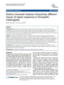
classes by maximizing the ratio of the inter-class variance to the intra-class variance. Fig. 1 shows a 3D projection of the features after undergoing PCA and LDA. From Fig. 1, we can observe the clustering and closeness of the three types of CA (AL, TTR, and TTRS), the features of the normal hearts that are dispersed (this is normal for control cases that may differ
II. CURRENT WORK Cardiac hypertrophy can be the result of cardiomyocyte hypertrophy, such as in sarcomeric hypertrophic cardiomyopathy (HCM) or the infiltration of the extracellular matrix, such as in Cardiac Amyloidosis (CA). A database of HCM and 3 types of CA has been collected in collaboration with Hôpital Henri Mondor: Amyloid Light-chain Amyloidosis (AL), Transthyretin-related hereditary Amyloidosis (TTR), and Senile Transthyretin Amyloidosis (TTRS). Twelve patients per disease type have been considered, in addition to 11 control cases of normal hearts. Frames of a complete heart cycle were studied for every patient, only regions of interest (ROI) isolating the septum were considered, as it is a part of the heart that gets affected by the diseases. We applied the Gabor Wavelets of size 39×39 with 3 decomposition levels and 8 orientations. Nine features were extracted for every image resulting in 216 total features: mean, variance, entropy, skewness, and kurtosis in addition to the co-occurrence matrix features as energy, contrast, homogeneity, and correlation. All the 216 extracted features then underwent Principal Component Analysis (PCA) and Linear Decomposition Analysis (LDA). PCA is used to reduce the number of feature vector dimensions and to identify and classify patterns in the data according to the amount of information stored in them. LDA, on the other hand, helps in the separation of the studied
Figure 1. 3D projection of the features after undergoing PCA and LDA. AL is in red, TTR in dark blue, TTRS in black, CMH in green, and control in light blue.
due to sex, age, and population), and the CMH which falls in between the two previous cases. III. CONCLUSION AND PERSPECTIVES Gabor wavelets are studied on different scales, with different filter sizes and orientations in order to classify directional speckle patterns due to cardiac hypertrophy presence in the septum. Textural features are then extracted from the wavelet filter response. The separation quality of the class features for each set of parameters after PCA and LDA is evaluated with scatter matrices and their indices. Moreover, feature selection algorithms are applied in order to identify the most coherent ones. The results of this study will be used to compare other wavelet-based texture methods such as curvelets. REFERENCES [1]
V. Damerjian is with the Laboratoire d’Images, Signaux et Systèmes Intelligents, Université Paris-Est Créteil, France. (phone: 0033652778044; e-mail:
[email protected]). O. Tankyevych and E. Petit are with the Laboratoire d’Images, Signaux et Systèmes Intelligents, Université Paris-Est Créteil, France. (e-mail:
[email protected] and
[email protected]). A. Guellich and T. Damy are with the Heart Failure Unit / Department of Cardiology, Henri Mondor Hospital, Créteil, France. (e-mail:
[email protected] and
[email protected]).
[2] [3] [4]
Ifeachor EC, Jervis BW. Digital signal processing: a practical approach.Pearson Education; 2002. Mala K, Sadasivam V, Alagappan S. Neural network based texture analysis of liver tumor from computed tomography images. Int J Biol MedSci 2007;21:33–41. Watson GH, Watson SK. Wavelet transforms on vector spaces as a method of multispectral image characterisation. IEEE Proc Vision Image Signal Process 1997;144(2):89–97. Zhang J, Tan T. Brief review of invariant texture analysis methods. Pattern Recognit 2002;35(3):735–47.