Clinical Case Study
Clinical Chemistry 55:4 827–832 (2009)
Genetic Testing for Developmental Delay: Keep Searching for an Answer David T. Miller,1,2,4* Yiping Shen,1,4 David J. Harris,2,4 Bai-Lin Wu,1,4 and Magdi M. Sobeih3,4
CASE A 6-year-old girl of Irish, English, and French ancestry was referred to a pediatric neurologist for evaluation of developmental delay. She presented with expressive language delay with disarticulation. She did not speak in phrases until age 3, and formal testing revealed a language equivalent of 3 years 4 months when she was 5 years 6 months old (Clinical Evaluation of Language Fundamentals–Preschool) and an IQ of 64 (Wechsler Preschool & Primary Scale of Intelligence). Gross motor development was also delayed; she first walked at age 17–18 months. She never had any developmental regression. There was a family history of learning disability in her mother and a maternal uncle, and a maternal first cousin once removed was born with myelomeningocele. She was delivered at term after an uncomplicated pregnancy that was conceived by in vitro fertilization. At 10 days of age, she was diagnosed with atrioventricular (A-V)5 canal malformation and coarctation of the aorta. She underwent surgical repair of her coarctation at age 10 days and of her A-V canal defect at age 4 months. She had a bifid uvula, a finding often associated with presence of a submucous cleft palate. Modified barium swallow demonstrated a poorly coordinated swallow reflex leading to poor feeding and aspiration. As an infant, she had true vocal cord paralysis, believed to be a complication from intubation. She had gastroesophageal reflux disease, treated with ranitidine (Zantac), and was diagnosed with mild vesicoureteral reflux after a urinary tract infection at age 7 months. She has not had any episodes suggesting seizures or any features of autism other than language delay.
Department of Laboratory Medicine, 2 Division of Genetics, and 3 Neurolinguistics Clinic/Behavioral Neurology in Department of Neurology, Children’s Hospital Boston, Boston, MA; 4 Harvard Medical School, Boston, MA. * Address correspondence to this author at: Department of Laboratory Medicine, Children’s Hospital Boston, 300 Longwood Ave., Boston, MA 02115. Fax: 617-730-0338; e-mail
[email protected]. Received October 16, 2008; accepted January 5, 2009. DOI: 10.1373/clinchem.2008.119438 5 Nonstandard abbreviations: A-V, atrioventricular; FISH, fluorescence in situ hybridization; aCGH, array comparative genomic hybridization; BAC, bacterial artificial chromosome; ST-FISH, subtelomere FISH; MLPA, multiplex ligation and probe amplification; NAHR, nonallelic homologous recombination.
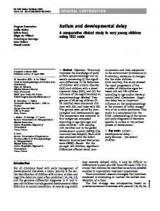
Notable physical exam features include widely spaced eyes (hypertelorism), bulbous nasal tip, high arched palate, and fifth finger clinodactyly. Her neurologist ordered an MRI that showed mild thinning of the corpus callosum with prominence of the lateral and third ventricles, all nonspecific findings. Multiple genetic tests were ordered during infancy to determine the cause of her cardiac anomalies and developmental delays. The history of A-V canal malformation and aortic coarctation raised suspicion for microdeletion of chromosome 22q11.2, also called velocardiofacial syndrome. Fluorescence in situ hybridization (FISH) for chromosome 22q11.2 was normal. G-banded karyotype, PTPN11 gene sequencing for Noonan syndrome, and fragile X DNA testing results were also normal. At age 3 years, array comparative genomic hybridization (aCGH) was ordered from an outside laboratory. This whole-genome array of 2600 bacterial artificial chromosome (BAC) clones (Spectral Genomics, Inc.) spaced 1 Mb apart showed a gain in copy number of BAC clones extending from clone RP11–1K11 at 8p23.2 (chr8: 4 596 114 – 4 755 793; human genome build 18) to clone RP11–23H1 at 8p22 (chr8: 15 027 287–15 191 603; hg18) indicative of an approximately 10.6-Mb duplication at 8p22p23.2. Neither parent was a carrier of the 8p22p23.2 duplication based on FISH testing, indicating a de novo copy number change. Two years later, the patient’s neurologist ordered high-resolution whole genome oligonucleotide microarray (244K array G4411B; Agilent Technologies), taking advantage of this new technology to gather more information. This test again identified the 8p22p23.2 duplication, and defined the lesion more precisely as an 11.5-Mb duplication (chr8: 3 969 033–15 475 755; hg18). In addition, a 3.0-Mb duplication was identified at chromosome 22q11.2 (chr22: 17 086 001–20 131 661; hg18) (Fig. 1).
1
DISCUSSION The evaluation of children with developmental delay, dysmorphic features, and even autism spectrum disorders has improved tremendously as a result of clinical laboratory testing. Many children with developmental delay do not have physical exam features or medical histories specific enough for a clear clinical diagnosis. 827
Clinical Case Study
Fig. 1. Copy number changes identified by aCGH (Agilent 244k). (A), 8p22p23.2 duplication (arrowhead) compared to duplications reported in other studies. (B), 22q11.2 duplication (arrowhead) compared to typical recurrent 3-Mb deletion/duplication region. Chromosomal position is indicated relative to the centromere (cen) and telomere (tel) of each chromosome. Chromosome band number increases moving away from the centromere. Scale in megabases (Mb).
Among such patients, laboratory testing can be extremely helpful and is an integral component of the diagnostic evaluation. Typical recommendations include a G-banded karyotype, fragile X molecular genetic testing, aCGH, and neuroimaging (1 ). Microarray-based CGH to detect changes in genomic copy number, more than any other single test, has dramatically increased the rate of diagnosis among individuals with unexplained developmental delay and mental retardation. G-banded karyotypes have typically identified abnormalities in 3%– 4% of individuals with idiopathic mental retardation (2 ). Subtelomere FISH (ST-FISH) for submicroscopic deletions and duplications adds to the diagnostic yield. In the largest study of ST-FISH, presumed pathogenic changes were found in 2.6% of 11 688 unselected cases with previously normal karyotype (3 ). Multiple studies now support the conclusion that CGH with broad genomic coverage, so-called chromo828 Clinical Chemistry 55:4 (2009)
somal microarray, has a higher diagnostic yield than G-banded karyotype and ST-FISH in the evaluation of patients with developmental delay and mental retardation, detecting abnormalities in up to 8% of patients with previously normal karyotypes using arrays targeted to clinically relevant areas of the genome (4, 5 ). This detection rate will increase as more laboratories implement arrays with whole-genome coverage. No other clinical laboratory test has a comparable clinical sensitivity for patients with a diagnosis of developmental delay and mental retardation. We would have expected a G-banded karyotype to detect the large 8p duplication, but we know of other clinical aCGH cases where equally large events were not detected. We also would have expected the earlier BAC array to detect the 22q11.2 duplication. The laboratory that reported those results only reported a positive result if 2 or more adjacent BAC clones showed a consistent copy number change, and there may have been some technical limi-
Clinical Case Study tation with 1 of the probes in that region or with performing or interpreting that array. In this patient, 2 relatively large chromosomal duplications were identified. Based on the first aCGH result, it was reasonable to assume that the large duplication of 8p22p23.2 could account for the learning disabilities seen in this patient. Two recurrent large duplication events involving 8p23.1– 8p23.2 overlap with the distal duplication in this patient. The more distal overlapping duplication is associated with speech delay, autism, and learning difficulties (6 ). The more proximal, and smaller, overlapping duplication has been described in individuals with learning disabilities, but also in healthy family members (7 ). A-V canal malformations have also been reported in some patients with 8p22p23.2 duplication. Deletions of chromosome 22q11.2 are the most common genetic cause for A-V canal malformations like that seen in this patient. Typically, these cases have been tested by metaphase FISH. However, duplications will usually be located adjacent to the original position on the chromosome and would be difficult to discern without performing interphase FISH. Newer hybridization-based methods such as multiplex ligation and probe amplification (MLPA) and aCGH can readily detect such duplications. In fact, the advent of these technologies has led to the discovery of numerous duplication syndromes that were not previously recognized, including the 22q11.2 duplication syndrome (8 ). The 22q11.2 duplication syndrome shows variable penetrance and expressivity, with shared features of DiGeorge/velocardiofacial syndrome. Typical reported features include hypertelorism, broad nasal bridge, epicanthal folds, fifth finger clinodactyly, urogenital anomalies, hypotonia, scoliosis, seizures, and/or abnormalities on electroencephalogram. To date, at least 65 patients have been reported (9 ), and the phenotype is widely variable, including several individuals with no detectable symptoms. As this patient entered formal schooling, her difficulties became more apparent. The history of significant language delay evolved into learning disability. Behavioral problems emerged with questions of attention deficit disorder because of her difficulties in retaining learned material. In addition, symptoms of anxiety in anticipation of new activities surfaced. Further characterization of these behaviors suggested they were not primary but secondary to intellectual disability, prompting further testing revealing IQ in the mild to moderate mental retardation range. These behavioral symptoms could be secondary to the cognitive impairment, but behavioral problems such as short attention span, hyperactivity, impulsivity, and aggression have been described in
patients with 22q11.2 duplication syndrome. Consideration of the underlying genetic disorder is important in clinical decision-making about treatment with psychotropic medications. Both deletions and duplications of 22q11.2 are mediated by recombination between nearby, segmentally duplicated sequences via a mechanism termed nonallelic homologous recombination (NAHR) (10 ). The broad phenotypic variability observed in this condition has not been explained, but could be attributable to several factors, including the influence of other genes on the expression of genes within 22q11.2 region, epigenetic factors that modify gene expression, or even environmental factors. With a correct diagnosis, genetic counseling for recurrence risk to future pregnancies was updated. The parents were also counseled that recurrence risk for the de novo 8p22p23.2 duplication would be minimal, approximately 2%–3%, due to possible germline mosaicism. They subsequently had twins under the assumption of a low recurrence risk. Further parental testing revealed the same 22q11.2 duplication in the patient’s mother, with the result of a 50% recurrence risk. This case underscores the impact of improvements in diagnostic genetic testing on genetic counseling for patients and their families.
POINTS TO REMEMBER • Lack of clinical correlation with test results should prompt further evaluation. Also, patients evaluated in the past for developmental delay may benefit from updated testing, especially if no diagnosis has been made. • Chromosomal microarrays more easily detect genomic duplications than metaphase FISH. FISH 22q11.2 was unable to detect the duplication in this case (interphase FISH would have been required). • The lower-density BAC array also missed the 22q11.2 duplication, underscoring the value of high-density oligonucleotide arrays with full genome coverage. • Many unnecessary tests were performed, placing a burden on the patient and family, and delaying the diagnosis. • Owing to variable penetrance for 22q11.2 deletions and duplications, a parent may not have obvious symptoms. • Identifying multiple imbalance events in a single patient provides a unique opportunity to look for genetic modifying effects and could help genotype–phenotype correlation studies.
Clinical Chemistry 55:4 (2009) 829
Clinical Case Study
Author Contributions: All authors confirmed they have contributed to the intellectual content of this paper and have met the following 3 requirements: (a) significant contributions to the conception and design, acquisition of data, or analysis and interpretation of data; (b) drafting or revising the article for intellectual content; and (c) final approval of the published article. Authors’ Disclosures of Potential Conflicts of Interest: No authors declared any potential conflicts of interest. Role of Sponsor: The funding organizations played no role in the design of study, choice of enrolled patients, review and interpretation of data, or preparation or approval of manuscript.
References 1. Moeschler JB. Medical genetics diagnostic evaluation of the child with global developmental delay or intellectual disability. Curr Opin Neurol 2008;21: 117–22. 2. Shevell M, Ashwal S, Donley D, Flint J, Gingold M, Hirtz D, et al. Practice parameter: evaluation of the child with global developmental delay: report of the Quality Standards Subcommittee of the American Academy of Neurology and the Practice Committee of the Child Neurology Society. Neurology
2003;60:367– 80. 3. Ravnan JB, Tepperberg JH, Papenhausen P, Lamb AN, Hedrick J, Eash D, et al. Subtelomere FISH analysis of 11 688 cases: an evaluation of the frequency and pattern of subtelomere rearrangements in individuals with developmental disabilities. J Med Genet 2006;43:478 – 89. 4. Shaffer LG, Kashork CD, Saleki R, Rorem E, Sundin K, Ballif BC, Bejjani BA. Targeted genomic microarray analysis for identification of chromosome abnormalities in 1500 consecutive clinical cases. J Pediatr 2006;149:98 –102. 5. Lu X, Shaw CA, Patel A, Li J, Cooper ML, Wells WR, et al. Clinical implementation of chromosomal microarray analysis: summary of 2513 postnatal cases. PLoS ONE 2007;2:e327. 6. Glancy M, Barnicoat A, Vijeratnam R, de Souza S, Gilmore J, Huang S, et al. Transmitted duplication of 8p23.1– 8p23.2 associated with speech delay, autism and learning difficulties. Eur J Hum Genet 2009;17:37– 43. 7. Barber JC, Maloney VK, Huang S, Bunyan DJ, Cresswell L, Kinning E, et al. 8p23.1 duplication syndrome: a novel genomic condition with unexpected complexity revealed by array CGH. Eur J Hum Genet 2008;16:18 –27. 8. Slavotinek AM. Novel microdeletion syndromes detected by chromosome microarrays. Hum Genet 2008;124:1–17. 9. Courtens W, Schramme I, Laridon A. Microduplication 22q11.2: a benign polymorphism or a syndrome with a very large clinical variability and reduced penetrance? Report of two families. Am J Med Genet A 2008;146A: 758 – 63. 10. Lupski JR. Genomic disorders: structural features of the genome can lead to DNA rearrangements and human disease traits. Trends Genet 1998;14:417–22.
Commentary Sau Wai Cheung
Array comparative genomic hybridization technology is now being used to provide a genome-wide screen for unexpected genomic imbalances. As a result, this technique has become a valuable clinical diagnostic test and is enabling rapid identification of microdeletions and microduplications in much larger numbers of patients, many of whom present with no obvious clinical diagnosis. One of the major impacts from this revolutionary technology is the ability to identify microduplications, thus enabling medical geneticists to correlate the findings with the clinical assessment, as with the patient described by Miller et al. However, discovery of microduplications also poses a challenge to clinicians owing to the lack of clinical description and wide phenotypic variability that appears to be characteristic of microduplications. Oligo-based arrays have further improved the diagnostic capabilities of this test over the previous artificial-chromosome– based arrays derived from
Department of Molecular and Human Genetics, Baylor College of Medicine, Houston, TX. Address correspondence to the author at: Department of Molecular and Human Genetics, Baylor College of Medicine, One Baylor Plaza, NAB 2015, Houston, TX 77030. Fax 713 798 4998; e-mail
[email protected]. Received January 8, 2009; accepted January 29, 2009. DOI: 10.1373/clinchem.2008.122713
830 Clinical Chemistry 55:4 (2009)
bacterial artificial chromosome/P1. The notable advantages of oligo-based arrays are: (a) better design flexibility, which allows avoidance of repetitive sequences and ability to select oligos with good performance; (b) increased robustness because of enhanced dynamic ranges (signal to background); and (c) higher reproducibility and greater precision in mapping of aberrations. The genome contains many low-copy repeats that can lead to these rearrangements, primarily as a result of nonallelic homologous recombination. Chromosome 22q11.2 is such a region, it is gene rich and contains multiple region-specific low-copy repeats. These low-copy repeats are known to mediate genomic disorders in this region, including DiGeorge syndrome/velocardiofacial syndrome, cat-eye syndrome, der(22) syndrome, and the 22q11.2 microduplication syndrome. Although DiGeorge syndrome/velocardiofacial syndrome is the most frequently identified genomic disorder of the 22q11.2 region, with an estimated frequency of 1 in 4000 live births, only a small number of patients with microduplication of this region have been described to date, likely owing to the highly variable and mild phenotype that may escape syndromic identification. Although FISH analysis can potentially identify microduplication in the DiGeorge syndrome/velocardiofacial syndrome critical region, this analysis method requires accurate clinical assess-
Clinical Case Study ment of the phenotype by the clinician, who must specifically request evaluation of interphase cells. Our experience indicates that microduplications are also more likely than microdeletions to be inherited, making the identification of these copy- number changes even more important for accurate recurrencerisk counseling for these families. We are practicing in an exciting time, because many more new diseases will be described on the basis of copy-number variants identified through this important clinical test.
Author Contributions: All authors confirmed they have contributed to the intellectual content of this paper and have met the following 3 requirements: (a) significant contributions to the conception and design,
acquisition of data, or analysis and interpretation of data; (b) drafting or revising the article for intellectual content; and (c) final approval of the published article. Authors’ Disclosures of Potential Conflicts of Interest: Upon manuscript submission, all authors completed the Disclosures of Potential Conflict of Interest form. Potential conflicts of interest: Employment or Leadership: S.W. Cheung, Department of Molecular and Human Genetics, Baylor College of Medicine. Consultant or Advisory Role: None declared. Stock Ownership: None declared. Honoraria: None declared. Research Funding: None declared. Expert Testimony: None declared. Role of Sponsor: The funding organizations played no role in the design of study, choice of enrolled patients, review and interpretation of data, or preparation or approval of manuscript.
Commentary Nelson L.S. Tang
Miller et al. report a case of developmental delay with microduplication of 22q11.2. This case illustrates several important issues related to structural aberration in the genome and shows that resolution of cytogenomic analysis is important in clinical genomic disorders. In this patient diagnosis was made with only a moderately dense 244K array, whereas the older 2.6K bacterial artificial chromosome array failed to detect this 3-Mb duplication. New chips with much higher resolution are entering the market, such as the one million single nucleotide polymorphism genotyping chips and chips with probes targeted for tens of thousands of copynumber variations (CNVs). These high-density platforms enable detection of submicroscopic structural variants that could not previously be revealed. On the other hand, the ability to scan the genome at this high resolution also leads to new challenges. We now know that a vast number of such structural variations are present in the human genome, and it has been estimated that up to a thousand CNVs may be found between any 2 individuals (1 ). At the moment there is insufficient understanding about the biological role and clinical significance of gain or deletion in most of these CNVs. As in the patient described by Miller et al.,
Laboratory for Genetics of Disease Susceptibility, Li Ka Shing Institute of Health Sciences, The Chinese University of Hong Kong. Address correspondence to the author at: Department of Chemical Pathology, Faculty of Medicine, The Chinese University of Hong Kong, Shatin, Hong Kong SAR, China. Fax ⫹852 30059108; e-mail
[email protected]. Received January 11, 2009; accepted February 2, 2009. DOI: 10.1373/clinchem.2008.122739
it is not certain what phenotypic role is played by the de novo 8p22 duplication. Although there is no good answer to the question of why genomic copy-number changes are common causes of developmental delay and mental retardation, CNVs have also been implicated in other neurodevelopment disorders, including autism and schizophrenia (2, 3 ). Duplication or gain of CNVs at 22q11.2 was found to be one of the recurrent structural changes among autistic patients. This structural change was inherited from the father in one case and sporadically with de novo duplication in another. A variable penetrance was observed in the family with inherited 22q11.2 gain, and the father who carried the same duplication was not affected. The latest research studies were carried out with a high-density single-nucleotide polymorphism– genotyping array, and the results suggested that CNV could be defined at an even higher resolution on these platforms. We are now facing a new dilemma. On one hand, it is tempting to apply the latest technology in clinical diagnostics. On the other hand, such an application will generate results that are so new that we do not have any understanding of their biological significance. Many more studies with large sample sizes and functional genetic experiments will be required to answer these questions.
Author Contributions: All authors confirmed they have contributed to the intellectual content of this paper and have met the following 3 requirements: (a) significant contributions to the conception and design, acquisition of data, or analysis and interpretation of data; (b) drafting
Clinical Chemistry 55:4 (2009) 831
Clinical Case Study or revising the article for intellectual content; and (c) final approval of the published article. Authors’ Disclosures of Potential Conflicts of Interest: No authors declared any potential conflicts of interest. Role of Sponsor: The funding organizations played no role in the design of study, choice of enrolled patients, review and interpretation of data, or preparation or approval of manuscript.
832 Clinical Chemistry 55:4 (2009)
References 1. Lee C, Iafrate AJ, Brothman AR. Copy number variations and clinical cytogenetic diagnosis of constitutional disorders. Nat Genet 2007;39:S48 –54. 2. Walsh T, McClellan JM, McCarthy SE, Addington AM, Pierce SB, Cooper GM, et al. Rare structural variants disrupt multiple genes in neurodevelopmental pathways in schizophrenia. Science (Wash DC) 2008;320:539 – 43. 3. Marshall CR, Noor A, Vincent JB, Lionel AC, Feuk L, Skaug J, et al. Structural variation of chromosomes in autism spectrum disorder. Am J Hum Genet 2008;82:477– 88.