Brain Research and Rehabilitation Center Neuron and Department of Neurology ..... Talairach JA, Tournoux P: Co-planar stereotaxic atlas of the human brain, ...
INTERNATIONAL JOURNAL OF BIOELECTROMAGNETISM, VOLUME 1, NUMBER 1
___________________________________________________________________________________
IJBEM 1999, 1(1), 11-16 • www.tut.fi/ijbem/
High-resolution Recording and Processing of Event-related Potentials Ina M. Tarkka Brain Research and Rehabilitation Center Neuron and Department of Neurology and Neuroscience, University of Kuopio, Kuopio, Finland ____________________________________
Abstract Scalp-recorded EEG is a noninvasive tool for studying both normal and dysfunctional human neurophysiology with unsurpassed temporal resolution. In order to enhance the spatial resolution of EEG recordings the high-resolution EEG has been developed. High-resolution EEG is necessary for topographic analysis of cerebral activity and for as accurate as possible source localization of EEG and event-related potential (ERP) data. Recently we have started to use a 128-channel recording system for cognitive ERP studies. A saline/detergent soaked novel sensor net is applied and electrode impedance in the 10 - 40 kΩ range is obtained in 15 minutes. In the present paper, as an example, one successful experiment using high-resolution EEG is explained. A version of contingent negative variation task was used to elicit an anticipatory activity related to either social-affective object recognition (familiar face) or neutral abstract pattern recognition in healthy humans. The brain activity was continuously recorded with 128-channels of EEG, preprocessed into 4350 ms ERP epochs, transferred into BESA program and finally the sources generating the ERP were analyzed in three separate windows. Spatio-temporal multiple dipole models of the first analyzed window, from the cue onset to the appearance of the first face/pattern picture, are reported. The models suggest six similar and two different active brain regions for this process of anticipation of a face or a pattern recognition task.
1. ERPs in Understanding Brain Function in Health and Disease Functional neuroimaging techniques are promising to revolutionize research on neural bases of cognitive processes. There are significant technical differences among the neuroimaging techniques and even though multimodality imaging is mostly recognized as the goal of research, still each imaging technique needs to have its own techniques further developed. There are at least three major advantages in using EEG over metabolic imaging techniques (such as PET or fMRI) to study the functions of the human brain. These are: electromagnetic recording reflects actual nerve cell activity (there is no estimated metabolic delay), the brain processes can be followed from millisecond to millisecond (no averaging over seconds and consequently smearing of data) and last but not least, putting up an electrophysiologic laboratory and maintaining it is really inexpensive compared to building and running a metabolic imaging laboratory. It should also be remembered that delivering stimuli and communicating with a subject during recording are both uncomplicated details in electromagnetic studies.
11
INTERNATIONAL JOURNAL OF BIOELECTROMAGNETISM, VOLUME 1, NUMBER 1
___________________________________________________________________________________ Although scalp recording of brain electrical activity as ongoing EEG has provided interesting insights to cognitive functions, such as the studies of various oscillatory and resonant frequencies, event-related potentials (ERPs) have provided most evidence relating neuropsychologic events to neurophysiologic events. Well-known evoked potentials (EPs), such as VEPs, BAEPs or SSEPs, are reliable diagnostic tools themselves but they are often a part of an ERP recording as well. Some of the facts on which the clinical utility of EPs is based applies to ERPs as well. ERPs also demonstrate abnormal function when sufficient control material is available, and ERPs can be helpful in monitoring objectively changes occurring in time in a disease or a developmental process. Last but not least, using relevant tasks, ERPs allow us to understand cognitive processes in healthy brain. The technical questions of obtaining high-resolution ERP data are discussed in the following.
2. Recording of High-resolution EEG and ERPs Facial skin and scalp abrasion has been a necessary part of electrode attachment in regular or highresolution EEG. It is both time consuming and a demanding task to attach e.g. 64 electrodes on the head when each site needs to be abraded. We needed even more than 64 recording sites for appropriate source localization and thus something new was searched with a sufficient sensor density together with a technique fast to apply to a subject’s head. We also needed the sensors to cover the entire surface of the head. Recently we have started to use a 128-sensor net EEG recording system in cognitive ERP studies.
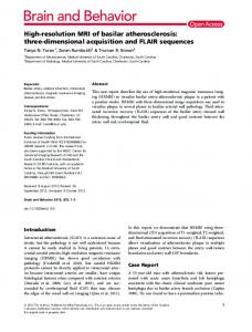
2.1. Recording of raw data The electrode attachment has been a well-known obstacle in developing high-resolution EEG recording. The flexible net (Electrical Geodesics, Inc.) we obtained has a special tension structure, which distributes all the sensors uniformly across the surface of the head and holds them against the head (see Fig. 1.a.). Each electrode is embedded in a small sponge and the whole net is soaked in a saline/detergent electrolyte for about 30 minutes before applying it on the subject’s head. The application of the net takes 10 - 15 minutes in most cases. The software to check electrode impedance is convenient. We regularly record continuous EEG with most electrodes below 20 kΩ impedance at the same time allowing up to 60 kΩ incidental individual electrode impedance. The goal of the traditional scalp abrasion has been to obtain below 5 kΩ sensor impedance, however, it is evident that the high input impedance amplifiers provide good quality EEG recordings with 10 - 15 times higher impedance. Our interest lies in the fairly low frequency signals of cognitive ERPs and for us the higher than commonly used impedance is no problem. In order to understand the contamination of non-neural potentials in recordings utilizing the net, which covers the whole head, we analyzed the extent of blink and other eye movement appearance. We observed that both eye blinks and voluntary eye movements activate a large frontal region (observable up to 39 sensor sites) and a string of neck electrodes. Phase reversals are observed around the eyes and in the frontotemporal regions. International 10 - 20 System electrode locations would definitely fail to pick up the maximum amplitudes of these non-neural potentials.
12
INTERNATIONAL JOURNAL OF BIOELECTROMAGNETISM, VOLUME 1, NUMBER 1
___________________________________________________________________________________
a) The 128-sensor electrode net on a subject’s head. b) The segmented, baseline corrected, filtered, and averaged signals of one healthy subject performing a face recognition task. Only 13 recording sites are shown for clarity, the numbers on the left of the wave forms refer to the numbers in the montage in Fig.2. A complete recording window, 4350 ms, is shown. The 3 vertical lines refer to the onset of 3 consecutive pictures in the task, first the cue, then the first face/pattern picture followed by the second face/pattern picture.
2.2. Data preprocessing Continuous EEG of 128 electrode sites recorded for the complete duration of the task performance is first segmented according to the stimulus. Usually a prestimulus baseline of about 100 ms is obtained and this is followed by a window including the full single trial task performance, which in our studies so far may vary from 500 ms to 4250 ms. The segmented data are then baseline corrected and filtered, if filtering is desired (more often than not). Next the data are averaged. In this process we also clean the data from blink, eye movement, and muscle artefacts setting automatic rejection values for sweeps unacceptable for averaging. After averaging the ERP appears even though single trial ERP is not uncommon to see in individual cases (see Fig. 1.b. for an example of a cognitive ERP). Grand average wave forms describing all subjects’ data or averages picking only specific parts of tasks can be then created.
2.3. Data transfer and analysis The averaged and filtered data are transferred into Brain Electromagnetic Source Analysis (BESA) program for topographic analysis, spline mapping and current source density (c.s.d.) mapping and, of course, source estimation. The grand averages of data may also be created at the BESA. The principal component analysis (PCA) can be performed next to guide in choosing the number of dipoles in the source model development (see Fig. 2. for an example of topographic mapping and PCA). Then a multiple dipole model is developed using prior anatomical and physiological knowledge and iterative fitting procedure. Spatio-temporal multiple dipole modeling assumes that each equivalent electric dipole represents a small region of firing parallel neurons. A detailed recent review concerning different issues in dipole modeling can be found (Achim 1995). In BESA it is assumed that each dipole has both a stationary location and orientation but changes its moment with time. The moment of the dipole is represented in the source activity, which varies in amplitude throughout the analyzed window and reflects dipole strength as a function of time. These point dipoles are only approximations to regional sources and describe many simultaneously active and somewhat spatially separated neurons. A reliable model has a low residual variance, is stable when tested by adding a new dipole, most of its sources are temporally and spatially separated from each other and, finally, the model makes physiological sense.
13
INTERNATIONAL JOURNAL OF BIOELECTROMAGNETISM, VOLUME 1, NUMBER 1
___________________________________________________________________________________
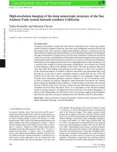
Steps of data processing before source localization. a) The electrode montage used for reading data into source analysis program. b) Examples of topographic maps (spline and current source density maps) created for each time-point. Here a visual evoked potential is shown at 201 ms. c) Principal component analysis of the data can be performed for guiding in the process to find the correct number of dipoles in the source model. After developing source models for the data the location and orientation coordinates and the activation patterns of the sources are analyzed. If an MRI or a CT is available from the subject’s head the coordinates of the dipoles are transferred into the image for naming the active brain regions, if no anatomic individual information is available the stereotaxic atlas (Talairach and Tournoux 1988) can be used to locate the active regions.
3. Example: data from familiar face recognition task The purpose of the study to be described was to test if electrical anticipatory activity in the human frontal cortex may be spatially separated in correspondence with different visual tasks. An earlier preliminary finding in a spatial versus central pattern recognition task study suggested that indeed the frontal cortex shows functional specialization (Tarkka and Basile 1998). Nineteen healthy subjects were tested each in one session of a face/pattern recognition task. Stimuli consisted of pictures of faces and dot patterns superimposed on them shown on a computer screen. One experimental session consisted of 4 series, 50 trials in each. Continuous EEG was recorded with 128 electrodes. The data were sampled each 4 ms (250 Hz A/D), with a low pass filter of 100 Hz, and with a time constant (high pass filter) of 0.01. Each subject’s EEG was segmented, filtered, baseline corrected, cleaned, and averaged off-line. Noninvasive source localization of the data was performed with the Brain Electromagnetic Source Analysis (BESA, version 3.0) algorithm (Scherg and von Cramon 1986; Scherg and Picton 1992; Berg and Scherg 1994, Scherg and Berg 1998). A 4-shell spherical head volume conductor model was used to develop equivalent electric dipole models and a head radius of 85 mm was used. First, grand averaged wave forms were created from individual subjects’ data for the face and the pattern recognition tasks. Spatio-temporal multiple dipole models were independently developed for both sets of data. In order to ascertain that the number of dipoles in the models was sufficient, one test dipole was added as recommended by Scherg and Berg (1998). If a new dipole failed to attract any activity, i.e. its source potential was negligible with no decline in the residual variance (RV), it was removed and the number of dipoles was judged sufficient.
14
INTERNATIONAL JOURNAL OF BIOELECTROMAGNETISM, VOLUME 1, NUMBER 1
___________________________________________________________________________________
Spatio-temporal multiple dipole source analysis of the face recognition task. An 8-dipole model is shown for grand averaged data of 19 subjects in an anticipation task to familiar face recognition. On the left are the source activities and on the right 3 different views of the model. Below is the box for temporal variation of the residual variance (RV) of the model. Time window for the source activities and the RV is 1500 ms from the onset of the cue. After descriptive data analysis the source models were developed. First the VEP elicited by the cue was separately analyzed as well as the CNV (two windows: 0-400 ms and 400-1200 ms, respectively) and then the models were combined. The face and pattern wave form VEPs were similar up to 300 ms and were both modeled successfully with 5 dipoles. The peak amplitude of the CNV occurred at 1090 ±79 and 1095 ±83 for face and pattern wave forms, respectively, and 3 dipoles were the minimum number to model the CNV window data. The final models suggested 7 active brain regions for the tasks in question. The reason was that the right prefrontal CNV source consisted of two dipoles with different orientations. VEP sources were very similar in the two tasks. Frontal source for pattern recognition CNV was more lateral and dorsal compared to face recognition CNV source, which was more medial. Both CNVs had frontal sources in both hemispheres however the anticipation for face recognition produced fairly even bilateral activation. Clearly differing from that the right prefrontal dipole moment was threefold in comparison to its left counterpart in the models for pattern recognition CNV. The model explaining the sources of the anticipation of the face recognition is shown in Fig. 3. Source modeling of CNV data of this demanding visual task indicated not only frontal sources for human CNV but that specific time windows of the CNVs of these two conditions had their sources in different regions in the right prefrontal cortex. This can be taken to indicate functional specialization in the frontal lobe, i.e. that different prefrontal areas of the human brain are involved in attentive anticipation of different kinds of visual stimuli. Thus our example shows that utilizing high-resolution EEG and a carefully designed visual task new information of specifically human cognitive function can be obtained non-invasively.
15
INTERNATIONAL JOURNAL OF BIOELECTROMAGNETISM, VOLUME 1, NUMBER 1
___________________________________________________________________________________
References 1. 2. 3. 4. 5.
6. 7.
Achim A: Cerebral source localization paradigms: Spatiotemporal source modeling, Brain and Cognition 27:256-287, 1995. Berg P, Scherg M: A fast method for forward computation of multiple-shell spherical head models, Electroencephalography and clinical Neurophysiology 90:58-64, 1994. Scherg M, Berg P: Brain Electromagnetic Source Analysis, Version 3.0, Megis, Münich, 1998. Scherg M, von Cramon D: Evoked dipole source potentials of the human auditory cortex, Electroencephalography and clinical Neurophysiology 65:44-360, 1986. Scherg M, Picton TW: Separation and identification of event-related potential components by brain electric source analysis, Electroencephalography and clinical Neurophysiology Suppl. 42:24-37, 1992. Talairach JA, Tournoux P: Co-planar stereotaxic atlas of the human brain, Thieme Verlag, New York, 1988. Tarkka IM, Basile LFH: Electric source localization adds evidence for task-specific CNVs, Behavioural Neurology 11:21-28, 1998.
16