arXiv:1607.04122v1 [q-bio.QM] 14 Jul 2016
iMet: A computational tool for structural annotation of unknown metabolites from tandem mass spectra Antoni Aguilar-Mogas∗ Miriam Navarro†‡
Marta Sales-Pardo∗
Ralf Tautenhahn§
Roger Guimer`a∗¶k
Oscar Yanes†‡k
Abstract Untargeted metabolomic studies are revealing large numbers of naturally occurring metabolites that cannot be characterized because their chemical structures and MS/MS spectra are not available in databases. Here we present iMet, a computational tool based on experimental tandem mass spectrometry that allows the annotation of metabolites not discovered previously. iMet uses MS/MS spectra to identify metabolites structurally similar to an unknown metabolite, and gives a net atomic addition or removal that converts the known metabolite into the unknown one. We validate the algorithm with 148 metabolites, and show that for 89% of them at least one of the top four matches identified by iMet enables the proper annotation of the unknown metabolite. iMet is freely available at http://imet.seeslab.net.
1
Introduction
The great success in the characterization of genes, transcripts and proteins is a direct consequence of two factors. First, such molecules result from the concatenation of a small set of known monomers, namely, nucleotides and amino acids. Second, existing technologies and bioinformatic tools allow for the amplification and subsequent accurate characterization of the sequence of ∗ Departament
d’Enginyeria Qu´ımica, Universitat Rovira i Virgili, Tarragona, Spain. for Omic Sciences, Universitat Rovira i Virgili, Reus, Spain. ‡ Metabolomics Platform, Spanish Biomedical Research Center in Diabetes and Associated Metabolic Disorders (CIBERDEM), Madrid, Spain. § Scripps Center for Metabolomics and Mass Spectrometry, The Scripps Research Institute, La Jolla, CA, USA. ¶ Instituci´ o Catalana de Recerca i Estudis Avan¸cats (ICREA), Barcelona, Spain. k To whom correspondence should be addressed:
[email protected];
[email protected] † Centre
1
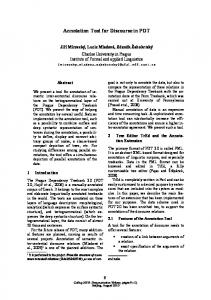
monomers. Metabolomics, in contrast, aims to identify and elucidate the structure of metabolites, which are not sequences of monomers and do not result from a residue-by-residue transfer of information. Instead, the large diversity of metabolites in living organisms results from series of chemical transformations catalyzed mainly by enzymes. As for the identification of proteins in proteomics, structural annotation of metabolites in complex biological mixtures relies on tandem mass spectrometry (MS/MS) analysis. However predicting MS/MS spectra for metabolites is much more challenging than for peptides. In practice, therefore, annotating metabolites relies on their MS/MS spectra being present in reference databases [1, 2, 3, 4]. In the simplest situation, the sample metabolite and its MS/MS spectra are already included in a reference database, so that the metabolite is annotated by matching both the intensities and the mass-to-charge values of each fragment ion to values from pure standard metabolites in the database. Unfortunately, MS/MS spectra of a large number of known metabolites are not described in databases [5]. To assist the structural annotation of such metabolites, efforts have emerged recently to heuristically predict fragmentation patterns in silico and compare these to experimental MS/MS spectra [6, 7, 8, 9, 10, 11, 12, 13]. Other methods are not based on MS/MS data and instead use the accurate mass of MS peaks. These methods often require additional information about the sample metabolite (e.g. pathways in which they participate), and the use of high precision instruments and techniques such as the Fourier Transform Ion Cyclotron Resonance (FTICR) or Orbitrap Fourier transform MS [14, 15, 16]. Despite these efforts, the false positive rate of these methods is still too high to use them in untargeted metabolomics analysis. Finally, in the most challenging case (which, arguably, is also very common), the sample metabolite is completely unknown, that is, the metabolite is not described in databases [17]. Existing approaches for this situation use neutral losses and characteristic fragment ions as signatures for unique chemical functional groups. These approaches have proved to be effective for classifying very specific lipid structures such as acyl-carnitines (e.g., fragments at m/z 85,02 and 60,08), glycerolipids, glycerophospholipids (e.g., fragment m/z 184.07) and sphingolipids [18, 19, 20]. However, there is not a general tool that allows structural annotation of unknown metabolites from their MS/MS spectra. To help in the annotation of completely unknown metabolites we have developed iMet, a computational tool designed to fill that gap (Fig. 1; iMet is avilable online at http://imet.seeslab.net). Its two only inputs are the ESI QTOF MS/MS spectra and the exact mass of an unknown metabolite (i.e., a metabolite that is not yet annotated in any database). Optionally, to increase the accuracy, the isotopic pattern of the intact unknown ion can also be supplied. Given these inputs, the algorithm identifies metabolites in a reference database that are likely to be structurally very similar to the unknown metabolite. Finally, iMet produces a sorted list of candidates, ranked by their similarity to the unknown metabolite. The algorithm also suggests the chemical transformation that is most likely to separate each of the candidates from the unknown metabolite. 2
Figure 1: The iMet algorithm. For each unknown metabolite, the algorithm takes as input its experimental MS/MS spectra (for different collision energies), its exact mass, and (optionally) its isotopic pattern. Then, iMet compares these inputs to a reference database containing experimental MS/MS spectra of known compounds (see text and Fig. 5 for details). The algorithm outputs a sorted list of candidate neighbors of the unknown metabolite. For each candidate neighbor, iMet gives a chemical transformation that converts the candidate into the unknown metabolite. The accuracy of the prediction is defined as a numeric score (s), whose value goes from 0 for the lowest reliability to 1 for the highest reliability.
3
2
Basic principle of iMet
Metabolites can be represented as nodes in a network; two metabolites A and B are connected, that is, are neighbors, if one can obtain the chemical structure of B by a chemical transformation of A, and vice versa (see Fig 2A). By a chemical transformation here we mean the addition or removal of a moiety, or a conformational change. By definition, neighbor metabolites are structurally more similar than a typical pair of non-neighbor metabolites, and this structural similarity should be reflected in their MS/MS spectra because the fragmentation pattern of a metabolite highly depends on its chemical structure. Therefore, from the MS/MS spectrum of a metabolite that is not annotated in the network, a trained algorithm should be able to locate possible neighbors on the basis of spectral similarity. To probe this idea, we built a network of neighbor metabolites on the basis of 814 “reactant pairs” (RP) defined in the KEGG database [21]. In KEGG, substrates and products of a known biochemical reaction are paired according to their chemical structure using graph theory [22] (see Fig 2A). By construction, the two metabolites in a RP are neighbors in the network, so we use RPs as ground truth for neighborhood. (Note, however, that not all neighbor metabolite are annotated as RPs in KEGG. This occurs, for example, when there is no described biochemical reaction that can transform one into the other, even though they are structurally very similar. Thus, the network of RPs in KEGG and our dataset of neighbor metabolites is a subgraph of the full network of neighbor metabolites that potentially exist in nature.)
2.1
Neighbor metabolites share structural similarities
To assess the structural similarity between neighbor metabolites, we computed the Dice coefficient [23] between ECFP4 molecular fingerprints [24] of pairs of metabolites. We obtained the molecular fingerprint for 5,060 different metabolites from public databases, including 932 metabolites listed in KEGG as forming a RP with another metabolite (and thus, being effectively a neighbor of another metabolite in the network). We compared all the possible pairs of structures and computed the receiver operating characteristic (ROC) curve [25], to check if structural similarity could be used to discriminate between neighbors and nonneighbor metabolites (Fig 2B). An overall measure of the discriminatory power is the area under the ROC curve (AUC statistic), which indicates how often RP (and in general, neighbor metabolites) have a higher structural similarity than metabolites that are not RPs. The value of the AUC found in this case was of 0.96, indicating that in the vast majority of cases, two metabolites that are neighbors (that are one chemical tranformation away from each other) have a more similar structure than those that are not. We also found that 95% of the RPs have a Dice coefficient higher than 0.22. With the aim of establishing the best threshold in a scale of 0 to 1 to separate between RPs and non RPs, we looked for the Dice coefficient value that minimizes the classification errors. To do so, we calculated the False Positive 4
Rate (FPR) and the False Negative Rate (FNR) at each value of the Dice coefficient. In this context, the FPR corresponds to the proportion of non RPs that have a higher Dice coefficient value than a given threshold, while the FNR represents the ratio of RPs that have a Dice coefficient value lower than that same threshold (Fig 2C). Adding these two curves into a third curve, we obtain the missclassification ratio, that is the ratio of pairs of metabolites that would be incorrectly classified if we used a certain value of their Dice coefficient to discriminate between RPs and non RPs. We found that the missclassification ratio is minimal for a Dice coefficient value of 0.32, with a FPR of 0.09 and a FNR of 0.09, for a total missclassification ratio of 0.18 (highlighted in Fig 2B and C). By using this value as a threshold to separate between RPs and non RPs the classification error is minimum.
2.2
Neighbor metabolites have similar MS/MS spectra
In order to numerically quantify the similarities between two MS/MS spectra, we use the cosine similarity, as this method is both time efficient and accurate (see Supplementary Data and Supplementary Table S1 for a discussion). In particular, we discretize each spectrum in equal intervals of width δm. In this way, for each spectrum we can construct an intensity vector v in which element vi corresponds to the relative intensity of m/z values in the interval [mi , mi +δm]. (Note that we use δm = 0.01Da, and we disregard relative intensity values below 1% of the highest m/z value.) Then, the cosine similarity c between spectra v and u is simply the dot product of the two vectors divided by the product of their norms. P vi ui c= i (1) kvkkuk To validate the hypothesis that spectral similarity is indicative of neighborhood in the network, we quantified to which extent metabolites that are neighbors have similar MS/MS spectra (Fig. 3). To this end, we considered all metabolites in KEGG for which we had the experimental MS/MS spectra from public databases (HMDB [26], MassBank [27] and METLIN [28]), which corresponds to 1,763 metabolites, and compared their spectra using the cosine similarity (Fig. 3A and B). We used the ROC curve to quantify the power of spectral similarity to distinguish pairs of metabolites that are RPs in KEGG (and, therefore, neighbors) from those that are not (Fig. 3C, D and E). In this case, the area under the ROC curve indicates how often RP metabolites have a higher spectral similarity than metabolites that are not RPs. For the three collision energies 10V, 20V and 40V in negative ionization mode, we found AUC10V = 0.81, AUC20V = 0.83 and AUC40V = 0.80, which indicates that the similarity between MS/MS spectra is useful to identify neighbor metabolites. Comparing MS/MS spectra in positive ionization mode gave similar AUC values. Note that these metrics
5
A
O O
OH HO
HO
O
HO
HO
OH OH
N
N
O O
O
P OH
HO
OH
P OH
N
O
O O
O
P
OH
O
H2N
OH
O
N N
N
OH
Adenosine triphosphate
glu
glu6P
O
P OH
HO
ATP
ADP
O O
P
OH
OH
OH
Adenosine diphosphate C
B
1.0 Missclassification Rate
1 True Positive Rate
OH
OH
OH
N N
P
Glucose-6-Phosphate
Glucose H 2N
O
0.5
0 0
0.5 False Positive Rate
1
0.8 0.6 0.4 0.2 0.0 0.0
0.2
0.4 0.6 Dice Coefficient
0.8
1.0
Figure 2: Neighbor metabolites. (A) An example of neighbor metabolites. Phosphorylation of glucose. Glucose (glu) is transformed into glucose-6-phosphate (glu-6P), while adenosine triphosphate (ATP) is dephosphorylated into adenosine diphosphate (ADP). Following the definition of reactant pairs in KEGG, glu and glu-6P are one chemical transformation (phosphorylation) away from each other. The same applies to ATP and ADP. (B) ROC curve of the structural similarity of neighbor and non-neighbor metabolites on the basis of the Dice coefficient. The AUC is of 0.96, indicating that neighbor metabolites have higher structural similarity than non-neighbor metabolites. The blue circle marks the maximum discrimination point. (C) False Negative Rate, i.e. ratio of RPs with a Dice coefficient below (green) a certain Dice coefficient value, and False Positive Rate, i.e. ratio of non RPs with a Dice coefficient above (red) a certain Dice coefficient value, as a function of that value. Black line corresponds to the sum of the other two curves. The black circle indicates the minimum of this curve, which corresponds to the maximum discrimination point.
6
Rel. Intensity
100 80 60 40 20 0 20 40 60 80 100
O OH
Arachidonic acid m/z O OH
OH
Hydroxyeicosatetraenoic acid
C True Positive Rate
B
c = 0.972
c = 0.011 O
100 80 60 40 20 0 20 40 60 80 100
OH
Rel. Intensity
A
Arachidonic acid m/z O OH
Lauric acid
D
E
1.0
0.5 10V 0.0 0.0
0.5
40V
20V 1.0 0.0 0.5 1.0 0.0 False Positive Rate
0.5
1.0
Figure 3: Similarity of MS/MS spectra discriminates between neighbor and non-neighbor metabolites. (A) MS/MS spectrum similarity for two neighbor metabolites (spectral similarity 0.972), and (B) for two non-neighbor metabolites (spectral similarity 0.011). (C-E) Classification power of the cosine similarity. We show the ROC curve for the cosine similarity when discriminating between RPs and non RPs in KEGG for different collision energies (10, 20 and 40V) in negative ionization mode, with a total area under the curve of (C) 0.81, (D) 0.83, and (E) 0.80, respectively. The three highlighted symbols correspond to cosine similarities of 0.5 (blue dot), 0.1 (purple diamond), and the first non-zero value (red triangle).
7
quantify the predictive power of the spectral similarity when comparing only two MS/MS spectra with the same collision energy and ionization mode. In general, MS/MS spectra acquired at high collision energies have higher sensitivity but lower specificity, and conversely for low collision energies. This implies that comparing two spectra obtained at high collision energies results in a highly conservative classification method, discarding pairs that are actually neighbor metabolites (low specificity or true negative rate), but assuring that most of the metabolites classified as neighbor metabolites are real neighbors (high sensitivity or true positive rate). In contrast, spectral similarity becomes a poorer classification method at low collision energies: while most of the neighbor metabolites are correctly classified as such, some non-neighbor metabolites are also labeled as neighbors. Finally, the analysis reveals that information is usually non-redundant–some pairs of metabolites have high spectral similarity at high collision energies and low similarity at low energies, whereas for other pairs the opposite is true. These results indicate that, indeed, spectral similarity at a fixed collision energy is predictive to some extent. As we show next, however, the predictive power of spectral similarity can be increased by considering spectra at different collision energies simultaneously, and combining them with mass difference, and, optionally, the isotopic pattern of the unknown metabolite (i.e., precursor ion).
2.3
Neighbor metabolites have well-defined mass differences, which correspond to well-defined chemical transformations
To complement the information obtained from the spectral similarity, we study the differences in exact mass between neighbor metabolites (Fig. 4). The mass difference between two metabolites corresponds to the mass of the group of atoms added to (or removed from) one of the metabolites to convert it into the other. As before, we take the mass difference, ∆m, between KEGG RPs as ground truth. We considered 5,060 different metabolites including 814 RPs. Even though the KEGG database comprises more RPs, the limiting factor is how many of these RPs have MS/MS spectra in public databases. We are convinced that adding more metabolites (165 by December 2015) to the analysis (out of a total of 5,060 metabolites currently considered) would not alter the results presented other than increasing its accuracy. Calculating the mass difference for every pair of metabolites in our database and plotting the proportion of RPs for each value of the mass difference yields a histogram that we assume reflects the probability of two metabolites being neighbors given their mass difference, although specific systems may deviate from this general (average) pattern [29]. As we show in Fig. 4A, this distribution displays well-defined maxima at specific ∆m values. Therefore, it is much more likely that two metabolites are neighbors if their ∆m corresponds to one of the maxima of the distribution in Fig. 4A. To understand what these ∆m represent, we extracted 202 distinct
8
A 0.12
p (RP | ∆ m)
0.1
0.08
0.06
0.04
0.02
0 0
20
40
60
80
100
120
140
160
180
200
∆ m [ Da ]
B
12
O H2
% of all reactant pairs
10 8
HPO3
CH2 6
CO2 4
H 2O
C6H10O5
2 0 1
10 100 Mass difference [ Da ]
Figure 4: Probability of neighborhood and of chemical transformation. (A) Probability of RP given a mass difference between two metabolites. We show the fraction of RPs that have associated a mass difference within a specific interval. We constructed the histogram using all compounds listed in KEGG, with bins of 0.01 Da. (B) Percentage of all reactant pairs in KEGG with a certain mass difference. The most frequent mass differences correspond to well defined moieties. The seven most frequent moieties (highlighted in the Figure) account for 46% of all reactant pairs. 9
chemical transformations from a total of 814 reactant pairs, (see Supplementary Table S2). Since each chemical transformation implies a well-defined mass difference, the distribution of mass differences among RPs is localized around certain values that correspond to the most common interconversions of atoms (Fig. 4B). For example, 11.3% of all reactant pairs correspond to the net addition of an oxygen atom (∆m=15.995 Da), 10.1% to the net addition of H2 (∆m=2.016 Da), and 6.9% to the addition of a phosphate group (∆m=79.966 Da). In summary, a relatively small number of transformations account for a large number of the observed RPs (for example, the seven most common transformations account for 46% of the pairs).
2.4
A random forest classifier identifies neighbor metabolites on the basis of mass difference and MS/MS spectral similarity
Taken together, our results indicate that MS/MS spectral similarity and mass difference are both predictive of neighborhood between two metabolites. Therefore, given an unknown metabolite and a library with MS/MS spectra, we propose that it is possible to identify the metabolites in the library that are most likely to be neighbors of the unknown one. To demonstrate this, we implemented a random forest classifier [30] to identify potential neighbors. The random forest classifier has the advantage of automatically taking care of the non-monotonic relationship between mass difference and probability of neighborhood, as well as the complex non-linear similarity patterns between MS/MS spectra at different collision energies. We trained the classifier using 50,000 metabolite pairs, including all RPs for which we have MS/MS spectra and completed with randomly chosen pairs among a library of 5,060 compounds with MS/MS data in databases such as HMDB [26], MassBank [27], and METLIN [28]. Increasing the size of the training set slowed the training to the point of making it unfeasible, and did not result in an increase of the accuracy of the classifier (because adding more pairs of unrelated compounds did not provide new useful information to the model). Based on the accurate mass-to-charge (m/z) measurement of a protonated (M+H)+ or deprotonated (M-H)- precursor ion of the unknown metabolite (mass error