© 2002 Oxford University Press
Nucleic Acids Research, 2002, Vol. 30, No. 12 e52
Improved detection of small deletions in complex pools of DNA Mark Edgley, Anil D’Souza1, Gary Moulder1, Sheldon McKay, Bin Shen, Erin Gilchrist, Donald Moerman and Robert Barstead1,* Biotechnology Laboratory and Department of Zoology, University of British Columbia, Vancouver, BC V6T 1Z4, Canada and 1Department of Molecular and Cell Biology, Oklahoma Medical Research Foundation, 825 N.E. 13th Street, Oklahoma City, OK 73104, USA Received December 14, 2001; Revised and Accepted April 16, 2002
ABSTRACT About 40% of the genes in the nematode Caenorhabditis elegans have homologs in humans. Based on the history of this model system, it is clear that the application of genetic methods to the study of this set of genes would provide important clues to their function in humans. To facilitate such genetic studies, we are engaged in a project to derive deletion alleles in every gene in this set. Our standard methods make use of nested PCR to hunt for animals in mutagenized populations that carry deletions at a given locus. The deletion bearing animals exist initially in mixed populations where the majority of the animals are wild type at the target. Therefore, the production of the PCR fragment representing the deletion allele competes with the production of the wild type fragment. The size of the deletion fragment relative to wild type determines whether it can compete to a level where it can be detected above the background. Using our standard conditions, we have found that when the deletion is 600 bp, they do not work well to detect mutants with smaller deletions. Here we report a new strategy to detect small deletion alleles in complex DNA pools. Our new strategy is a modification of our standard PCR based screens. In the first round of the nested PCR, we include a third PCR primer between the two external primers. The presence of this third primer leads to the production of three fragments from wild type DNA. We configure the system so that two of these three fragments cannot serve as a template in the second round of the nested PCR. The addition of this third primer, therefore, handicaps the amplification from wild type template. On the other hand, the amplification of mutant fragments where the binding site for the third primer is deleted is unabated. Overall, we see at least a 500-fold increase in the sensitivity for small deletion
fragments using our new method. Using this new method, we report the recovery of new deletion alleles within 12 C.elegans genes. INTRODUCTION Until recently, most genes in the nematode Caenorhabditis elegans were known through their mutant phenotypes. Now, however, more C.elegans genes are known through sequencing than through genetics (1); of the approximately 19 000 genes in C.elegans, only about 1300 have mutant alleles. As genetics is one of the most important tools in the arsenal of C.elegans biologists, we and others have devised methods to derive mutations in C.elegans genes known only through their sequence (2–6). Our present methods are conceptually simple. We treat worms at the L4 larval stage with a mutagen. The progeny of the mutagenized worms are subdivided into populations that are allowed to reproduce. We then extract the DNA from ∼30% of each population. The extracted DNA samples are pooled and subjected to PCR with nested primer sets (Fig. 1). Candidate populations are identified by the presence of a PCR product that is smaller than the size predicted by the genomic DNA sequence. Each candidate population is subdivided and subjected to similar growth and PCR analysis. This process of sib selection continues until we recover a single individual with the deletion. Using this protocol, typically we can recover such an individual in three steps of growth and sib selection. In principle, our ability to detect a deletion should be a function of the resolution of the gels used for electrophoresis. In practice, however, gel resolution does not determine the size of the deletions that we identify; typically, we do not detect deletions that are less than ∼600 bp. This limitation is a consequence of our effort to reduce, as far as possible, the cost and time required to identify a candidate deletion. To accommodate the relatively low frequency at which deletions are induced by chemical mutagens (7), we pool the DNA template samples to minimize the numbers of PCRs that are needed to detect a deletion. Using our present DNA pooling strategy, for every copy of the deletion DNA we have approximately 12 000 copies of wild type DNA. This places the mutant template at a substantial disadvantage in PCR. Deletion fragments that are close to the size of wild type DNA apparently are not able to overcome this initial disadvantage.
*To whom correspondence should be addressed. Tel: +1 405 271 1766; Fax: +1 405 271 3153; Email:
[email protected]
e52 Nucleic Acids Research, 2002, Vol. 30, No. 12
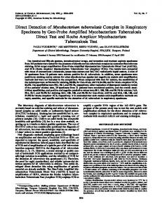
Figure 1. Protocol schematic. Our current protocols use nested PCR to identify smaller than normal PCR fragments derived from a mutagenized population of worms. Typically, we use two external (A and C) and two internal PCR primers (D and E). As shown, the poison primer method adds a primer (B) in the first round of the nested PCR.
However, recent work has shown that a significant proportion of deletions induced by trimethylpsoralen (TMP) treatment followed by UV irradiation are