RESEARCH ARTICLE
IRBIT controls apoptosis by interacting with the Bcl-2 homolog, Bcl2l10, and by promoting ER-mitochondria contact Benjamin Bonneau1*, Hideaki Ando1, Katsuhiro Kawaai1, Matsumi Hirose1, Hiromi Takahashi-Iwanaga2, Katsuhiko Mikoshiba1* 1
Laboratory for Developmental Neurobiology, RIKEN Brain Science institute, Wakoshi, Japan; 2Department of Anatomy, School of Medicine Hokkaido University, Sapporo, Japan
Abstract IRBIT is a molecule that interacts with the inositol 1,4,5-trisphosphate (IP3)-binding pocket of the IP3 receptor (IP3R), whereas the antiapoptotic protein, Bcl2l10, binds to another part of the IP3-binding domain. Here we show that Bcl2l10 and IRBIT interact and exert an additive inhibition of IP3R in the physiological state. Moreover, we found that these proteins associate in a complex in mitochondria-associated membranes (MAMs) and that their interplay is involved in apoptosis regulation. MAMs are a hotspot for Ca2+ transfer between endoplasmic reticulum (ER) and mitochondria, and massive Ca2+ release through IP3R in mitochondria induces cell death. We found that upon apoptotic stress, IRBIT is dephosphorylated, becoming an inhibitor of Bcl2l10. Moreover, IRBIT promotes ER mitochondria contact. Our results suggest that by inhibiting Bcl2l10 activity and promoting contact between ER and mitochondria, IRBIT facilitates massive Ca2+ transfer to mitochondria and promotes apoptosis. This work then describes IRBIT as a new regulator of cell death. DOI: 10.7554/eLife.19896.001 *For correspondence: benjamin.
[email protected] (BB); mikosiba@ brain.riken.jp (KM) Competing interests: The authors declare that no competing interests exist. Funding: See page 23 Received: 27 July 2016 Accepted: 24 November 2016 Published: 20 December 2016 Reviewing editor: Richard S Lewis, Stanford University School of Medicine, United States Copyright Bonneau et al. This article is distributed under the terms of the Creative Commons Attribution License, which permits unrestricted use and redistribution provided that the original author and source are credited.
Introduction Elevation of intracellular Ca2+ concentration serves as a second messenger for numerous processes, including the cell cycle, fertilization or apoptosis (Berridge et al., 2003). At the endoplasmic reticulum (ER), Ca2+ signals are mainly generated by the Ca2+ channel inositol-1,4,5-trisphosphate receptor (IP3R) in response to IP3 binding. The cellular response to a Ca2+ signal depends on the amplitude and frequency of this signal. IP3R is then tightly regulated by post-translational modifications and interacting partners that modulate Ca2+ release according to cellular context (Mikoshiba, 2007; Foskett et al., 2007). In particular, several Bcl-2 family proteins have been reported to regulate IP3R activity (Bonneau et al., 2013). These proteins are well known for their role in apoptosis through the control of outer mitochondrial membrane permeabilization, cytochrome c release, and subsequent activation of caspases (Youle and Strasser, 2008). However, Bcl-2 family proteins are also involved in Ca2+-induced apoptosis. The correct functioning of mitochondria requires Ca2+, which is supplied by some portions of ER that are in physical contact with the mitochondria (called MAMs for mitochondria-associated ER membranes). In MAMs, IP3R associates with the mitochondrial voltage-dependent anion channel (VDAC), allowing a direct transfer of Ca2+ into the mitochondria (Szabadkai et al., 2006). However, if the amount of Ca2+ transferred is too high, it induces cytochrome c release and apoptosis. MAMs are then acknowledged to be an essential component of Ca2+-induced apoptosis (Giorgi et al., 2009). In this regard, some Bcl-2 family proteins localize at ER and modulate Ca2+ release (Bonneau et al., 2013). Several antiapoptotic members notably interact with IP3R, each one
Bonneau et al. eLife 2016;5:e19896. DOI: 10.7554/eLife.19896
1 of 27
Research article
Cell Biology
regulating channel activity by a distinct mechanism. For example, Bcl-2 interacts with the central part of IP3R and reduces Ca2+ release to inhibit proapoptotic Ca2+ signals (Hanson et al., 2008; Rong et al., 2009). By contrast, Bcl-xL interacts with the most C-terminal domain of IP3R and stimulates pro-survival Ca2+ transfer to the mitochondria (White et al., 2005). Recently, Nrz, a Bcl-2 homolog in zebrafish, was shown to interact with the N-terminal IP3-binding domain of IP3R and to decrease ligand binding on the receptor, thus reducing Ca2+ release from ER (Bonneau et al., 2014). Only a few regulators of IP3R interact with the IP3-binding domain (IP3BD) or act by interfering with ligand fixation. Among them, IRBIT acts in a manner similar to Nrz. IRBIT by interacting with the residues of directly competes with IP3 the IP3 binding domain (IP3BD) that are involved in IP3 binding (known as the IP3-binding pocket). This increases the threshold of IP3 required for IP3R opening and then decreases Ca2+ release (Ando et al., 2006). In addition to IP3R, IRBIT interacts with various partners, such as ion transporters and exchangers including NBCe1-B (Shirakabe et al., 2006), NHE3 (He et al., 2008), and Slc26a6 (Park et al., 2013), the Cl channel CFTR (Yang et al., 2009), Fip1 (Kiefer et al., 2009), the ribonucleotide reductase (RNR) (Arnaoutov and Dasso, 2014) and kinases including CaMKIIa (Kawaai et al., 2015), PIPKI, and IIa (Ando et al., 2015). All of these interactions involve the N-terminal region of IRBIT, which contains a serine-rich region with several phosphorylation sites (Ando et al., 2006; Devogelaere et al., 2007). In particular, phosphorylation of Ser68 is required for the subsequent sequential phosphorylation of Ser71, Ser74, and Ser77 by the casein kinase I (Ando et al., 2006; Devogelaere et al., 2007). This multiple phosphorylation of IRBIT is critical for its interaction with IP3R and most of its other partners. Nrz is the zebrafish ortholog of the mammalian antiapoptotic protein Bcl2l10 (also called Nrh, Bcl-B, or Diva/Boo). To date, little is known about Bcl2l10, particularly regarding its effect on Ca2+ signaling. In mammals, Bcl2l10 is mainly expressed in the ovary and testis, but also in the lung and the developing nervous system (Aouacheria et al., 2001; Inohara et al., 1998). Interestingly, IRBIT is also strongly expressed in these organs (Ando et al., 2003). Nrz and IRBIT both regulate IP3R activity by interacting with the IP3BD. However, they do not share a common binding site on the IP3BD as Nrz does not interact with the residues required for IP3 and IRBIT binding (Bonneau et al., 2014). This suggests that Nrz and IRBIT may interact with the IP3BD at the same time. In the present study, we showed that, like Nrz, Bcl2l10 interacts with the IP3BD of IP3R. We then analyzed how IRBIT and Bcl2l10 behave towards each other. We found that IRBIT and Bcl2l10 interacted together independently of IRBIT binding to IP3R and that they cooperated to regulate IP3R activity. Furthermore, we showed that these two proteins localized in MAMs, where they are part of a protein complex with IP3R and VDAC. Unexpectedly, we found that IRBIT promoted cell death through two mechanisms. First during apoptosis, IRBIT was dephosphorylated, and unphosphorylated IRBIT appeared to inhibit the antiapoptotic activity of Bcl2l10. Second, IRBIT appeared to promote contact between ER and mitochondria that may facilitate proapoptotic Ca2+ transfer. Considered collectively, our results suggested a strong relationship between IRBIT and Bcl2l10 and pointed out, for the first time, the implied involvement of IRBIT in cell death.
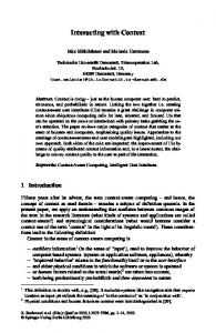
Results Bcl2l10 interacts with IP3BD and reduces Ca2+ release from IP3R The Bcl2l10 orthology group is highly divergent (Guillemin et al., 2011) and human Bcl2l10 only shares 28.4% identity with Nrz (Figure 1A). We therefore first investigated whether Bcl2l10 behaves like Nrz and interacts with IP3R. The three different isoforms of IP3R were immunoprecipitated from HeLa cells extract, and we found that endogenous Bcl2l10 interacted with the three isoforms, although the interaction with IP3R2 appeared weaker than that of IP3R1 or IP3R3 (Figure 1B). We next evaluated whether Bcl2l10 also interacts with the IP3BD (amino acids 224–604 of IP3R1) as does Nrz. Extracts of HeLa cells expressing FLAG-Bcl2l10 were subjected to GST-pulldown assay, and we demonstrated that Bcl2l10 interacts with recombinant GST-IP3BD (Figure 1C). Nrz as well as Bcl-2 and Bcl-xL were shown to interact with IP3R via their N-terminal BH4 domain (Bonneau et al., 2014; Monaco et al., 2012). The deletion of the BH4 domain of Bcl2l10 (DBH4Bcl2l10) suppressed its interaction with GST-IP3RDCD (a protein that has IP3R deleted from its channel domain) and GST-IP3BD (Figure 1C), demonstrating that Bcl2l10 interacted with IP3R via its BH4 domain.
Bonneau et al. eLife 2016;5:e19896. DOI: 10.7554/eLife.19896
2 of 27
Research article
Cell Biology
Figure 1. Bcl2l10 interacts with and regulates IP3R. (A) Clustal Omega alignment of human Bcl2l10 and zebrafish Nrz primary structures. Identical residues are boxed in blue. The positions of conserved Bcl-2 homology (BH) domains and of the C-terminal transmembrane (TM) domain are indicated. (B) Western blot of immunoprecipitation (IP) between the three endogenous IP3R isoforms (IP3R1, IP3R2 and IP3R3) and endogenous Bcl2l10. IgG antibodies are indicated by arrows. Arrow heads indicate bands corresponding to Bcl2l10 in IP. Western blots are representative of three independent experiments. (C) Western blot of GST-pulldown performed with GST, GST-IP3RDCD or GST-IP3BD on lysates of HeLa cells expressing FLAG-Bcl2l10 or FLAG-DBH4Bcl2l10. Arrows indicate the bands corresponding to GST, GST-IP3RDCD and GST-IP3BD. Western blot representative of three independents experiments. (D) Left panel: representative Ca2+ response curve of Fura-2-loaded cells stimulated with 1 mM ATP at the indicated times. Cells were transfected with empty vector or FLAG-Bcl2l10. Basal Fura-2 F340 nm/F380 nm = 0.61 ± 0.01 and 0.59 ± 0.01 for empty vector and Bcl2l10 cells, respectively. Right panel: Bar graph showing the mean amplitude (±SEM) of the ATP-induced Ca2+ peak (n: number of cells analyzed from five independent experiments). (E) Left panel: representative response curve of Fura-2-loaded cells treated with 1 mM thapsigargin at the indicated times. Right panel: Bar graph showing the mean area under curve (AUC) (±SEM) of the thapsigargin-induced Ca2+ peak (n: number of cells analyzed from three independent experiments). ***p0.05. DOI: 10.7554/eLife.19896.002
Bonneau et al. eLife 2016;5:e19896. DOI: 10.7554/eLife.19896
3 of 27
Research article
Cell Biology
Subsequently, we examined the effect of Bcl2l10 on IP3-induced Ca2+ release (IICR) in cultured cells. In these experiments, mouse embryonic fibroblasts (MEF) were used as they exhibit robust IICR in response to ATP stimulation. Expression of Bcl2l10 significantly reduced IICR following the treatment of cells with 1 mM ATP without affecting basal cytosolic [Ca2+], as measured with the cytosolic Ca2+-sensitive dye Fura-2 (Figure 1D). It has been shown that Bcl-2 family proteins can modify Ca2+ release from the ER by acting on the steady-state concentration of Ca2+ in the ER (Pinton and Rizzuto, 2006). To address a possible effect of Bcl2l10 on ER Ca2+ content, MEF cells were incubated with Fura-2 and treated with 1 mM thapsigargin, which induced the depletion of ER [Ca2+]. We found that expression of Bcl2l10 did not alter ER Ca2+ content (Figure 1E). Considered collectively, these results suggest that Bcl2l10 acts like Nrz in zebrafish and reduces Ca2+ release from the ER by interacting with the IP3BD of IP3R.
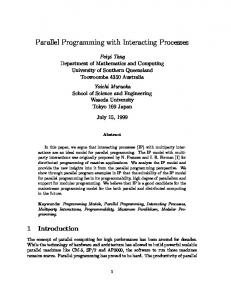
IRBIT and Bcl2l10 exert an additive inhibition of IP3R IRBIT and Bcl2l10 are among the few regulators of IP3R that interact with the IP3BD. This raises the possibility of cooperation or, alternatively, competition between these two proteins. To address this question, we first analyzed the effect of IRBIT and Bcl2l10 on IICR. Overexpression of IRBIT in cells expressing the endogenous protein has no effect on IICR (Ando et al., 2006). Thus, to avoid the contribution of endogenous IRBIT protein, we used MEF cells derived from IRBIT knockout (KO) mice (Kawaai et al., 2015). In these cells, expression of IRBIT or Bcl2l10 alone significantly reduced IICR elicited by ATP treatment compared to the response in the control without affecting basal cytosolic [Ca2+]. Interestingly, co-expression of IRBIT and Bcl2l10 had a stronger effect on IICR than expression of each protein alone (Figure 2A and B). This result suggested that IRBIT and Bcl2l10 cooperate to exert an additive inhibition of IP3R. To further explore the cooperation between IRBIT and Bcl2l10, we next studied whether each of these two proteins has an effect on the interaction of the other with the IP3BD. Extracts from cells expressing IRBIT alone or in combination with Bcl2l10 were subjected to GST-pulldown with recombinant GST-IP3BD. When expressed with Bcl2l10, IRBIT appeared to interact more with IP3BD (Figure 2C). In the same way, we performed a GST-pulldown assay with GST-IP3BD and a recombinant Bcl2l10 protein in the presence or absence of recombinant IRBIT produced in Sf9 cells. This production allows the phosphorylation of IRBIT, which is essential for the interaction of IRBIT with the IP3BD (Ando et al., 2006). Similarly, our results showed that in the presence of IRBIT, Bcl2l10 interacted more strongly with the IP3BD (Figure 2D). Thus, IRBIT and Bcl2l10 appeared to strengthen each other’s interaction with IP3R. IRBIT was first characterized as a protein released from IP3R by IP3 (Ando et al., 2003). Indeed, as IP3 and IRBIT share the same binding site, the binding of IP3 on IP3R occurs to the detriment of the interaction of IRBIT with IP3R. Thus, reduction of the IRBIT interaction with the IP3BD in the presence of an increasing concentration of IP3 reflected the binding of IP3 on the receptor (Figure 2E). However, in the presence of Bcl2l10, we observed that the effect of IP3 on IRBIT’s interaction with the IP3BD was attenuated (Figure 2E). This result confirmed the fact that Bcl2l10 strengthened the interaction of IRBIT with IP3R, and suggested that Bcl2l10 and IRBIT associated to interfere with IP3 binding on the receptor and then reduced Ca2+ release from the ER.
IRBIT and Bcl2l10 interact The above results suggest that IRBIT and Bcl2l10 can form a regulatory complex on IP3R. Consequently, the possibility that these two proteins interact was investigated. Endogenous IRBIT protein was immunoprecipitated from HeLa cells extract, following which we detected an interaction of this protein with endogenous Bcl2l10 (Figure 3A). To further characterize this interaction, we then searched for the domain with which Bcl2l10 and IRBIT are involved. The BH4 domain of Bcl-2 family members mediates their interaction with proteins outside of the Bcl-2 family, such as Raf-1 (Wang et al., 1996), calcineurin (Shibasaki et al., 1997) and VDAC (Shimizu et al., 2000). Co-immunoprecipitation between FLAG-tagged full length Bcl2l10 or DBH4Bcl2l10 and HA-IRBIT showed that the BH4 domain of Bcl2l10 was essential for its interaction with IRBIT, as the deletion mutant DBH4Bcl2l10 lost its ability to bind IRBIT (Figure 3B). The first 104 amino acids of IRBIT are required for its interaction with IP3R (Ando et al., 2003). In particular, phosphorylation of residues Ser71, Ser74, and Ser77 is essential for IRBIT binding on IP3R.
Bonneau et al. eLife 2016;5:e19896. DOI: 10.7554/eLife.19896
4 of 27
Research article
Cell Biology
Figure 2. IRBIT and Bcl2l10 cooperate to regulate IP3R activity. (A) Representative Ca2+ response curve of Fura-2-loaded IRBIT KO MEF cells stimulated with 1 mM ATP at the indicated times. Cells were transfected with empty vector or with a plasmid expressing either FLAG-Bcl2l10 or FLAG-IRBIT alone, or FLAG-Bcl2l10 and FLAG-IRBIT together. Basal Fura-2 F340 nm/F380 nm = 0.61 ± 0.01, 0.59 ± 0.01, 0.59 ± 0.01 and 0.58 ± 0.01 for empty vector, Bcl2l10, IRBIT and Bcl2l10+IRBIT, respectively. (B) Bar graph showing the mean amplitude (±SEM) of the ATP-induced Ca2+ peak (n: number of cells analyzed Figure 2 continued on next page
Bonneau et al. eLife 2016;5:e19896. DOI: 10.7554/eLife.19896
5 of 27
Research article
Cell Biology
Figure 2 continued from five independent experiments). (C) Western blot of GST-pulldown performed with GST or GST-IP3BD on lysates of HeLa cells expressing HA-IRBIT alone or in combination with FLAG-Bcl2l10. Quantification was performed from three independent experiments. (D) Western blot of GST-pulldown performed with GST-IP3BD on recombinant Bcl2l10 alone or in combination with recombinant IRBIT produced in Sf9 cells. Quantification was performed from three independent experiments.(E) Western blot of GST-pulldown performed with GST-IP3BD on lysates of HeLa cells expressing HAIRBIT alone or in combination with FLAG-Bcl2l10 in the presence of 0, 1 or 10 mM IP3. Quantification was performed from three independent experiments. *p