This article appeared in a journal published by Elsevier. The attached copy is furnished to the author for internal non-commercial research and education use, including for instruction at the authors institution and sharing with colleagues. Other uses, including reproduction and distribution, or selling or licensing copies, or posting to personal, institutional or third party websites are prohibited. In most cases authors are permitted to post their version of the article (e.g. in Word or Tex form) to their personal website or institutional repository. Authors requiring further information regarding Elsevier’s archiving and manuscript policies are encouraged to visit: http://www.elsevier.com/copyright
Author's personal copy
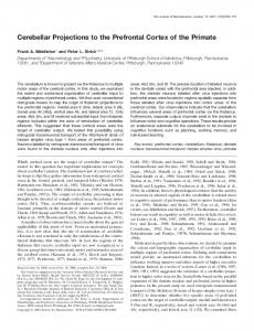
Neuropsychologia 46 (2008) 2056–2063
Lateralized contribution of prefrontal cortex in controlling task-irrelevant information during verbal and spatial working memory tasks: rTMS evidence Marco Sandrini a,∗ , Paolo Maria Rossini b,c , Carlo Miniussi a,d a
Cognitive Neuroscience Section, IRCCS ‘S. Giovanni di Dio-Fatebenefratelli’, via Pilastroni 4, Brescia, Italy b Clinical Neurology, University ‘Campus Biomedico’, Rome, Italy c Casa di Cura S. Raffaele Cassino & IRCCS S. Raffaele Pisana, Rome, Italy d Department of Biomedical Sciences and Biotechnologies, University of Brescia, Italy
Received 11 September 2007; received in revised form 31 January 2008; accepted 1 February 2008 Available online 8 February 2008
Abstract The functional organization of working memory (WM) in the human prefrontal cortex remains unclear. The present study used repetitive transcranial magnetic stimulation (rTMS) to clarify the role of the dorsolateral prefrontal cortex (dlPFC) both in the types of information (verbal vs. spatial), and the types of processes (maintenance vs. manipulation). Subjects performed three independent experiments (1-back and 2-back tasks) while rTMS was applied over dlPFC for 500 ms in the last period of the delay. In two experiments (1 and 2) physically identical stimuli (letters shown at different locations on a screen) under different domain conditions (letters or locations) were employed. Under these conditions, we discovered a double dissociation only in the 2-back task: during the letter condition, when applied to the right dlPFC, rTMS significantly delayed task performance, whereas, the same result was present during the location condition, but only when rTMS was applied to the left dlPFC. The other 2-back task (experiment 3), in which we had eliminated the task-irrelevant information (i.e. we used stimuli that varied only in one domain), did not show significant results. We propose that the functional dichotomy of the hemispheres may be due to mechanisms of cognitive control on interference, which resolve conflict through the inhibition of task-irrelevant information only during high WM load. In conclusion, these findings confirm the role of dlPFC in implementing top-down attentional control, and provide evidence for the theoretical suggestion that working memory serves to control selective attention in the normal human brain. © 2008 Elsevier Ltd. All rights reserved. Keywords: Dorsolateral prefrontal cortex (dlPFC); Lateralization; Working memory; Inhibition; Repetitive transcranial magnetic stimulation (rTMS)
1. Introduction It has been hypothesized that higher brain functions such as language, planning and problem-solving rely on working memory (WM), i.e., a system which acts to temporarily maintain and manipulate task-relevant information (Baddeley, 1986; Just & Carpenter, 1992; Shallice, 1988). According to Baddeley (1986, 2000), working memory is represented by a central executive that controls information in three storage buffers (the phonological loop, the visuo-spatial
∗
Corresponding author. Tel.: +39 030 3501594; fax: +39 030 3533513. E-mail address:
[email protected] (M. Sandrini).
0028-3932/$ – see front matter © 2008 Elsevier Ltd. All rights reserved. doi:10.1016/j.neuropsychologia.2008.02.003
sketchpad and the episodic buffer) that act as a workspace for the storage and manipulation of information. Evidence from neurophysiological (Fuster, 1989; GoldmanRakic, 1987), neuropsychological (Shimamura, 1994; Stuss, Eskes, & Foster, 1994), functional neuroimaging (see Fletcher & Henson, 2001, for a review) and single or repetitive transcranial magnetic stimulations (rTMS) (see Mottaghy, 2006, for a review) studies supports a role of the prefrontal cortex (PFC) in a wide variety of WM tasks. Nevertheless, even if PFC has been identified to play a key role in WM, till now there is no consensus on its functional organization in humans (Cabeza & Nyberg, 2000; Duncan & Owen, 2000). Investigators have raised the question of whether different PFC regions subserve different functional processes
Author's personal copy
M. Sandrini et al. / Neuropsychologia 46 (2008) 2056–2063
and/or different types of information. It has been suggested that dorsolateral PFC (dlPFC) and ventrolateral PFC (vlPFC) are associated, respectively, with manipulation/monitoring and maintenance/inhibition (D’Esposito, Postle, & Rypma, 2000; Owen, Evans, & Petrides, 1996; Petrides, 2000), or with spatial and non-spatial information (D’Esposito et al., 1998; GoldmanRakic, 1987, 1995; Haxby, Petit, Ungerleider, & Courtney, 2000). It has also been suggested that left and right PFCs are associated, respectively, with verbal and non-verbal information (Smith & Jonides, 1997), or that PFC is functionally organized by both process and type of information (Johnson, Raye, Mitchell, Greene, & Anderson, 2003). However, a recent alternative perspective portrays “working memory as a property that arises through the coordinated recruitment, via attention, of brain systems that have evolved to accomplish sensory-, representation-, or action-related functions”. One corollary of this emergent process view is that the contribution of PFC to working memory does not include the temporary storage of information (see Postle, 2006, for a review). Evidence from TMS studies supports this point showing that delay-period rTMS does not disrupt storage of verbal (Feredoes, Tononi, & Postle, 2007; Postle et al., 2006) or spatial (Hamidi, Tononi, & Postle, 2006) information. In particular, in one of these studies (Postle et al., 2006) subjects were presented two types of trials in random order in which they were required to either (1) maintain a sequence of letters across a delay period or (2) manipulate (alphabetize) this sequence during the delay in order to respond correctly to a probe. Their two-step procedure entailed first, acquiring fMRI data and second, delivering rTMS to fMRI-identified areas of the dlPFC and superior parietal lobe while the same subjects performed the same task. Although, rTMS of the dlPFC selectively disrupted manipulation, rTMS of the superior parietal lobe disrupted manipulation and shortterm retention at the same extent. In conclusion, their findings are consistent with the view that dlPFC contributes more importantly to the control of information in working memory than to its short-term retention. As it can be seen in many studies investigated the cortical structures activated in WM paradigms and tried to disentangle areas involved in the different aspect of these tasks like sensory analysis, temporary storage, retrieval and action programming. Nevertheless, results are highly contradictory about the role of the PFC. This is also due to the fact that until now many studies have used physically different stimuli for spatial and object/verbal WM tasks, and this has introduced some difficulties into the data interpretation process. Such approach might be problematic, since it cannot reliably distinguish between perceptual stimulus effects and domain-related processes per se. It is generally assumed that using physically identical stimuli and only varying instructions is the best way to rule out confounding factors in an experiment and as a result we can convincingly attribute differences in activation to WM processes. However, doing so, might actually introduce a new confound in that it might require inhibition of attention to variation in the irrelevant domain. Recently, Ellis, Silberstein, and Nathan (2006) have examined the temporal dynamics of the spatial WM n-back task
2057
using steady state visual evoked potentials. Authors identified three different time periods of significance during the spatial nback task—an early perceptual/encoding period (approximately 0–500 ms), an early delay period just following the stimulus disappearing from view (approximately 850–1400 ms), and a late period lasting the final second of the delay and anticipation of the new stimulus (approximately 2500–3500 ms). However, the main finding of this study was that the delay period was associated with two relatively distinct electrophysiological stages. In particular, during the last second of the delay period, both amplitude and latency were reduced. Although, the functional significance of such amplitude reduction in the late delay period is unknown, prefrontal amplitude reductions have previously been associated with cognitive set changes during the Wisconsin card sort test, a well-known test of executive function (Silberstein, Ciorciari, & Pipingas, 1995). Therefore, such reductions suggest that the frontal cortex is reallocated to executive (non-maintenance) aspects of the task (which may include manipulation of information, response preparation and anticipation of the new stimulus). In order to clarify the influence of both the types of information (verbal vs. spatial) and the types of processes (maintenance vs. manipulation) in WM, we performed two independent experiments (1-back and 2-back). Both experiments used variants of the n-back task and involved physically identical stimuli (letters shown at different locations on a screen) for the different domain conditions (letters or locations). Regarding the type of process, although the mechanisms underlying “maintenance” and “manipulation” in our conceptual framework remain somewhat underspecified at present, we expect that the 1-back task will be classified as a maintenance task, and that a 2-back task will be seen as involving manipulation in addition to maintenance. Regarding our task, the last second of the delay period could also be critical for executive aspects of the task, such as the inhibition of task-irrelevant information. Therefore, in order to test this suppression hypothesis we compared two different 2back tasks (stimuli varied in both domains vs. stimuli varied only in one). By means of repetitive rTMS, we decided to investigate the contribution of the dlPFC in these WM tasks. By inducing brief electric currents circulating within the brain areas immediately beneath the coil, rTMS provides the unique opportunity of transiently and non-invasively manipulating the brain activity of selected neural networks as an independent variable and, therefore, of investigating their influence on the performance of different cognitive tasks within a controlled experimental design. Imaging studies can reveal the brain regions which are active during the execution of a given task, but not which areas are essential for the performance of that task. With rTMS it is possible to interact with specific cortical areas at specific in time epochs, so that it can be used to establish the role of a given brain region in a particular task. In the present study, rTMS was applied on target scalp areas (right and left dlPFCs) for 500 ms at the end of the delay period of the task. Thus, varying WM load and matching this with the stimulus properties, it was possible to identify the influence
Author's personal copy
2058
M. Sandrini et al. / Neuropsychologia 46 (2008) 2056–2063
of both the types of information and the types of processes. Moreover, comparing tasks (2-back) in which we used stimuli that were bivalent or not, we were able to identify whether this final period was crucial for the inhibition of task-irrelevant information. 2. Materials and methods 2.1. Subjects Nine healthy volunteers (4 males and 5 females, age 24–35 years) participated in the experiment 1 (1-back task), 14 healthy volunteers (6 males and 8 females, age 21–37 years) participated in the experiment 2 (2-back task) and nine healthy volunteers (4 males and 5 females, age 24–36 years) participated in this experiment 3 (2-back task with stimuli that varied only in one domain). One subject was excluded from experiment 2 due to neurological problems, revealed several weeks after the experimental session. Informed consent was obtained prior to the experiment, and the protocol was approved by the local ethics committee for research with human subjects. All participants were righthanded (mean score on the Edinburgh handedness inventory = 92.9) and had normal or corrected-to-normal visual acuity.
2.2. Experiments 1 and 2 These experiments used the n-back task, in which subjects view a continuous sequence of stimuli, deciding for each stimulus whether it matches the stimulus shown n stimuli earlier in the sequence (Gevins & Cutillo, 1993). In the experiment 1 (1-back condition), the target was any stimulus that was identical to the one immediately preceding it (i.e., one trial back). In the experiment 2 (2-back condition), the target was any stimulus that was identical to the one presented two trials back, respectively. Therefore on each trial subjects must (1) maintain in working memory the current stimulus and the last two stimuli, (2) evaluate the current stimulus with the n − 2 stimulus, (3) answer ‘yes’ or ‘no’, (4) dump the n − 2 stimulus, and (5) continue maintaining the n − 1 and n stimuli for the next trial. It is clear that this task requires wide manipulation and updating of information, rather than simple selection and maintenance of information. Physically identical stimuli (letters shown at different locations on a screen) were used in all conditions, differing only in the order of appearance. Therefore,
we used the same set of stimuli, and differentiated between letter WM and location WM simply through task instructions. This ensured that subjects received identical displays in both conditions, and eliminated potential confounds, providing the opportunity to equate performance between stimulus types, so to minimize the chance that one type can strongly activate some regions, due to increased effort. Subjects performed two versions of the task. In a verbal version (letter task), subjects were required to remember the identity of the visual stimulus presented; in a spatial version (location task), they were required to remember the stimulus position on the screen. The order of task presentation was counterbalanced across subjects. Letters were presented in a 40 points Arial font, in randomly chosen upper or lowercase. Subjects were told not to distinguish between upper and lowercase presentations of the same letter. This mixing of cases was intended to encourage subjects to encode and rehearse letter stimuli as verbal phonemes, instead of as visual letter forms. The letter stimuli were chosen from a set of 16 letters (all consonants except J, W, and Y). The locations of the letter presentation were chosen from a set of 16 positions evenly spaced around the circumference of an imaginary 6 cm diameter circle centred on the display. Sixteen positions were used to discourage a verbal coding for locations by decreasing the configural familiarity and ease of naming each of the locations, as might occur with a more familiar configuration like eight locations (akin to a compass) or 12 locations (like the face of a clock). The stimuli were presented on a 17 inch monitor as white against a grey background. The experiment was controlled by a PC running the Superlab Pro software (Cedrus Corporation, San Pedro, CA, USA), Version 2.0.4, with a RB-400 response pad. Participants were seated facing a computer-monitor at a distance of 70 cm. Each block started with the relative instruction (“letter” task or “location” task), followed by 36 stimuli. Matches occurred in 33% of the trials. Stimuli were presented for 250 ms, once every 3000 ms. A fixation point appeared in the centre of the screen, 1500 ms before the stimulus (see Fig. 1). Before the rTMS experiment, subjects were trained in a block of 54 stimuli for each type of task. Subjects were included in the study only after reaching a criterion level of performance (>75% accuracy). Participants were instructed to respond with both hands (index finger) by pressing on the two buttons (e.g., left finger, target stimuli; right finger, non-target stimuli) of a response pad, as quickly as possible after the presentation of the stimulus. The assignment of the response buttons was counterbalanced across subjects. Any potential bias at the level of visual input or motor output can be excluded because the stimuli occurred equally often in the left and right visual hemifields, and subjects responded equally often with their left and right hands.
Fig. 1. Task conditions with an example of 2-back condition.
Author's personal copy
M. Sandrini et al. / Neuropsychologia 46 (2008) 2056–2063
2.3. Experiment 3 In this experiment the paradigm was equivalent to experiment 2 (2-back), but without task-irrelevant information (stimuli that varied only in one domain). Therefore, in the letter task letters were presented centrally, and in the location task there were white dots (diameter, 8 mm) presented in the same 16 different locations. Each experiment and therefore each task included three rTMS blocks, counterbalanced across subjects as site of stimulation (rTMS to left dlPFC, right dlPFC or sham).
2.4. Application of rTMS rTMS was applied by using a Magstim super rapid magnetic stimulator and a figure-of-eight coil having an outer winding diameter of 70 mm (Magstim Company Limited, Whiteland, UK). Before the experiment, the individual resting motor excitability threshold of stimulation was established. The criterion was defined as the lowest stimulation intensity induced over the primary motor cortex and resulting in a visible contraction in the right first interosseous dorsalis muscle on at least three out of six consecutive stimulations. The average motor threshold was 55.6 ± 3.9% of the maximum stimulator output for experiment 1, 57.6 ± 11.5% for experiment 2 and 52.5 ± 6.1% for experiment 3. The stimulation intensity used during the experiment was set at 90% of the individual threshold. Two sites, which were estimated – as detailed in the next paragraph – to overlie the left and right dlPFCs, were stimulated by placing the anterior end of the junction of the coil wings. The handle of the coil was angled backwards at about 45◦ away from the midline and its correct positioning was repeatedly checked. The coil was supported and fixed in place by a mechanical arm. The subject’s head was relatively immobilized by using a chin and a forehead rest. In each trial, rTMS was applied using a train of six pulses at 10 Hz frequency for a total duration of 500 ms in the last period of the delay. The same parameters were used for the sham-TMS conditions, but – in this case – the Magstim Placebo Coil was employed, and centred on the vertex (Cz). This coil was designed to replicate the standard figure-of-eight coil, and provides slight sensory stimulation and discharge noise quite similar to the real TMS without stimulating cortical tissue, since its magnetic field output is about tenfold lower than that delivered by the standard coil (maximal magnetic field strength, 0.2 T for the Placebo Coil vs. 2.2 T for the Standard Coil). All participants tolerated rTMS well and did not report any adverse effects.
2059
Talairach coordinates of cortical sites underlying coil locations were automatically estimated for each subject by the SofTaxic evolution navigator system (E.M.S., Bologna, Italy). This frameless stereotaxic neuronavigational system registered the relative positions of landmarks on the head, and the position of the stimulation site, which then can be identified in the individual brain MRIs. The system consists of a graphic user interface and a 3D Fastrak digitizer (Polhemus Inc., Colchester, VT, USA) having four receivers and one stylus (Static accuracy: 0.03 inc. RMS for the X, Y, or Z position; 0.15◦ RMS for receiver orientation; Resolution 0.0002 inc. per inch of transmitter and receiver separation; 0.025◦ orientation). Three of these receivers were placed solidly on the subject’s head by means of a dedicated helmet, in order to rule out the inaccuracy due to head movements. The fourth receiver was accurately positioned on the TMS coil, in order to measure its position (X, Y, and Z Cartesian coordinates) and orientation (azimuth, elevation, and roll). The stylus was instead used to register craniometric landmarks on the subject’s head. The accuracy of this neuronavigation system, based on a MF digitizer, is millimetric (Bastings et al., 1998). Furthermore, the SofTaxic navigator system permits the computation of an estimated volume of MRIs of the head, in order to guide the TMS coil positioning in subjects for whom MRIs were previously unavailable. The estimated MRIs are automatically calculated by means of a warping procedure, by operating on a generic MRI volume (template) on the basis of a set of about 40 points digitized from the subject’s scalp. These digitized points are used to compute a subsequent set of reference points which are analogous to a set of points prelocalized on the scalp of the template. Finally, the warping procedure is done by using such two corresponding sets of reference points. With respect to individual MRIs, the mean (±S.D.) accuracy of the estimated MRIs is 4.06 (±1.54) mm, i.e., comparable to the spatial resolution of TMS at motor threshold intensity (Herwig et al., 2001). This method represents a good compromise among localization accuracy, high economic demands relating to neuronavigation devices, and availability of the single-subject MRI. It should be noted that we can compute the location of the coil with very high precision, but this does not strictly imply that we know with the same precision the width of the brain areas directly or indirectly influenced by the magnetic field, independently of the presence of single subject MRI. Therefore, we can only assume that, during our experiment, we were predominantly stimulating the estimated cortex site underling the figure-of-eight coil centre. Based on these estimated MRIs, on average the location of the stimulating points (see Fig. 2) was centred on Talairach coordinates X = ±40, Y = 32, Z = 30 (BA 46/9) (Talairach & Tournoux, 1988), and we chose to stimulate this area on the basis of a meta-analysis of normative neuroimaging studies on the n-back WM paradigm (Owen, McMillan, Laird, & Bullmore, 2005).
Fig. 2. Coronal, axial, sagittal (left) and lateral (right) views of the stimulated site depicted on a standard template from MRIcro (v1.39; http://www.mricro.com). This template is used for display purposes only; for details on the precise coil location, see the Section 2. The cross hairs indicate the estimated right site stimulated by TMS. The Talairach coordinates of this site, which were calculated by the Softaxic navigator software, are X = 40, Y = 32 and Z = 30, corresponding to dlPFC (BA 46/9).
Author's personal copy
2060
M. Sandrini et al. / Neuropsychologia 46 (2008) 2056–2063
Table 1 Reaction times (RTs) and error rate (ER) of all experiments for letter and location tasks at the three sites of stimulation (rTMS to left dlPFC, sham or right dlPFC) 1-Back (experiment 1)
2-Back (experiment 2)
2-Back (experiment 3)
RTs ± S.E. (ms)
ER ± S.E.
RTs ± S.E. (ms)
ER ± S.E.
RTs ± S.E. (ms)
ER ± S.E.
Letter task Left Sham Right
608 ± 41 584 ± 45 601 ± 46
0.8 ± 0.3 3.5 ± 1.0 0.9 ± 0.6
790 ± 21 803 ± 28 834 ± 21
8.4 ± 2.3 5.5 ± 1.3 7.5 ± 1.5
583 ± 57 559 ± 59 580 ± 49
4.3 ± 1.4 4.0 ± 1.7 5.4 ± 2.1
Location task Left Sham Right
535 ± 41 536 ± 39 554 ± 45
6.6 ± 1.5 5.2 ± 1.5 5.9 ± 0.9
792 ± 25 737 ± 19 728 ± 18
9.0 ± 0.8 10.8 ± 2.1 10.3 ± 2.2
560 ± 38 591 ± 55 598 ± 43
11.2 ± 4 9.9 ± 1.8 10.5 ± 2.3
S.E. means standard errors.
3. Results The number of errors (ER) and the mean reaction time (RT) were calculated for each block of trials. RTs that fell below or above two S.D. from each individual’s average were eliminated. RTs and errors were analyzed separately. Statistical analysis was performed using ANOVA for repeated measures with a 3 × 2 factorial design: site of stimulation (rTMS to left dlPFC, sham or right dlPFC) and task (letter vs. location) as within-subjects factors. RTs and ER for each experiment are shown in Table 1. 4. Experiment 1: 1-back 4.1. Reaction times The overall RTs were significantly faster for location (541 ms) than for letter (598 ms) task [F(1, 8) = 6.532; p < .005]. The main effect site of stimulation [F(2, 16) = .217, ns] as well as the interaction task × site of stimulation [F(2, 16) = 1.041, ns] were not significant. 4.2. Error rate The ER for the location (5.9%) was significantly larger than letter (2.2%) task [F(1, 8) = 14.517; p < .005]. The main effect site of stimulation [F(2, 16) = .465, ns] as well as the interaction task × site of stimulation [F(2, 16) = 2.100, ns] were not significant.
contrary, during the location task, RTs after left dlPFC stimulation were significantly slower than sham and right stimulation (all’s p < .005) (see Fig. 3). 5.2. Error rate The ER for the location (10.1%) was significantly larger than letter (7.1%) task [F(1, 12) = 7.846; p < .005]. Nevertheless, the main effect site of stimulation [F(2, 24) = .115, ns] as well as the interaction task × site of stimulation [F(2, 24) = .856, ns] were not significant. 6. Experiment 3: 2-back task without task-irrelevant information 6.1. Reaction times The main effects of task [F(1, 8) = .250, ns] and site of stimulation [F(2, 16) = .284, ns], as well as their interaction task × site of stimulation [F(2, 16) = 2.299, ns] were not significant. 6.2. Error rate The ER for the location (10.5%) was significantly larger than letter (4.5%) task [F(1, 8) = 15.735; p < .005]. The main effect site of stimulation [F(2, 16) = .155, ns], as well as the interaction task × site of stimulation [F(2, 16) = .277, ns] were not significant.
5. Experiment 2: 2-back task 5.1. Reaction times The overall RTs were significantly faster for location (753 ms) than for letter (809 ms) task [F(1, 12) = 6.617; p < .005]. The main effect site of stimulation [F(2, 24) = .606, ns] was not significant. However, the interaction between task and site of stimulation was significant [F(2, 24) = 6.526; p < .005]. Direct post hoc comparison (t-tests) showed that, during the letter task, RTs after right dlPFC stimulation were significantly slower than sham and left stimulation (all’s p < .005). On the
Fig. 3. Effects of TMS on mean reaction times (RTs) across sites of stimulation and task (2-back). During the letter task, rTMS, when applied to the right dlPFC, significantly delayed task performance, whereas, during the location task, rTMS, when applied to the left dlPFC, significantly delayed task performance. Vertical bars represent standard errors of the mean; *p < .05.
Author's personal copy
M. Sandrini et al. / Neuropsychologia 46 (2008) 2056–2063
In summary, the data from these three experiments showed an effect on RTs only in experiment 2 (2-back task with stimuli that varied simultaneously in both domains). Specifically, we found a double dissociation between the hemispheres: during the letter task, rTMS to the right dlPFC interfered with the performance, while interference was found during the location task only after rTMS to the left dlPFC. 7. Discussion In this study, we demonstrated that rTMS interference on subject performance (RTs) occurred only during experiment 2 (stimuli that varied in both domains). Specifically, we found a significant interaction between type of task and site of stimulation. During the letter task, an effect was present after stimulation of the right dlPFC, while during the location task an effect was present after stimulation of the left dlPFC. Experiment 1 (1-back task) and experiment 3 (2-back task in which we eliminated the task-irrelevant information) did not show any significant findings. These results could be interpreted as an interference effect on the central executive (control mechanisms) and, consequently, on its role in the suppression of task-irrelevant information, only during high WM load. Moreover, the comparison between performances in sham condition of experiment 2 (stimuli that varied in both domains) vs. 3 (stimuli that varied only in one domain) indicate a superior execution in the latter, reinforcing the idea that experiment 3 required fewer demands on control. However, effects on n-back task performance, particularly during dlPFC stimulation, have been reported by other authors using TMS (Mottaghy et al., 2000; Mottaghy, Gangitano, Krause, & Pascual-Leone, 2003; Mull & Seyal, 2001). All these previous studies used a verbal n-back task with stimuli presented centrally (they varied only in one domain). Mull and Seyal (2001) applied a single pulse TMS 400 ms after each letter presentation, during a 3-back task, and found an impairment after left, and not after right, dlPFC stimulation. Mottaghy et al. (2000) applied rTMS at 4 Hz continuously during a 2-back task, and an increased error rate was observed, when stimulating the right or left dlPFC, compared to vertex stimulation or without stimulation. In another study, Mottaghy et al. (2003) applied single pulse TMS over dlPFCs at 10 different time points 140–500 ms into the delay period of the task (2-back). Interference with task accuracy was induced by TMS in dlPFC, and was earlier over the right than over the left hemisphere. Significant interference with reaction time was observed after 180 ms with left PFC stimulation. Finally, Oliveri et al. (2001) using a variant of the 2-back task, investigated the role of dlPFC in visuo-object and visuo-spatial information. Their results showed an effect on both WM tasks, in term of both accuracy and RTs. These effects were evident when single pulse TMS was applied after a delay of 600 ms, but not after a delay of 300 ms. Our study differs from previous TMS studies in that we used the same set of physically identical stimuli (letters shown at different locations on a screen) in both task conditions, namely, we differentiated letter WM and location WM simply through task instructions. Moreover, we applied TMS during the final
2061
delay period of the task, whereas in other studies TMS was administered throughout (Mottaghy et al., 2000) or during the first period of the task (Mottaghy et al., 2003; Mull & Seyal, 2001). In these latter cases, the initial delay period was used not only for retention, but also for responding, and therefore TMS could have disrupted response processing. All these differences in the experimental setting can account for different results. To succeed with this variant of the 2-back task (experiment 2), it is necessary not only to monitor, to update and to manipulate information, but also to suppress task-irrelevant information (verbal or spatial). In order to make task-related decisions about stimuli, subjects had to focus attention on relevant information (e.g., letter identity) and to inhibit irrelevant one (e.g., letter location). It would be possible that interfering with rTMS over dlPFC at this specific time (last period of the delay) would interfere with the inhibition of the task-irrelevant information that is spatial in the letter task (right dlPFC interference), and verbal in the location task (left dlPFC interference). In support of this hypothesis, there is the null result of experiment 3 in which the task-irrelevant information was eliminated suggesting that experiment 2’s results were due to rTMS interference with central executive functioning. All these data confirm that, during the final period of delay, the frontal cortex is allocated to executive aspects of the task (Ellis et al., 2006). Previous neuroimaging studies, in agreement with the present interpretation, showed that PFC is distinctly organized with respect to materials or information domains—verbal (left PFC) vs. non-verbal (right PFC) (Smith & Jonides, 1997), and that task-irrelevant information increases the involvement of the PFC in top-down attentional control (MacDonald, Cohen, Stenger, & Carter, 2000; Milham, Banich, & Barad, 2003). Moreover, there is a bulk of neuropsychological data suggesting that dlPFC is crucial for gating of distracting information during delay tasks (Chao & Knight, 1995). A number of imaging studies have emphasized the role of vlPFC in behavioural inhibition (e.g., Konishi et al., 1999), rather than that of dlPCF. However, some of them (Bunge, Ochsner, Desmond, Glover, & Gabrieli, 2001; Garavan, Ross, & Stein, 1999; Leung, Skudlarsky, Gatenby, Peterson, & Gore, 2000) have found interference-related activations of equal or greater extent in dorsolateral as compared to ventrolateral regions. Moreover, the role of dlPFC in the selection or inhibition of non-selected information has also been demonstrated recently by Johnson et al. (2005, exp. 5) in a study where they compared refreshing a single item with refreshing one of three items. The authors argue that refreshing can be viewed as a method of selection and, therefore, a greater dlPFC activity is associated with selecting an item from among a number of possibilities. However, in our study we only found an rTMS interference effect for the 2-back task, and not for the 1-back task. Although we know that PFC is important for working memory, it may be not important for storage per se. There is direct evidence that supports this point, using the same method and the same stimulus domains, showing that delay-period rTMS to PFC disrupts manipulation of verbal information (Postle et al., 2006). There is also indirect evidence coming from an fMRI study (Cohen
Author's personal copy
2062
M. Sandrini et al. / Neuropsychologia 46 (2008) 2056–2063
et al., 1997) where the authors reported no activation of dlPFC while subjects were performing tasks involving low control on memory load (i.e., for a single item) like in a 1-back memory condition. Kane and Engle (2003) argue that the ability to control attention underlies the ability to both inhibit irrelevant processing and switch between competing tasks. Other models of attention have also grouped these functions; for example, Baddeley (1986) attributes them to the central executive component of his tripartite WM model, and Shallice and Burgess (1996), to their Supervisory Attentional System. Among these models, the suggestion that the ability to control attention is influenced by and related to WM. For instance, Kane, Bleckley, Conway, and Engle (2001) argue that controlling attention is, among a number of things, the ability to maintain a stimulus or goal in the face of interference. WM is important to this process because it contributes to the active maintenance of task goals and information relevant to these goals. Moreover, some researchers have suggested that increasing WM load has a deleterious influence on executive function (Baddeley, Chincotta, & Adlam, 2001; De Fockert, Rees, Frith, & Lavie, 2001; Hester & Garavan, 2005; Lavie, Hirst, De Fockert, & Viding, 2004; Mitchell, Macrae, & Gilchrist, 2002; Roberts, Hager, & Heron, 1994). All these studies indicate that performance, either in the switching between tasks, or exerting inhibitory control, declined as a function of the WM load. In one of these studies (De Fockert et al., 2001), participants had to maintain digits in WM during a selective attention task. In this setup, high WM load impaired attention selectivity, so that suppression of irrelevant visual stimuli was less effective. However, this effect was observed in experimental designs where the WM task was added to a selective attention task and consequently might interfere with selection, rather than directly influence the processing of the irrelevant stimuli. Recently, Rose, Schmid, Winzen, Sommer, and B¨uchel (2005) have examined the consequences of load manipulation on the processing of the irrelevant object images using a surprise recognition task after the study phase. During the study phase, participants performed the n-back task (1-back and 2-back) on centrally presented letters overlaid on the task-irrelevant background images. The letters were overlaid on images that were noisy due to various degree of scrambling (0%, 25%, 50%, 75% and 100%). The degree of object encoding of the irrelevant background images was estimated in a surprise recognition task. Increasing the visibility resulted in increasing memory but later memory was impaired when images were seen under high WM load (2-back). Therefore, WM load can directly suppress processing of irrelevant background stimuli. The importance of this study is in the fact that WM load is imposed on the relevant stimulus and does not represent an additional task to attentional selection. Thus, both studies show that WM load reduces resources available for another task. Although, it is clear that PFC is important for higher cognitive skills, particularly in humans, it is unknown how it achieves these functions. Researchers have proposed a number of theories on the PFC function, many of which are focused around PFCmediated representations or processes. We believe that our data fit well with the adaptive coding model postulated by Duncan
(2001). He proposes that WM, attention and cognitive control are subserved by a common underlying process. This is due to the highly adaptable nature of PFC neurons in coding task-relevant information to provide a temporary, task-specific, context-dependent operating space. The operating space is a temporary state, as the same neurons will code different aspects of a situation if the task or context changes and provide a mechanism for selective attention, or a selective emphasis on relevant inputs and the filtering out of irrelevant inputs. By selecting the inputs that are most task-relevant, PFC focuses processing in posterior cortical regions on task-relevant representations. Duncan proposes that there is a variation in the flexibility of PFC neurons in coding particular types of information, and that not all neurons can represent all task features to the same extent. Better, he suggests that overlapping regions of PFC are selective to different task demands. In conclusion, the functional dichotomy found in this experiment may be due to interference with cognitive control mechanisms, whose correlates have been localized to dlPFC sites (MacDonald et al., 2000) capable of resolving conflict through inhibition of task-irrelevant information only during high WM load and manipulation processes (2-back task). Therefore, these findings confirm the role of dlPFC in implementing top-down attentional control, and provide evidence for the theoretical suggestion that working memory serves to control selective attention in the normal human brain. Acknowledgement The authors wish to thank Lars Nyberg and Brad Postle for helpful comments and discussion. References Baddeley, A. D. (1986). Working memory. New York: Oxford University Press. Baddeley, A. D. (2000). The episodic buffer: A new component of working memory? Trends in Cognitive Sciences, 4, 417–423. Baddeley, A., Chincotta, D., & Adlam, A. (2001). Working memory and the control of action: Evidence from task switching. Journal of Experimental Psychology: General, 130, 641–657. Bastings, E. P., Gage, H. D., Greenberg, J. P., Hammond, G., Hernandez, L., Santago, P., et al. (1998). Co-registration of cortical magnetic stimulation and functional magnetic resonance imaging. Neuroreport, 9, 1941–1946. Bunge, S. A., Ochsner, K. N., Desmond, J. E., Glover, G. H., & Gabrieli, J. D. E. (2001). Prefrontal regions involved in keeping information in and out of mind. Brain, 124, 2074–2086. Cabeza, R., & Nyberg, L. (2000). Imaging cognition II: An empirical review of 275 PET and fMRI studies. Journal of Cognitive Neuroscience, 12, 1–47. Chao, L. L., & Knight, R. T. (1995). Human prefrontal lesions increase distractibility to irrelevant sensory inputs. Neuroreport, 6, 1605–1610. Cohen, J. D., Perlstein, W. M., Braver, T. S., Nystrom, L. E., Noll, D. C., Jonides, J., et al. (1997). Temporal dynamics of brain activation during a working memory task. Nature, 386, 604–608. D’Esposito, M., Aguirre, G. K., Zarahn, E., Ballare, D., Shin, R. K., & Lease, J. (1998). Functional MRI studies of spatial and nonspatial working memory. Cognitive Brain Research, 7, 1–13. D’Esposito, M., Postle, B. R., & Rypma, B. (2000). Prefrontal cortical contributions to working memory: Evidence from event-related fMRI studies. Experimental Brain Research, 133, 3–11. De Fockert, J. W., Rees, G., Frith, C. D., & Lavie, N. (2001). The role of working memory in visual selective attention. Science, 291, 1803–1806.
Author's personal copy
M. Sandrini et al. / Neuropsychologia 46 (2008) 2056–2063 Duncan, J. (2001). An adaptive coding model of neural function in prefrontal cortex. Nature Neuroscience, 2, 820–829. Duncan, J., & Owen, A. M. (2000). Common regions of the human frontal lobe recruited by diverse cognitive demands. Trends in Neurosciences, 23, 475–483. Ellis, K. A., Silberstein, R. B., & Nathan, P. J. (2006). Exploring the temporal dynamics of the spatial working memory n-back task using steady state visual evoked potential (SSVEP). Neuroimage, 31, 1741–1751. Feredoes, E., Tononi, G., & Postle, B. R. (2007). The neural bases of the short-term storage of verbal information are anatomically variable across individuals. Journal of Neuroscience, 27(41), 11003–11008. Fletcher, P. C., & Henson, R. N. A. (2001). Frontal lobes and human memory. Insights from functional neuroimaging. Brain, 124, 849–881. Fuster, J. M. (1989). The prefrontal cortex: Anatomy, physiology and neuropsychology of the frontal lobe. New York: Raven Press. Garavan, H., Ross, T. J., & Stein, E. A. (1999). Right hemispheric dominance of inhibitory control: An event-realted functional MRI study. Proceedings of the National Academy of Sciences USA, 96, 8301–8306. Gevins, A. S., & Cutillo, B. C. (1993). Neuroelectric evidence for distributed processing in human working memory. Electroencephalography and Clinical Neurophysiology, 87, 128–143. Goldman-Rakic, P. S. (1987). Circuitry of primate prefrontal cortex and regulation of behavior by representational memory. In F. Plum & V. Mountcastle (Eds.), Handbook of physiology—the nervous system (pp. 373–417). Bethesda: American Physiology Society. Goldman-Rakic, P. S. (1995). Architecture of the prefrontal cortex and the central executive. In J. Grafman, K. J. Holyoak, & F. Boller (Eds.), Structure and functions of the human prefrontal cortex (pp. 71–83). New York: New York Academy of Sciences. Hamidi, M., Tononi, G., & Postle, B. R. (2006). Role of frontal and parietal regions in retention of information in spatial working memory: A repetitive transcranial magnetic stimulation study. Annual Meeting of the Society for Neuroscience, Program # 568.5. Haxby, J. V., Petit, L., Ungerleider, L. G., & Courtney, S. M. (2000). Distinguishing the functional roles of multiple regions in distributed neural systems for visual working memory. Neuroimage, 11, 145–156. Herwig, U., Schonfeldt-Lecuona, C., Wunderlich, A. P., von Tiesenhausen, C., Thielscher, A., Walter, H., et al. (2001). The navigation of transcranial magnetic stimulation. Psychiatry Research, 108, 123–131. Hester, R., & Garavan, H. (2005). Working memory and executive function: The influence of content and load on the control of attention. Memory & Cognition, 33, 221–233. Johnson, M. K., Raye, C. L., Mitchell, K. J., Greene, E. J., & Anderson, A. W. (2003). fMRI Evidence for an organization of prefrontal cortex by both type of process and type of information. Cerebral Cortex, 13, 256–273. Johnson, M. K., Raye, C. L., Mitchell, K. J., Greene, E. J., Cunningham, W. A., & Sanislow, C. A. (2005). Using fMRI to investigate a component process of reflection: Prefrontal correlates of refreshing a just-activated representation. Cognitive Affective & Behavioural Neuroscience, 5, 339–361. Just, M. A., & Carpenter, P. A. (1992). A capacity theory of comprehension: Individual differences in working memory. Psychological Review, 99, 122– 149. Kane, M. J., & Engle, R. W. (2003). Working memory capacity and the control of attention: The contribution of goal neglect, response competition, and task set to stroop interference. Journal of Experimental Psychology: General, 132, 47–70. Kane, M. J., Bleckley, M. K., Conway, A. R., & Engle, R. W. (2001). A controlled attention view of working-memory capacity. Journal of Experimental Psychology: General, 130, 169–183. Konishi, S., Nakajima, K., Uchida, I., Kikyo, H., Kameyama, M., & Miyashita, Y. (1999). Common inhibitory mechanism in human inferior prefrontal cortex revealed by event-related functional MRI. Brain, 122, 981–991. Lavie, N., Hirst, A., De Fockert, J. W., & Viding, E. (2004). Load theory of selective attention and cognitive control. Journal of Experimental Psychology: General, 133, 339–354. Leung, H. C., Skudlarsky, P., Gatenby, J. C., Peterson, B. S., & Gore, J. C. (2000). An event-related functional MRI study of the Stroop color word interference task. Cerebral Cortex, 10, 553–600.
2063
MacDonald, A. W., III, Cohen, J. D., Stenger, V. A., & Carter, C. S. (2000). Dissociating the role of the dorsolateral prefrontal and anterior cingulate cortex in cognitive control. Science, 288, 1835–1838. Milham, M. P., Banich, M. T., & Barad, V. (2003). Competition for priority in processing increases prefrontal cortex’s involvement in top-down control: An event-related fMRI study of the stroop task. Cognitive Brain Research, 17, 212–222. Mitchell, J. P., Macrae, C. N., & Gilchrist, I. D. (2002). Working memory and the suppression of reflexive saccades. Journal of Cognitive Neuroscience, 14, 95–103. Mottaghy, F. M. (2006). Interfering with working memory in humans. Neuroscience, 139, 85–90. Mottaghy, F. M., Krause, B. J., Kemna, L. J., Topper, R., Tellmann, L., Beu, M., et al. (2000). Modulation of the neuronal circuitry subserving working memory in healthy human subjects by repetitive transcranial magnetic stimulation. Neuroscience Letters, 280, 167–170. Mottaghy, F. M., Gangitano, M., Krause, B. J., & Pascual-Leone, A. (2003). Chronometry of parietal and prefrontal activations in verbal working memory revealed by transcranial magnetic stimulation. Neuroimage, 18, 565–575. Mull, B. R., & Seyal, M. (2001). Transcranial magnetic stimulation of left prefrontal cortex impairs working memory. Clinical Neurophysiology, 112, 1672–1675. Oliveri, M., Turriziani, P., Carlesimo, G. A., Tomaiuolo, F., Panella, M., & Caltagirone, C. (2001). Parieto-frontal interactions in visual-object and visual–spatial working memory: Evidence from transcranial magnetic stimulation. Cerebral Cortex, 11, 606–618. Owen, A. M., Evans, A. C., & Petrides, M. (1996). Evidence for a twostage model of spatial working memory processing within the lateral frontal cortex: A positron emission tomography study. Cerebral Cortex, 6, 31–38. Owen, A. M., McMillan, K. M., Laird, A. R., & Bullmore, E. (2005). N-back working memory paradigm: A meta-analysis of normative functional neuroimaging studies. Human Brain Mapping, 25, 46–59. Petrides, M. (2000). Frontal lobes and human memory. In F. Boller & J. Grafman (Eds.), Handbook of neuropsychology (pp. 67–84). Amsterdam: Elsevier. Postle, B. R. (2006). Working memory as an emergent property of the mind and brain. Neuroscience, 139(1), 23–38. Postle, B. R., Ferrarelli, F., Hamidi, M., Feredoes, E., Massimini, M., Peterson, M., et al. (2006). Repetitive transcranial magnetic stimulation dissociates working memory manipulation from retention functions in prefrontal, but not posterior parietal, cortex. Journal of Cognitive Neuroscience, 18, 1712–1722. Roberts, R. J., Hager, L. D., & Heron, C. (1994). Prefrontal cognitive processes: Working memory and inhibition in the antisaccade task. Journal of Experimental Psychology: General, 123, 374–393. Rose, M., Schmid, C., Winzen, A., Sommer, T., & B¨uchel, C. (2005). The functional and temporal characteristics of top-down modulation in visual selection. Cerebral Cortex, 15, 1290–1298. Shallice, T. (1988). From neuropsychology to mental structure. Cambridge University Press. Shallice, T., & Burgess, P. (1996). The domain of supervisory processes and temporal organization of behaviour. Philosophical Transactions of the Royal Society B: Biological Sciences, 351, 1405–1411. Shimamura, A. P. (1994). Memory and frontal lobe function. Cambridge: MIT press. Silberstein, R. B., Ciorciari, J., & Pipingas, A. (1995). Steady-state visually evoked potential topography during the Wisconsin card sorting test. Electroencephalography and Clinical Neurophysiology, 96, 24–35. Smith, E. E., & Jonides, J. (1997). Working memory: A view from neuroimaging. Cognitive Psychology, 33, 5–43. Stuss, D. T., Eskes, G. A., & Foster, J. K. (1994). Experimental neurospychological studies of frontal lobe function. In F. Boller & J. Grafman (Eds.), Handbook of neuropsychology. Amsterdam: Elsevier. Talairach, J., & Tournoux, P. (1988). Co-planar stereotaxic atlas of the human brain: 3Dimensional proportional system. An approach to cerebral imaging. New York: Thieme.