Machine Learning based Evaluation of Functional Index for Coronary Lesion Severity Duc Minh Tran
Minh Tuan Nguyen
Sang-Wook Lee
University of Ulsan 93 Daehak-ro Namgu Ulsan, Korea, 44610 82-52-259-1613
École Polytechnique de Montréal
P.O. Box 6079, Montréal (Québec) Canada, H3C 3A7 82-52-259-2765
University of Ulsan 93 Daehak-ro Namgu Ulsan, Korea, 44610 82-52-259-2765
[email protected]
[email protected]
[email protected]
ABSTRACT
physiology based functional index has been required [1].
One of the physiology based clinical indices for coronary lesion severity, fractional flow reserve (FFR)is currently the gold standard for identifying the ischemia-causing stenosis in coronary circulation and for deciding revascularization of the clogged artery. In this study, we newly propose a machine learning based FFR prediction approach from geometric features of stenotic lesion and circulation conditions. We generated total 1,116 anatomic vessel models with various geometric features of a stenosis. FFR data were computed by 3D-0D coupled blood flow dynamics simulations. We employed a fully connected deep neural network model with four hidden layers and a sigmoidal activation function. The input layer has six neurons corresponds to geometric features of stenotic lesion as well as aortic pressure.
Pressure-wire-based fractional flow reserve (FFR), which is the ratio of the mean coronary pressure measured distal to the stenosis and the mean aortic pressure in hyperemic condition is currently the gold standard for identifying the ischemia-causing stenosis in coronary circulation and for deciding revascularization of the clogged artery[2]. However, invasive nature of the procedure impedes wider application clinically. Recently, with remarkable advancement of computing resources and medical image processing techniques, noninvasive FFR evaluation using multiscale computational fluid dynamics(CFD) with computed tomography (CT) angiography has evidenced high diagnostic and prognostic accuracy. Since the 3D CFD simulation is, however, involved in numerically solving partial differential equations with millions of degrees of freedom, typically more than a few hourcomputing time even in high performance parallel cluster is required. Therefore, an alternative approach is demanded despite its promising potentials of CFD based FFR prediction.
This novel data-driven approach for near-real time assessment of coronary lesion severity has promising potential in on-site routine clinical practices.
CCS Concepts
Itu et al. [3] introduced a reduced-order model (1D) for patientspecific coronary blood flow dynamics and demonstrated at least two order of magnitude faster performance compared to 3D CFD model. In addition, they developed machine learning (ML) algorithm to evaluate the FFR by training the model with the database from the 1D model computations[4]. However, this 1D approach intrinsically has limitation to use an empirical model for pressure drop across the stenosis.
• Applied computing➝Computer-aided design.
Keywords Coronary artery; Stenotic lesion, Functional index, Computational fluid dynamics, Fractional flow reserve, Geometric features, Machine learning, Deep neural network model
1. INTRODUCTION Coronary artery disease is the most common cause of mortality worldwide. Plaque build-up in a coronary artery, narrows vessel lumen and blocks oxygen-rich blood flow to the myocardium, which may result in ischemic symptoms and eventually myocardial infarction. The lack of accurate diagnostic measure of lesion severity makes clinicians difficult to decide treatment direction. Morphology based marker of stenotic lesion significance, mainly, the percentage reduction of arterial diameter from coronary angiography was considered best previously. However, this was revealed to have limited accuracy and a
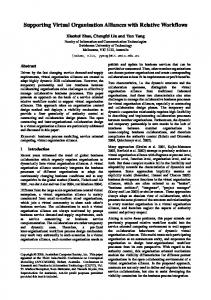
Figure 1. Schematic of geometric parameters of stenotic lesion. In this study, we propose a new ML based FFR prediction . approach from geometric features of stenotic lesion and circulation conditions. This approach uses training dataset from multi-scale (3D-0D coupled) CFD simulation results with synthesized straight vessel models. We demonstrated that this technique can effectively predict the FFR in near-real time.
Permission to make digital or hard copies of all or part of this work for personal or classroom use is granted without fee provided that copies are not made or distributed for profit or commercial advantage and that copies bear this notice and the full citation on the first page. Copyrights for components of this work owned by others than the author(s) must be honored. Abstracting with credit is permitted. To copy otherwise, or republish, to post on servers or to redistribute to lists, requires prior specific permission and/or a fee. Request permissions from
[email protected]. ICMLSC 2018, February 2–4, 2018, Phu Quoc Island, Viet Nam © 2018 Copyright is held by the owner/author(s). Publication rights licensed to ACM. ACM ISBN 978-1-4503-6336-5/18/02…$15.00
2. METHOD 2.1 Geometric Features of Coronary Stenosis We generated total 1,116 anatomic vessel models by synthetically designed straight conduits with various geometric parameters of a stenosis as shown in Fig. 1. Three different mean aortic pressures were also considered as a variable because flow rate as well as the cardiac pressure play an important role in coronary flow dynamics.
https://doi.org/10.1145/3184066.3184079
1
Details of geometric parameters and aortic pressure conditions considered in this study were presented in Table 1. The ranges of parameters were determined based on the in vivo data typically observed in patients.
𝜌 Step 1:
Values
Description
%DS
stenotic degree
40
50
𝐿𝑠
total length
1.0D
2.0D
𝐿𝑖
entrance length
0.25D 0.5D
1.0D
-
𝐿𝑜
exit length
0.25D 0.5D
1.0D
-
𝜖
eccentricity
0
10
40
-
𝑃𝑚𝑒𝑎𝑛
mean aortic pressure
85
95
105
-
60
,𝑗
− 𝑢𝑖 = 𝑝,𝑖𝑛 , Δ𝑡
𝜌
Step 3:
𝑝𝑛+1 = ,𝑖𝑖
Step 4:
𝜌
𝜌
(4)
𝑢∗𝑖,𝑖 ,
(5)
𝑢𝑖𝑛+1 − 𝑢𝑖∗ = − 𝑝,𝑖𝑛+1 . Δ𝑡
(6)
Δ𝑡
where Δ𝑡 is the time increment,the superscript 𝑛 indicates the time level and 𝑢𝑖 and 𝑢∗𝑖 are intermediate velocities. In the present study, a P2P1 finite element scheme is employed to achieve higher accuracy efficiently. Whereas a conventional P1P1 element allocates both velocity and pressure at vertex nodes, the P2P1 element allocates the pressure at vertex nodes and the velocity at both vertex and mid nodes.
To obtain hemodynamic information and FFR data which are required for training the ML algorithm, 3D-0D coupled blood flow dynamics simulations for the stenoticvessel models were conducted[5].
In order to accommodate the specific time-varying pressure-flow characteristics of coronary flow, the 0D LPN modelling technique for coronary circulation is necessary to be integrated into 3D CFD model at all the coronary outlets. The LPN model represents the coronary circulatory system by combining the resistance and the capacitance elements, based on the similarity concept between a hydraulic and electric system. Total resistances and capacitance for distal coronary bed at the resting condition as well as hyperemic condition, which is maximum flow condition by minimizing the microvascular resistance, are determined based on the study of Sankaran [7] and Sharma et al. [8].
For 3D CFD model, the Navier-Stokes equations and the continuity equations which describe the mass and momentum conservation for unsteady incompressible flow were considered. These equations are written using Einstein notation as follows: ,𝑗
(3) ,
70
3.0D 4.0D
2.2 Computational Fluid Dynamics
𝜌𝑢𝑖 + 𝜌𝑢𝑗 𝑢𝑖,𝑗 = − 𝑝,𝑖 + τ𝑖𝑗
𝑢𝑖∗
Step 2: Table 1. Geometric parameter values considered for training model Parameter
𝑢𝑖 − 𝑢𝑖𝑛 1 𝑛 +𝜌 𝑢 𝑢 + 𝑢𝑗𝑛 𝑢𝑖,𝑗 Δ𝑡 2 𝑗 𝑖,𝑗 1 = − 𝑝,𝑖𝑛 + 𝜏𝑖𝑗 + 𝜏𝑖𝑗𝑛 2
,
𝑢𝑖,𝑖 = 0.
(1) (2)
Ra
Pa
Ra-micro
Rv-micro
𝑅𝑎 : coronary arterial resi
𝐶𝑎 : coronary arterial com Ca
Cim
Rv Pim
𝑅𝑎−𝑚𝑖𝑐𝑟𝑜 : coronary micro arterial resistance
𝐶𝑖𝑚 : intra-myocardial com
𝑃𝑖𝑚 : intra-myocardial pre
Figure 2. Schematic for multi-scale computational fluid dynamics.
𝑅𝑣−𝑚𝑖𝑐𝑟𝑜 : coronary micro venous resistance
We employed MPI parallel algorithm to speed up the computing𝑅𝑣 : coronary venous resi time. The P2P1 unstructured meshes of whole computational domain are decomposed into multiple subdomains using k-way partition method supported by Metis library 5.1 [9].The computing work for each subdomain is distributed equally to the corresponding processor for parallel computing.
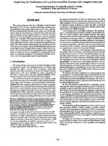
2.3 Machine Learning Model
Figure 3. Fully connected machine learning network for FFR prediction.
The ML model was trained by deep neural network using TensorFlow software[10]. We employed a fully connected network model with four hidden layers. The input layer has six neurons corresponds to the geometric parameters of stenosis and aortic pressure. Each hidden layer has 256, 64, 16 and 4 neurons, with a sigmoidal activation function as shown in Fig. 3. The model was optimized using a mean-squared loss function with a batch gradient descent algorithm. For validation of the ML algorithm, the dataset was randomized and divided in a 5:1 ratio for training and testing sets, which results in the number of data of 971 for the training set.
These governing equations are split into four steps for time advancement based on a fully implicit fractional step method [6] as follows:
2
3. RESULTS AND DISCUSSION
1.0
We validated the developed ML model with randomly selected testing set. FFRML and FFRCFD were compared and it produces excellent correlation as shown in Fig. 4(R = 0.9998, P< 0.001).
0.8
Invasive-CFD* Invasive-ML
FFR 0.6 C CFD/ML
We also applied the trained ML model to predict FFR index for patient-specific coronary models. Geometric parameters were currently extracted from coronary angiographic images manually.
CFD
/ 0.4 FFR
1.0
ML
CFD-ML
0.2
0.8 0.0
CFD/ML
0.0
FFR
0.2
0.6
0.4 0.6 Invasive FFR Invasive
0.8
1.0
Figure 6. Comparison of FFRInvasive and FFRML / FFRCFDfor patient-specific coronary artery
ML
0.4
4. CONCLUSION We demonstrated the development of a near-real time ML algorithm for FFR prediction by using multi-scale (3D-0D coupled) CFD simulation output data with synthetically generated stenotic coronary models. This may be useful in on-site routine clinical practices for fast assessment of coronary lesion severity. In order to improve the accuracy, further study with larger database will be necessary.
0.2
0.0 0.0
0.2
0.4 0.6 Invasive FFR
0.8
1.0
CFD
Figure 4. Comparison of FFRCFD and FFRMLfor synthetically designed stenotic vessel model
5. ACKNOWLEDGMENTS This work was supported by the National Research Foundation of Korea(NRF) grant (No. 2017R1 D1A1B03034932).
6. REFERENCES [1] Toth, G., Hamilos, M., Pyxaras, S., Mangiacapra, F., Nelis, O., De Vroey, F., Di Serafino, L., Muller, O., Van Mieghem, C., Wyffels, E., Heyndrickx, G., Bartunek, J., Vanderheyden, M., Barbato, E., Wijns, W., and De Bruyne, B. 2014. Evolving concepts of angiogram: fractional flow reserve discordances in 4000 coronary stenoses. Eur. Heart J. 35 (Oct, 2014), 2831–2838. [2] Tonino, P. 2009. Fractional flow reserve versus angiography for guiding percutaneous coronary intervention.N. Engl. J. Med. 360 (Mar, 2009), 213-224. [3] Itu, L., Sharma, P., Mihalef, V., Kamen, A., Suciu, C., and Comaniciu, D. 2012. A patient-specific reduced-order model for coronary circulation. In: 2012 9th IEEE International Symposium on Biomedical Imaging (ISBI). Barcelona, Spain: IEEE, 832–835.
Figure 5. A case example of geometric features measurement and FFRCFD for a patient-specific coronary artery Fig. 5 shows a sample case for manually measuring of geometric features of stenotic lesion and FFRCFD from CT image based CFD simulation in a patient-specific coronary artery.
[4] Itu, L., Rapaka, S., Passerini, T., and Georgescu, B. 2016. A machine-learning approach for computation of fractional flow reserve from coronary computed tomography. J. Appl. Physio. 121 (Jul, 2016), 42-52.
The ML predicted FFR value (FFRML) was compared with invasively measured FFR (FFRInvasive) in the corresponding patient as shown in Fig. 6and showed fairly good correlation(R = 0.727, P< 0.001) and diagnostic accuracy. Thetraining and the prediction time in FFRML were about one and a half hours and less than a second, respectively.
[5] Bertoglio, C. 2013. Fractional-step schemes for the coupling of distributed and lumped models in hemodynamics. SIAM J. Sci. Comp. 35 (May, 2013), B551-B575. [6] Choi, H. 1997. A fractional four-step finite element formulation of the unsteady incompressible Navier-Stokes equations using SUPG and linear equal-order element methods.Comp. Meth. App. Mech. Eng. 143 (Apr, 1997), 333-348. [7] Sankaran, S., Esmaily Moghadam, M., Kahn, A., Tseng, E., Guccione, J., and Marsden, A.2012. Patient-specific
3
multiscale modeling of blood flow for coronary artery bypass graft surgery. Ann. Biomed. Eng. 40(Oct, 2012), 2228-2242. [8] Sharma, P.,Itu, L., Zheng, X., Kamen, A., Bernhardt, D., Suciu, C., and Comaniciu, D.2012. A framework for personalization of coronary flow computations during rest and hyperemia. Conf. Proc. IEEE Eng. Med. Biol. Soc.66656668. [9] Karypis, G. and Kumar, V. 1998. A Fast and High Quality Multilevel Scheme for Partitioning Irregular Graphs. SIAM J. Sci. Comput.20 (Aug, 1998), 359-392. [10] McClure, N.2017.TensorFlow machine learning cookbook. Packt Publishing Ltd.
4