Advances in Alzheimer’s Research, Vol. 2, 2014, 291-374
291
CHAPTER 12 Methods for Studying and Structure–Function Relationships of Non-Fibrillar Protein Assemblies in Alzheimer's Disease and Related Disorders Farid Rahimi1 and Gal Bitan2,* 1
Research School of Biology, Division of Biomedical Science and Biochemistry, College of Medicine, Biology, and Environment, The Australian National University, Canberra, ACT, Australia, and 2Department of Neurology, David Geffen School of Medicine, Brain Research Institute, and Molecular Biology Institute, University of California at Los Angeles, USA Abstract: Several neurodegenerative diseases, including Alzheimer's, Parkinson's, Huntington's, and prion diseases, are characterized by intra- and/or extracellular deposition of fibrillar proteinaceous aggregates, and by extensive, neuron loss. Related non-neuropathic systemic diseases, e.g., light-chain and senile systemic amyloidoses, and other organ-specific diseases, such as dialysis-related amyloidosis and type-2 diabetes, also are characterized by deposition of aggregated proteins. It is debated whether these hallmark lesions are causative. Substantial evidence suggests that the aggregates are the end state of protein misfolding whereas the actual culprits likely are transient, non-fibrillar assemblies preceding the aggregates. The non-fibrillar, oligomeric assemblies are believed to initiate pathogenesis, leading to synaptic dysfunction, neuron loss, and pathognomonic brain atrophy. It is hypothesized that nonfibrillar assemblies or fibril-derived fragments may promote anatomical progression of pathology, or even disease transmissibility, akin to misfolded prions. Amyloid β-protein (Aβ), which is believed to cause Alzheimer's disease, is considered an archetypal amyloidogenic protein. Intense studies have led to nominal, functional, and structural descriptions of oligomeric Aβ assemblies. However, the dynamic and metastable nature of Aβ oligomers renders their study difficult. Different results generated using different methodologies under different experimental settings further complicate this complex area of research and identification of the exact pathogenic assemblies in vivo seems daunting. In this chapter we review structural, functional, and biological experiments used to produce and study non-fibrillar Aβ assemblies, and highlight similar studies of proteins involved in related diseases. We discuss challenges that contemporary researchers are facing and future research prospects in this demanding, yet highly important, field. *Corresponding author Gal Bitan: David Geffen School of Medicine, University of California at Los Angeles, Neuroscience Research Building 1, Room 451, 635 Charles E. Young Drive South, Los Angeles, CA 90095-7334, USA; Tel: 310-2062082; Fax: 310-2061700; E-mail:
[email protected] D.K. Lahiri (Ed) All rights reserved-© 2014 Bentham Science Publishers
292 Advances in Alzheimer’s Research, Vol. 2
Rahimi and Bitan
Keywords: Amyloid, neurodegeneration, Alzheimer's disease, amyloid β-protein, protein misfolding, non-fibrillar assemblies, oligomers, toxicity, prion INTRODUCTION The “amyloid cascade hypothesis” [1], suggesting that amyloid β-protein (Aβ) fibril formation and plaque deposition lead to neuronal dysfunction, dementia, and death in Alzheimer's disease (AD), had guided scientific research into discovery of etiologic and pathogenic mechanisms of AD. However, this hypothesis has been contentiously debated because: 1) fibrillar amyloid burden does not correlate well with neurological dysfunction [2], 2) cognitive impairment in transgenic murine models of AD is observed before and/or independently of amyloid plaque formation [3], 3) plaque-independent pathology can be explained by the neurotoxicity of soluble Aβ assembly intermediates, 4) oligomer-induced memory dysfunction occurs before neuronal death, and 5) brain, plasma, and cerebrospinal fluid (CSF) concentrations of soluble Aβ oligomers correlate with neurodegeneration better than those of fibrils [4]. These observations have led to a burgeoning yet encompassing alternative paradigm hypothesizing that soluble non-fibrillar protein assemblies, rather than mature fibrillar deposits, act as proximate neurotoxins that cause synaptic dysfunction, neuron loss, dementia, and death [4-11]. This new hypothesis has been supported by discovery of toxic nonfibrillar protein assemblies involved in other protein-misfolding diseases, such as, Parkinson's disease (PD), Huntington's disease (HD), transmissible spongiform encephalopathies (TSEs), amyotrophic lateral sclerosis, polyglutamine diseases, type-2 diabetes mellitus (T2D), and systemic amyloidoses [7, 12-15]. Diversity and sometimes inaccuracy in nominal definitions, and in structural/functional descriptions of soluble non-fibrillar Aβ assemblies, along with different methodologies to generate and study these assemblies, are confounding factors in this already vast and complex research area. Various forms of soluble non-fibrillar Aβ assemblies (reviewed in [10, 16-18]) including monomeric Aβ conformers [19], secreted cell- and brain-derived low-order oligomers [20-25], Aβ-derived diffusible ligands (ADDLs) [26, 27], protofibrils (PF) [28-30], Aβ*56 [31], paranuclei [32-34], amylospheroids [35], annular assemblies [36], amyloid pores [18, 36, 37], toxic fibrillar oligomers [38], and
Protein Assemblies in Alzheimer’s Disease
Advances in Alzheimer’s Research, Vol. 2 293
βamy balls [39] have been described Protein Asseblies. However, despite a global concerted scientific effort, the relationships amongst these Aβ-derived assemblies and their relevance to AD pathogenesis are unclear and the fundamental quest for a unanimous pathogenic “equivalent” active in AD-afflicted brain is ongoing [9]. We begin our discussion in the first step of studying non-fibrillar Aβ assemblies—protein preparation. SOURCES AND METHODS OF Aβ PREPARATION The low physiological concentration and the difficulty of procuring highly pure and homogeneous tissue-derived Aβ have precluded its routine use in experimental studies in vitro. Therefore, synthetic Aβ preparations have emerged as alternatives. Synthetic Aβ is produced either by standard solid-phase peptide synthesis (SPPS) [40, 41] or by recombinant DNA technology [42-45]. The Aβ sequence is recognized as a “difficult” target for SPPS owing to its high hydrophobicity and innate propensity to aggregate [46-48]. To overcome these issues, various deprotecting agents [46], novel solvent systems for coupling [47], solid-support modifications [49], or microwave-assisted SPPS [50] have been used to augment synthetic yield and improve purity of crude Aβ peptides. Application of an O–N acyl migration reaction, also called “O-acyl isopeptide” chemistry was proposed as an efficient alternative SPPS route for generating Aβ with increased solubility and purity [51, 52]. Similarly, tailored recombinant or semisynthetic expression systems have been used to produce Aβ in high yields. A strategy to increase solubility and facilitate purification is co-expression of Aβ with Aβ-specific affibodies [53] or as fusions with sequences affording high solubility, followed by cleavage of the fusion protein and purification by high-performance liquid chromatography (HPLC) [4245, 54-56]. A semisynthetic procedure for generating full-length Aβ has been reported, producing 8–10 mg of pure Aβ(1–40) per liter of bacterial culture [57]. Because self-association of Aβ is believed to be central in the pathogenesis of AD, extensive research has been dedicated to developing methodologies to characterize Aβ assemblies structurally and biologically [58]. Multiple studies
294 Advances in Alzheimer’s Research, Vol. 2
Rahimi and Bitan
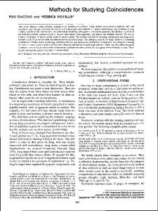
have shown that many of the neurotoxic effects of Aβ assemblies can be recapitulated by synthetic Aβ in vitro or in vivo [59]. However, differences in peptide quality, presence of trace contaminants in Aβ preparations from different sources, and compositional variation of Aβ preparations, even from the same source, have been serious problems leading to irreproducible or discrepant reports and study outcomes [60-62]. For example, under identical conditions, an Aβ oligomer-specific monoclonal antibody was shown to react only with oligomers derived from recombinant Aβ but not those derived from chemically synthesized Aβ [63]. In our hands, Photo-Induced Cross-linking of Unmodified Proteins (PICUP) using Aβ obtained from different sources, but prepared identically, yielded distinct results (Fig. 1).
Figure 1: Comparison of photo-cross-linking using Aβ peptides from different sources. Synthetic Aβ from Global Peptide (G) and the UCLA Biopolymers Laboratory (U), and recombinant Aβ from rPeptide (R) were prepared in 10 mM sodium phosphate, pH 7.4, at 2 mg/mL nominal concentration and filtered through a 10-kDa molecular-weight cut-off filter [106]. Each filtered peptide was cross-linked using PICUP [98]. The resulting cross-linked oligomers were subjected to SDS-PAGE and silver-staining. The data suggest that Aβ40 from Global peptide contained contaminants that prevented cross-linking and that Aβ42 from rPeptide aggregated during the filtration step and was hardly detectable.
Neither SPPS nor recombinant methods can produce 100% pure Aβ. Failure sequences, oxidation of Met35, and racemization may occur during various SPPS steps [58]. Similarly, Met35 oxidation may occur in bacterial inclusion bodies [57]. In recombinant preparations where a fusion protein is enzymatically cleaved to release Aβ, it is important to verify that the cleavage product is not contaminated with the uncleaved fusion protein, the cleaving enzyme, or adventitious proteolytic fragments [58]. Practically, it is important that the researcher verifies chemical purity of the preparation and ensures removal of residual components which could complicate solvation and stock preparation,
Protein Assemblies in Alzheimer’s Disease
Advances in Alzheimer’s Research, Vol. 2 295
potentially alter the biophysical and biological characteristics of the peptide, and render concentration measurements error-prone [58]. METHODS USED TO STUDY NON-FIBRILLAR Aβ ASSEMBLIES Important biological functions of oligomeric Aβ assemblies have spurred extensive efforts to characterize them structurally. The non-crystalline nature of the oligomers and their slow tumbling time in aqueous solutions preclude highresolution structural determination by X-ray crystallography and solution-state NMR [64], respectively. Moreover, the metastable nature of Aβ oligomers and their existence in rapidly changing mixtures have made their structural characterization particularly difficult [65, 66]. To address these issues, multiple low-resolution methods have been used to assess various structural features of non-fibrillar Aβ assemblies. Here, we outline some of the key methods used to provide structural and biophysical information on non-fibrillar Aβ assemblies. Atomic-Resolution Techniques NMR Spectroscopy Solution-state NMR is a powerful high-resolution technique for determining peptide and protein structure in solution [67]. Typically, the structure is calculated based on distances and angles obtained through measurements of nuclear Overhauser effect and spin–spin scalar coupling interactions as constraints for computer-generated models. As mentioned above, peak broadening due to slow tumbling times [64] currently precludes solving structures of Aβ assemblies using solution-state NMR. However, multiple NMR studies have assessed structural properties of Aβ monomers. For instance, studies by Lee et al. introduced the concept of “plaque competence”, which defines the propensity of nearphysiological concentrations of soluble Aβ to deposit onto authentic amyloid plaques in vitro [68]. The plaque competence assay identified a central 26-residue fragment (Tyr10–Met35) which was deemed necessary to mimic plaque-deposition characteristics of the full-length Aβ [68]. Preliminary NMR conformational analyses showed that this 26-residue fragment had a different conformation from a plaque-incompetent fragment (Asp1–Lys28) [68]. Further NMR studies also
296 Advances in Alzheimer’s Research, Vol. 2
Rahimi and Bitan
confirmed that the central 26 residues of Aβ were sufficient to mimic amyloidogenic properties of Aβ40 and Aβ42 [69]. It was reported that the central hydrophobic cluster of full-length Aβ, and Aβ(10–35), both adopted well-defined, albeit irregular, conformations in solution, whereas the C- and N-terminal flanking regions of the full-length Aβ were partially disordered [69]. NMR studies also have highlighted differences between Aβ40 and Aβ42. Solution-phase NMR studies of non-oxidized [70] or Met-oxidized [71, 72] Aβ40 and Aβ42 showed that the C-terminus of Aβ42 is more rigid compared to that of Aβ40, likely due to the extended hydrophobic C-terminus of Aβ42. Similarly, a study combining molecular dynamics and NMR experiments, showed that the Cterminus of Aβ42 is more structured than that of Aβ40 [73]. NMR studies also revealed that common C-terminal peptide segments within Aβ40 and Aβ42 have distinct structures, which may be relevant to the strong disease association of elevated Aβ42 production [74]. Various types of NMR applications to studying of oligomeric assemblies of amyloid proteins have been reviewed recently by Bemporad et al. [75]. X-Ray Crystallography X-ray crystallography examines atomic structures of crystals by X-ray diffraction techniques (reviewed in [76]). The signal is intensified by the coherent alignment and repetitive lattice of crystal structures. The wavelength of radiation used in Xray crystallography is usually ~1.5 Å, about the length of a C–C bond. Use of Xrays with this wavelength theoretically allows resolution of individual atoms. Sawaya et al. reported that 33 peptide segments derived from 14 different amyloidogenic proteins formed amyloid-like fibrils, microcrystals, or both [77] and used X-ray crystallography to examine the atomic organization of molecules within microcrystals of these peptides. Microcrystals of three Aβ segments were resolved, Gly37–Ala42, Met35–Val40, and Val36–Ala42. The authors suggested that the structural organization of these peptides within the crystals is similar to those of Aβ fibrils and concluded that the fundamental unit of amyloid-like fibrils is a steric zipper arrangement formed by two tightly interdigitated β-sheets [77].
Protein Assemblies in Alzheimer’s Disease
Advances in Alzheimer’s Research, Vol. 2 297
Hydrogen–Deuterium Exchange Hydrogen–deuterium exchange is a powerful technique for probing protein structure and dynamics. The method involves studying of exchange rates of labile protons in proteins with deuterons from the solvent, typically D2O. Labile protons are those bonded to nitrogen, sulfur, or oxygen. These protons can exchange with solvent hydrogen or deuterium cations. Labile protons that are solvent-exposed and are not involved in hydrogen bonding exchange rapidly, whereas buried or hydrogen-bonded protons exchange at substantially slower rates. This makes hydrogen–deuterium exchange sensitive to structural rearrangements occurring during protein aggregation. Thus, amide protons buried in the core of oligomeric and high-order assemblies or hydrogen-bonded in helices and sheets do not exchange readily with solvent deuterons. The exchange rate is detected using NMR and/or mass spectrometry [67]. For study of rapidly changing assemblies, mass spectrometric detection of exchange may be advantageous because NMR requires longer times (hours) to record the spectra, making the study of short-lived oligomers difficult. In addition, NMR requires prior assignment of protons and generally is limited to proteins smaller than 25 kDa. An additional advantage of mass spectrometric detection of exchange is requirement of substantially smaller amounts of proteins. However, assignment of specific exchanging protons using mass spectrometry requires tandem mass spectrometry and can be a daunting task, whereas for a previously assigned NMR spectrum, identification of exchanging protons is straightforward. Hydrogen–deuterium exchange coupled with mass spectrometry was used to map structural differences in Aβ PF and fibrils [78]. Determination of Oligomer Size Distribution Sodium Dodecyl Sulfate–Polyacrylamide Gel Electrophoresis (SDS-PAGE) SDS-PAGE is a routine and inexpensive method enabling separation of proteins based on their electrophoretic mobility, which is affected by a combination of primary, secondary, tertiary, and quaternary structures of proteins. In this method, protein mixtures are electrophoresed after treatment with SDS. SDS binds proteins via its hydrophobic dodecyl tail, leaving its sulfate group solventexposed, thus creating a negatively charged envelope that “coats” protein molecules [79]. SDS binds different polypeptides at an approximately constant
298 Advances in Alzheimer’s Research, Vol. 2
Rahimi and Bitan
mass ratio—1.4 g SDS per gram of polypeptide (reviewed in [67]). In most cases, SDS binding denatures secondary and non-disulfide-linked tertiary structures, negatively charging proteins approximately uniformly and proportionally to their mass. Under these conditions, electrophoretic migration of proteins through the gel matrix is governed directly by the molecular mass of the protein. Without SDS, different proteins of the same mass may electrophorese distinctly due to differences in overall charge (different isoelectric points) and folding. It has been shown that at an appropriate protein–SDS molar ratio, SDS readily induces aggregation of diverse proteins at a pH two units below their respective isoelectric points (pI) with no aggregation at a pH two units above pI [80]. Electrostatic interactions were observed to play a leading role in the induction of amyloid under these conditions [80]. Importantly, the effect of SDS on all proteins is not equivalent [81]. Different proteins, different conformations of the same protein [82], or truncated versions of certain proteins [83] may not bind stoichiometric amounts of SDS. In many cases, SDS can induce or stabilize secondary and quaternary structures rather than denaturing them [82, 84, 85]. SDS may cause dissociation of some protein assemblies or conversely induce oligomerization and aggregation [85-88]. Aβ42-derived “globulomers”, for example, are oligomeric assemblies produced by incubating Aβ42 with 0.2% SDS [89]. Therefore, resolution of apparently monomeric or low-molecular-weight (LMW) oligomeric components in a protein mixture does not necessarily indicate existence of such components under native conditions, i.e., without SDS. Aβ is an amphipathic protein known to form SDS-stable oligomers [90]. Indeed, SDSinduced assembly of Aβ into insoluble aggregates has been capitalized on to purify Aβ from brain homogenates [91]. When treated with SDS, Aβ assembles rapidly into high-molecular-mass aggregates [63]. Furthermore, circular dichroism (CD) spectroscopy, thioflavin T fluorescence, nuclear magnetic resonance, and Fourier-transform infrared (FTIR) spectroscopy experiments have shown that Aβ40 undergoes structural transitions when treated with SDS or lithium dodecyl sulfate [92]. In this study, in solutions without SDS, Aβ40 yielded CD signals consistent with a random coil or a “statistical coil” structure [93], whereas at SDS concentration of ~4 mM (53-fold excess detergent to protein),
Protein Assemblies in Alzheimer’s Disease
Advances in Alzheimer’s Research, Vol. 2 299
this signal disappeared [92]. At 67-fold excess SDS, CD showed a typical β-sheet spectrum. At SDS concentrations of >10 mM (133-fold excess detergent), Aβ40 secondary structure was mainly α-helical [92]. Thus, the concept of “SDS stability” [94] in its essence is questionable in view of the fact that SDS has been used as amyloid [80, 92] or as α-helix inducer [92]. During electrophoresis of Aβ40, aggregates dissociate completely and only a monomer is observed following detection, whereas electrophoresis of Aβ42 yields apparent trimeric and tetrameric components [32]. Essentially identical monomer–trimer–tetramer distributions are observed when different preparations of Aβ42, including “monomeric”, oligomeric, and fibrillar Aβ42 are analyzed by SDS-PAGE [95]. In a urea-containing SDS-PAGE system, Aβ and truncated versions thereof do not obey the law of mass–mobility relationship, likely because Aβ–SDS binding is proportional to the sum of the hydrophobicity indices, rather than the number, of constituent amino acids [83]. Thus, despite its wide use, SDS-PAGE is not a reliable method for characterization and size determination of non-covalently associated Aβ assemblies (see also [96] for a discussion of other analytical challenges in studying amyloid proteins). Photo-Induced Cross-Linking of Unmodified Proteins (PICUP) PICUP is a method originally developed to study stable protein homo- and heterooligomers [97]. PICUP was used by us and others to study oligomer size distribution of Aβ [66] and a variety of other proteins, including amyloidogenic proteins [98]. PICUP generates covalent bonds between closely interacting polypeptide chains within ≤1 s exposure to visible light without pre facto chemical modifications of the native sequence and without using spacers. The cross-linking is induced by rapid photolysis of a tris-bipyridyl Ru(II) complex in the presence of an electron acceptor. Illumination causes generation of a Ru(III) ion, which subsequently abstracts an electron from, and produces a carbon radical within, the polypeptide. The radical reacts rapidly with adjacent susceptible groups and forms covalent bonds. Therefore, PICUP stabilizes oligomer populations by covalent cross-linking, and “freezes” molecular interactions that exist before cross-linking. The mechanism, protocol, and limitations of PICUP were discussed in detail elsewhere [97-100]. Recently, a continuous-flow reactor,
300 Advances in Alzheimer’s Research, Vol. 2
Rahimi and Bitan
an “oligomerator”, has been used to produce large-scale PICUP-stabilized amyloid oligomer mixtures [101]; however, purification of large quantities of single-order oligomers stabilized by PICUP still is a laborious and costly process. Size-Exclusion Chromatography (SEC) SEC (gel-permeation or gel-filtration chromatography) fractionates solutes based on their Stokes (hydrodynamic) radii, i.e., hydrodynamic volumes (which can be correlated with molar mass) [102]. SEC is used to determine molar mass averages and molar mass distributions of solutes. SEC separation is achieved using a stationary phase comprising a porous matrix. When solutes of different sizes pass through a SEC column packed with porous material, larger molecules spend less time interacting with the solid phase and elute faster, whereas smaller molecules diffuse into the pores and therefore spend more time interacting with the solid phase and elute later. SEC affords an SDS-independent separation mechanism and covers a molecular mass range of ~103–106 Da. However, SEC provides lower resolution than SDS-PAGE and estimations of polypeptide molecular masses may be inaccurate because the elution profiles are sensitive to protein conformation. SEC analysis of Aβ assemblies does not resolve LMW assemblies but can distinguish between PF and small oligomers [90]. At this resolution, SEC may be useful for studying the kinetics involved in conversion of LMW Aβ to PF (or dissociation of PF into LMW Aβ). In addition to its use as an analytical method [39], SEC has been used extensively to purify fractions of particular Aβ assemblies [30, 32, 37, 103-105]. Description of the basic instrumentation and utilization of SEC for preparation of aggregate-free Aβ was published previously [106]. Analytical Ultracentrifugation Analytical ultracentrifugation is a versatile technique used extensively to characterize the hydrodynamic and thermodynamic properties of proteins or macromolecules (reviewed in [67]). Conceptually, this method allows real-time quantitative analysis of spatial redistribution of soluble analytes under a high centrifugal force field. Unlabeled and unmodified proteins can be analyzed by this method in various excipients, obviating analyte–matrix interactions such as those occurring during SEC. Because molecular frictional forces oppose the centrifugal force, sedimentation depends, in addition to other parameters, on the hydrodynamic
Protein Assemblies in Alzheimer’s Disease
Advances in Alzheimer’s Research, Vol. 2 301
properties of the molecules, providing low-resolution information on the structure of protein complexes, thus enabling detection of conformational alterations. This technique combines an ultracentrifuge, a multicompartmental rotor, and an optical detection system for measuring analyte concentration inside the centrifuge cell during sedimentation. The optical detection system is triggered by the revolution of the rotor, allowing data acquisition only during the extremely short intervals when a particular sample assembly aligns within the optical path. Coupled with data-analysis software, analytical ultracentrifugation can determine sample purity and molecular mass in the native state, measure sedimentation and diffusion coefficients, characterize assembly–disassembly mechanisms of complex analytes, determine subunit stoichiometry, detect and characterize macromolecular conformational changes, and measure equilibrium constants and thermodynamic parameters for homo- or hetero-associating assemblies. Two types of experiments are commonly performed using ultracentrifugation— sedimentation velocity [107, 108] and sedimentation equilibrium [109]. Typically, sedimentation-velocity experiments are conducted at relatively high rotor speeds. Under these conditions, each protein species in a mixture forms a unique boundary and sediments at a characteristic speed governed by its molecular mass, size, and shape. The velocity and shape of the moving concentration boundary is used to estimate the sedimentation coefficient, diffusion coefficient, molecular mass, and equilibrium constants of interacting species. In contrast, sedimentationequilibrium experiments are performed at relatively low rotor speeds. The sedimentation of proteins under lower centrifugal forces than in sedimentationvelocity experiments is opposed by the diffusion, and eventually when equilibrium is reached, a time-invariant exponential concentration gradient is established throughout the centrifuge cell. The concentration gradient of proteins at equilibrium has been used widely to determine the molecular weight, stoichiometry, binding affinity, and virial coefficient of hetero-interacting or selfassociating systems. Sedimentation-equilibrium experiments can analyze a mixture of moieties of different molecular masses. After each analyte reaches its equilibrium, high-molecular-mass species locate towards the cell bottom, whereas low-molecular-mass species dominate at the top. The equilibrium data can be fitted to calculated models for solute distribution. Using this type of analysis,
302 Advances in Alzheimer’s Research, Vol. 2
Rahimi and Bitan
Huang et al. have reported that Aβ40 existed as an equilibrium mixture of monomers, dimers, and tetramers at neutral pH [110]. However, other equilibria, including monomer–dimer, monomer–trimer, or monomer–tetramer, produced equivalent residuals [110], hindering precise determination of the oligomerization state of the peptide. ADDLs, which are soluble, Aβ42-derived, non-fibrillar neurotoxic assemblies, have been shown to contain a high-molecular-mass component with an estimated mass of 175 kDa by sedimentation-velocity and 120–540 kDa by sedimentation-equilibrium experiments [95]. Dynamic Light Scattering (DLS) Spectroscopy DLS, also known as quasielastic light scattering, intensity-fluctuation spectroscopy, or photon-correlation spectroscopy, is a non-invasive, nondestructive, quantitative, optical analytical method for determining diffusion coefficients of particles (0.1–3000nm size range [111]) undergoing Brownian motion in solution (reviewed in [67]). DLS measures the temporal dependence of light scattering emanated from an analyte in solution over 10−7–1 s. Temporal fluctuations in intensity of the scattered light relate to rate of the Brownian motion which is correlated to the diffusion coefficients, D, and radii of particles according to the Stokes–Einstein equation [112, 113]: =
=
ℎ
,
where KB is Boltzmann's constant, T is the absolute temperature, η is the viscosity of the suspending liquid, and Rh and Dh are the hydrodynamic radius and diameter, respectively. Knowledge of diffusion coefficients of molecules allows determination of molecular features, including size and flexibility. Temporal changes in these parameters provide information about the kinetics and structural transitions occurring, e.g., during protein assembly–disassembly processes. In a polydisperse mixture, a distribution of diffusion coefficients is obtained. DLS is intrinsically biased for large aggregates because the intensity of the scattered light is proportional to the square of the particle mass [114]. Therefore, DLS is suited well to measure minute amounts of protein aggregates (