African Journal of Biotechnology Vol. 11(77), pp. 14132-14139, 25 September, 2012 Available online at http://www.academicjournals.org/AJB DOI:10.5897/AJB11.398 ISSN 1684-5315 ©2012 Academic Journals
Full Length Research Paper
Prevalence of viruses infecting cowpea in Uganda and their molecular detection Amayo, R., Arinaitwe, A. B., Mukasa, S. B.*, Tusiime, G., Kyamanywa, S., Rubaihayo, P. R. and Edema, R. School of Agricultural Sciences, Makerere University, P. O. Box 7062 Kampala, Uganda. Accepted 7 May, 2012
The main areas for cowpea cultivation in Uganda were surveyed in June and October 2006 for viruses affecting the crop. Seed and leaf samples from symptomatic and asymptomatic plants were collected from farmers’ fields and analysed for infecting viruses using double antibody sandwich enzyme-linked immunosorbent assay (DAS-ELISA). The viruses detected in the leaf and seed samples were: cucumber mosaic cucumovirus (CMV), cowpea mild mottle calarvirus (CPMMV), cowpea mottle carmovirus (CPMoV), Cowpea chlorotic mottle bromovirus (CCMV), Cowpea yellow mosaic comovirus (CYMV), cowpea severe mosaic comovirus (CPSMV), cowpea aphid-borne mosaic potyvirus (CABMV) and Southern bean mosaic sobemovirus (SBMV). CPMV was detected only in leaf samples. CMV and CABMV were later confirmed using reverse transcription polymerase chain reaction (RT-PCR). Of the viruses detected in leaf samples, 53.26% occurred as single infections, 24.46% dual and 22.28% multiple infections. Similarly, analysis of seed samples revealed infection of 40.6, 34.6 and 24.8% for single, dual and multiple infections, respectively. Multiple virus infections were associated with more disease severity and higher yield losses. The seed transmission levels of 23.0, 20.3 and 16.4% were recorded for CMV, CPMMV and CABMV, respectively. This study identified six more viruses in addition to what was previously reported in the country, of which eight were seed-borne. This necessitates the need for the production and use of virus-free seeds, development of virus resistant genotypes and adoption of efficient seed certification systems. Key words: Vigna unguiculata, disease incidence, seed-borne viruses, ELISA, (RT-PCR).
INTRODUCTION Cowpea, Vigna unguiculata (L) Walp, is an important legume crop in Uganda, ranking third by acreage and economic value only after beans and groundnuts (Adipala
*Corresponding author. E-mail:
[email protected]. Abbreviations: DAS-ELISA, Double antibody sandwich enzyme-linked immunosorbent assay; CMV, cucumber mosaic cucumovirus; CPMMV, cowpea mild mottle calarvirus; CPMoV, cowpea mottle armovirus; CCMV, cowpea chlorotic mottle bromovirus; CYMV, cowpea yellow mosaic comovirus; CPSMV, cowpea severe mosaic comovirus; CABMV, cowpea aphidborne mosaic potyvirus; SBMV, Southern bean mosaic sobemovirus; RT-PCR, reverse transcription polymerase chain reaction.
et al., 1999). The crop has a high nutritional value; the dry grains contain 23 to 38% protein, 1.3% fat, 56.9% carbohydrates, 3.9% fiber, 3.6% ash, 0.74% thiamine, 0.42% riboflavin, and 2.8% niacin (Steele et al., 1985). About 90% of the crop is grown in the Eastern and Northern regions of the country (Omongo et al., 1998) in the districts of Lira, Soroti, Palisa, Kumi, Nebbi and Arua mainly for its leaves which are eaten as ‘spinach’ and grains eaten as pulse (Adipala et al., 1999). Despite the importance of field-grown cowpeas in many areas of Uganda, only limited information is available on the occurrence and the incidence of viral diseases and their impact on cowpea production (Orawu, 2002). Viral diseases of cowpea have been reported to cause appreciable losses in yield if the plants are infected in early growth stages (Booker et al., 2005). Over 140
Amayo et al.
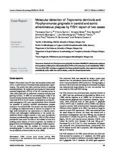
viruses worldwide have been reported to attack cowpea (Hughes and Shoyinka, 2003) and at least eleven of these occur in Africa (Hughes and Shoyinka, 2003). Hampton et al. (1997) listed nine viruses considered most damaging to cowpea in Africa, seven of which were reported as seed-borne; Blackeye cowpea mosaic potyvirus (BlCMV), CABMV, CMV, CPMV, CPSMV, SBMV, and CPMoV. The two non seed-borne viruses considered important by Hampton et al. (1997) are Cowpea golden mosaic geminivirus (CGMV) and CCMV. The other seed-borne cowpea virus is Cowpea mild mottle calarvirus (CPMMV) (EPPO/CABI, 1996). On the basis of their relative economic importance and ability to cause losses either alone or in combination, eight of the above viruses and (CYMV), a close relative of CPMV, were targeted in this study. Survey was conducted twice in the months of June and October 2006 in the major cowpea-growing areas to estimate the incidence and relative importance of viral diseases occurring in cowpea fields in Uganda. The seed transmission levels of the most common cowpea viruses were also determined. This was the first major survey conducted on viruses infecting cowpea in the country and thus, provides valuable information for the design of more sustainable control strategies of viral diseases on cowpea in farmers’ fields. MATERIALS AND METHODS Survey and sample collection Leaf and seed samples were collected from over 70 farms in two main cowpea growing regions of Uganda; the West Nile and Eastern regions. The survey was conducted twice during the cropping season of 2006 in the months of June and October. Four districts in the West Nile region (Nebbi, Arua, Yumbe and Moyo), and three districts in the Eastern region (Kumi, Pallisa and Soroti) (Figure 1) were targeted. For comparison, samples were also collected from experimental plots at the Makerere University Agricultural Research Institute Kabanyolo (MUARIK) in central region. In each district, 10 farms at least 2 km apart were randomly chosen and assessed for presence of viral symptoms. Incidence and severity of virus infection in each field was assessed by counting the number of plants showing virus-like symptoms, and scoring for level of damage on each plant accrued to virus infection (Gumedzoe et al., 1997), respectively. Following the method of Nutter (1997), a total of 80 plant samples (20 plants along each transect) per field was visually assessed for virus disease symptoms. Disease incidence per field was expressed as a percentage of symptomatic samples per total number of plants sampled (Madden and Hughes, 1999). In addition, fresh leaf and mature seed samples (100 per field) were collected from selected plants and taken into the laboratory for detection of the viruses involved. Some of the seed samples were grown in pots in screen houses at MUARIK to determine the seed transmission levels of the major viruses.
Serological virus detection Leaf and seed samples were bagged separately and kept on dry ice while in the field till they were brought into the laboratory for further
14133
analysis. Leaf samples to be analysed serologically were stored at 4°C and those for RT-PCR were kept at -80°C. While the seed samples collected were further air dried and kept at room temperature in paper bags in a moisture-free environment. The presence of CMV, CPMMV, CPMoV, CCMV, CYMV, CPSMV, CABMV, SBMV and CPMV were detected serologically by using the double antibody sandwich enzyme-linked immunosorbent assay (DAS-ELISA) (Clark and Adams, 1977) with the help of kits obtained from DSMZ (Germany) following the manufacturer's protocol. Seed samples were processed as described by Koenraadt and Remeeus (2006), whereas leaf samples were processed as per Clark and Adams (1977). The controls used consisted of a blank (extraction buffer without plant sap), negative control (healthy cowpea leaf samples) and positive control (leaf samples from infected cowpea plant). The last two were supplied together with the ELISA kits. After addition of p-nitrophenyl phosphate substrate (1 mg/ml in 10% diethanolamine, pH 9.8) and incubation for 2 h at room temperature in the dark, ELISA reactions (absorbencies) were measured using the Microplate Manager 5.0 PC (Bio-Rad Laboratories) at 405 nm. A sample was considered virus infected if its A405nm value was greater than twice that of negative control. Individual virus incidence was determined as a percentage of total infected samples over the total leaf or seed samples analyzed.
RT-PCR virus confirmation A modified RT-PCR protocol (Gillaspie et al., 2001) was used to confirm the presence of CMV, CPMMV and CABMV that were associated with more severe symptoms, following detection with ELISA. Total viral RNA were extracted from infected leaf samples kept at -80°C by using Trizol method (Invitrogen, Life Technologies) according to the manufacturer’s instructions. Complementary DNAs (cDNAs) were synthesized from total RNA extracts using 200 U/µl M-MLV reverse transcriptase (Promega, Madison WI USA) together with random primers (100 µg/µl, Promega Madison WI USA) and Poly-T primer for CABMV to a total volume of 25 µl. The cDNAs were diluted in two folds and amplified using polymerase chain reaction (PCR) thermocycler (GeneAmp PCR system 9700). The PCR reaction mixture consisted of 3 µl of the cDNA template, an equivalent of between 2 to 3 µg, 2.5 µl of 10X Polymerase buffer, 1.5 µl of 25 mM MgCl2, 1.0 µl of 10 mM of forward and reverse primers (Table 1), 0.5 µl of 10 mM deoxyribonucleotide (dNTP) mix and 0.5 µl Taq DNA polymerase (Promega, Madison WI USA) and topped up with Rnase free water to a total volume of 25 µl. The PCR conditions consisted of initial heating at 95°C for 2 min followed by 35 cycles of 95°C for 30 s, 53 to 55°C for 30 s and 72°C for 30 s, followed by incubation for 7 min at 72°C and the reactions were held at 4°C. The PCR products were separated by electrophoresis using 1% ethidium bromide stained agarose gel and the products visualised and photographed under ultraviolet (UV) light.
Symptom expression and yield loss The symptom expression and impact of the identified viruses on yield and yield components were assessed using healthy cowpea seeds of cultivars Ebelat, Ichirikukwai, Secow, and FE 69 planted at MUARIK in insect-free screenhouse cages. The plants were mechanically inoculated with single and multiple virus combinations at 10 days after planting (DAP). Inoculum of each virus or virus combination was prepared following the procedure of Taiwo and Akinjogunia (2006). Four plants were inoculated per virus or virus combination. Prior to inoculation, the plants were dusted and rubbed with carborundum to induce injury to the plant cells to ease virus entry into the cells. Plants were monitored for symptom
14134
Afr. J. Biotechnol.
SUDAN Y
N
M
A D R. C O N G O
N S K P K E N Y A
TANZANIA Location of MUARIK
Water bodies Figure 1 A = Arua District K = Kumi District M = Moyo District N = Nebbi District P = Palisa District S = Soroti District Y = Yumbe District
Location of MUARIK Water bodies
Figure 1. Map of Uganda showing the districts where leaf and seed samples for this study were collected.
development and data collected on symptoms two and 14 days after inoculation (DAI). Disease severity for each inoculated plants was scored using a scale of 1-5 (Gumedzoe et al., 1997). The relative area under disease progress curve (RAUDPC) was computed using the
severity scores according to Campbell and Madden (1990). Plant height, number of pods set and weight of the grains per plant were also recorded. The effects of either single- or multi-virus infections on disease severity and yield loss were estimated by calculating the percentage yield differences between grain yield of infected
Amayo et al.
14135
Table 1. Primer pairs used for confirmation of CMV, CABMV and CPMMV.
Virus CABMV
Primer name Forward Reverse
Sequence 5’-CGCTCAAACCCATTGTAGAA-3’ 5’-TATTGCTTCCCTTGCTCTTTC-3’
CMV
Forward Reverse Carla virus forward Carla virus reverse
CPMMV
Fragment size (bp)
Reference
221
Gillaspie et al. (2001)
5’-GGCATGGCTTTCCAAGGTA-3’ 5’-GGAAAGACACCAAAGCGGGA-3’
250
Dubey et al. (2010)
5’- GTCTTTAGRTTKTRAGAYTTA-3’ 5’-GCTCAAAAGTACTTTAAAAC-3’
500
Mukasa (2004)
Table 2. Sero-incidence of cowpea viruses detected in leaf and seed samples collected from farmers’ fields in Eastern and West Nile regions in Uganda.
Location
Virus Incidence (%)
Eastern region Pallisa 73.6 (42.8) Kumi 72.7 (37.0) Soroti 87.5 (58.8) West Nile Nebbi Arua Yumbe Moyo MUARIK F. Prob.
87.6 (0.0) 47.5 (36.4) 48.6 (85.7) 73.6 (50.0) 66.1 (96.7) < 0.01
Viruses detected SAMV CPMoV CYMV
CMV
CABMV
CPMMV
16(6) 27(3) 20(4)
4(2) 4(2) 4(7)
22(2) 24(1) 13(3)
0(2) 0(0) 0(2)
7(4) 11(8) 11(5)
11(0) 22(0) 2(0) 7(0) 29(23)
2(0) 0(4) 0(1) 11(3) 6(7)
4(0) 2(0) 2(2) 18(4) 8(31)
0(0) 0(0) 0(0) 0(0) 0(0)
7(0) 4(0) 2(0) 7(3) 17(0)
CPMV
CPSMV
CCMV
4(1) 13(1) 11(1)
0(0) 0(0) 0(0)
20(1) 18(1) 16(2)
16(2) 13(3) 11(3)
9(0) 4(0) 11(0) 9(0) 9(0)
0(0) 0(0) 0(0) 1(0) 1(0)
2(0) 20(0) 2(0) 2(0) 18(0)
7(0) 0(0) 0(2) 9(1) 10(1)
The figures in brackets () are percentage virus incidences in seed samples per district and number seed samples that tested positive for particular virus.
plants as a fraction of the total grain yield of control (uninfected) plant. Data collected were subjected to analysis of variance (ANOVA) using GenStat Discovery Edition 3.
Assessment of seed transmission levels of major viruses Fractions of seeds from seed samples that serologically tested positive for CMV, CPMMV and CABMV were planted in pots containing sterile soil in an insect-free screen house at MUARIK. Three plants were maintained per pot and the presence of viruses was determined serologically and later by using RT-PCR. Data obtained were used to calculate the seed transmission level expressed as percentage of total number of infected seedlings to the total number of seedlings assessed for each seed sample.
RESULTS Occurrence, distribution and incidence of viruses A total of nine cowpea viruses were detected in leaf and seed samples from farmers’ fields using DAS-ELISA. CPMV and SBMV were detected only in leaf and seed
samples, respectively. The viruses detected were found to be distributed throughout the districts surveyed. The most common viruses were CMV, CPMMV, CABMV, CCMV and CPMoV, while CPMV and SBMV was the least common in farmers’ fields. CMV was most prevalent and was detected in about 17% of the total number of samples per district surveyed (Table 2). Further analysis using RT-PCR technique confirmed the presence of CMV and CABMV. Single and multiple virus infections were detected in both leaf and seed samples with single virus infections being the most common followed by dual infections. The single virus infections constituted CMV, CPMMV, CABMV, CPMoV and CCMV. While in the double and triple infections, the most common combinations include CPMMV + CMV and CPMMV + CCMV, and CABMV + CPMMV + CPMoV and CABMV + CPMMV + CMV, respectively. In three seed samples, combinations of up to six different viruses, CABMV + CPMMV + CMV + CPMoV + CCMV + CYMV and CABMV + CPMMV + CMV + CPMoV + CCMV + CPSMV were detected.
14136
Afr. J. Biotechnol.
Virus and viral disease incidences High disease incidences were observed in the fields with some fields having as high as 100% disease incidence. Average disease incidence per district ranged between 47.5 and 87.6%. Relatively higher disease incidence were recorded in the Eastern region (77.9%) compared to the West Nile region (62.4%). CMV, CPMMV and CPSMV occurred in 50, 34 and 33% of the leaf samples, respectively. In seed samples, CPMMV had 37.8% incidence followed by CMV (32.3%), CAMBV (17.1%), and CPMMV (12.0%) (Table 2).
Viral disease symptoms and yield losses attributed to virus infection Several viral disease symptoms were observed in plants in both farmers’ fields and in the screenhouse at MUARIK. The common symptoms included; mottling, chlorosis, vein clearing, leaf distortion, leaf curling, mosaic and stunting. Mixed virus infections were found to be associated with higher disease severity and a reduction in plant height, number of pods and yield (Table 3). The reduction in plant growth and yield parameters ranged between 0 to 56.3% for plant height, number of pod (42.1 to 93.0%) and grain yield (18.7 to 95.4%). The disease severity based on RAUDPC ranged between 0.16 and 0.43 (Table 3). Most of the cultivars used in this study were found to be susceptible to the different virus infections. Cultivar Ichirikukwai, the most preferred cultivar, was severely affected (Table 4). Unlike the cultivar - virus interaction (P > 0.05), variation in mean yield and yield components considering cultivar and virus as source of variation were significant (P < 0.001).
Virus seed transmission levels Seed transmission levels were relatively high for the major viruses of CMV, CPMMV and CABMV (Table 5). The transmission levels varied between 16.0 and 23.0% with CMV having the highest percentage followed by CPMMV and CABMV. Seeds of the plants from dually or triply infected seed samples when assessed were found to carry two or three different viruses.
DISCUSSION The serological method used in this study was efficient in the detection of cowpea viruses in both leaf and seed samples. The results reveal occurrence of 9 viruses in cowpea fields in the country and with the exception of CPMV, the other viruses were found to be seed-borne. This is the first comprehensive report on occurrence and incidence of viruses affecting cowpea in Uganda, more
especially the seed-borne. Cowpea viruses, more importantly seed-borne viruses have been reported to have devastating effect on cowpea production causing stunting and plant deformation in early growth stage and not allowing the plants to reach their full potential (Booker et al., 2005; Hampton et al., 1997). The wide distribution of viruses observed in this study clearly highlights the potential of the viruses to affect the production of the cowpea in the country. If the losses due to viruses continue to occur, farmers may abandon cowpea cultivation. During the survey, some farmers in some of the districts surveyed were contemplating not to cultivate the crop again. These observations further underscore the need to initiate breeding programmes for developing resistant genotypes and mechanism for virus-free seed production systems. The most prevalent virus was found to be CMV, which was detected in most of the districts surveyed. CMV one of the most important and widely spread plant viruses that (Verma et al., 2006) has a wide host range and therefore could exist in a region where cowpea is not grown (Yang et al., 1997). Most of its alternate hosts are commonly cultivated crops and natural weeds in farmers’ fields (Sacristan et al., 2004) that act as readily available inoculum source posing great threat not only to cowpea but to other crops also. The presence of the major viruses was assessed with RTPCR and the results confirmed the presence CMV and CABMV in leaf samples. The expected band sizes of 250 bp for CMV and 221 bp for CABMV were observed. However, the Carlavirus primer pair used in the present study failed to amplify CPMMV suggesting the need to use more specific primers. This observation could also mean that the virus detected using serological method could be closely related to CPMMV. Although, single virus infections were dominant among samples analysed, the number and prevalence of multiple virus infections observed in this study indicate the presence of complex viral infections. Some seed samples were found to contain as high as six different viruses, thus, confirming the complexity of the viral disease problem in the farmers’ fields. Shoyinka et al. (1997) reported that cowpea can carry many seedborne viruses and multiple virus infections not only exacerbate disease severity, but also create challenges towards breeding for resistance. Data on disease incidence in the two regions (Eastern and West Nile) show that the Eastern region is more prone to viral disease than the West Nile region. A number of factors such as variation in the agronomic practices like high cropping intensity (Isubikalu et al., 2000), recycling of seeds from season to season by farmers, and the prevalence of insect vectors in the eastern region are major contributors to higher viral disease incidence. A high incidence of cowpea pests particularly aphids, whitefly and beetles have been reported in cowpea fields in the country (Isubikalu et al., 2000; Adipala et al., 1999)
Amayo et al.
14137
Table 3. Effect of single and multiple virus infections on height, number of pods, weight and the RAUDPC of cowpea plants grown in screen house at MUARIK.
Virus and combination Healthy (Control) CMV CPMMV CPMoV CCMV CYMV CPMV CABMV CMV+CPMMV CMV+CPMoV CMV+CCMV CMV+CABMV CCMV+CPMoV CABMV+CPMoV CCMV+CPMoV+CMV CMV + CPMoV + CABMV CMV + CPMMV + CCMV CPMMV+ CPMoV + CCMV CMV + CPMoV + CYMV + CCMV CCMV+CMV+CPMoV+CABMV CCMV+CYMV+CABMV+CMV F. probability Grand mean
Ht/cm at 5 WAE
Yield and yield components* Number of Number of pods/ plant seeds/ plant
59.75 46.67 54.50 49.92 65.25 63.42 57.92 39.75 51.58 42.17 50.25 43.58 49.67 50.50 45.00 48.92 52.83 47.50 52.33 43.67 31.67