520
IEEE Transactions on Ultrasonics, Ferroelectrics, and Frequency Control ,
vol. 56, no. 3,
March
2009
Multipulse Technique Exploiting the Intermodulation of Ultrasound Waves in a Nonlinear Medium Elena Biagi, Luca Breschi, Enrico Vannacci, and Leonardo Masotti Abstract—In recent years, the nonlinear properties of materials have attracted much interest in nondestructive testing and in ultrasound diagnostic applications. Acoustic nonlinear parameters represent an opportunity to improve the information that can be extracted from a medium such as structural organization and pathologic status of tissue. In this paper, a method called pulse subtraction intermodulation (PSI), based on a multipulse technique, is presented and investigated both theoretically and experimentally. This method allows separation of the intermodulation products, which arise when 2 separate frequencies are transmitted in a nonlinear medium, from fundamental and second harmonic components, making them available for improved imaging techniques or signal processing algorithms devoted to tissue characterization. The theory of intermodulation product generation was developed according the Khokhlov-Zabolotskaya-Kuznetsov (KZK) nonlinear propagation equation, which is consistent with experimental results. The description of the proposed method, characterization of the intermodulation spectral contents, and quantitative results coming from in vitro experimentation are reported and discussed in this paper.
I. Introduction
C
urrently many investigators are involved in the study of acoustic nonlinear properties of materials to develop new imaging techniques for clinical diagnostic and nondestructive testing (NDT) applications. Nonlinear ultrasound parameters represent a significant opportunity to improve the information available about mechanical and structural properties of matter. In biomedical applications of ultrasound such as echography, diagnostic methods based on nonlinear imaging achieve a high level of optimization and are in widespread use in many clinical environments. Harmonic imaging techniques exploit harmonic generation due to the nonlinear propagation of ultrasounds in tissue, which occurs when acoustic pressure is increased to very high levels [1]–[5] rejecting the tissue response in the fundamental band. Harmonic images of tissue provide a high level of clarity and reduction of clutter noise and artifacts due to lower lateral side lobes effects of the ultrasound beam; in addition, they show both a better quality grain, if com-
Manuscript received February 15, 2008; accepted June 18, 2008. This work was supported by Ministero Istruzione, Università e Ricerca (MIUR), Ente Cassa di Risparmio di Firenze and Fondazione Monte dei Paschi di Siena. The authors are with the University of Florence, Electronics and Telecommunications, Firenze, Italy (e-mail:
[email protected]). Digital Object Identifier 10.1109/TUFFC.2009.1069 0885–3010/$25.00
pared with the fundamental image, and an enhancement of lateral and axial resolution [6]. Nonlinear imaging is also employed for contrast imaging, thanks to the highquality images it provided [7]–[9]. The current generation of contrast agents based on microbubble suspension has a strong nonlinear scattering behavior when exposed to an ultrasound field. Portions of biological tissue having a high level of perfusion appear as a bright area in the harmonic B-mode because a strong harmonic content due to the microbubbles is present in the echo signals; in contrast, tissues with low perfusion exhibit a lack of harmonic generation in the echo signal, and hence, they appear as residual background in the final image. To achieve a very high contrast to tissue ratio (CTR) value, the tissue response in the fundamental band should be completely rejected. Furthermore, the harmonic bands should be well separated to avoid the superposition of fundamental spectral side lobes (primarily derived from linear tissue response). Removal of the tissue response in the fundamental band, while maintaining the broadband features of the ultrasound signal leading to high axial resolution, was a big challenge in the past, and many different excitation schemes have been developed to detect and extract harmonic contents. Pulse inversion (PI) [10] is a well-known technique in nonlinear imaging because it allows effective separation of the fundamental band from the second harmonic band due to contrast agent or nonlinear tissue response. In PI technique, a sequence of 2 ultrasonic pulses is transmitted for each line of sight; the second pulse is an inverted replica of the first one. The 2 received RF (radio frequency) echo signals are added together, and the result for a linear medium will be zero, whereas for a nonlinear medium the sum will be different from zero. Considering the harmonic response from a nonlinear medium, the PI method is able to suppress the odd bands, such as the fundamental one, and to reinforce the even harmonic components, such as the second harmonic band, producing a strong signal. PI imaging overcomes the limitation of harmonic imaging (i.e., overlapping of the fundamental and harmonic components and loss of axial resolution due to a narrowing of the transmitted and received bandwidths) by detecting nonlinear echoes with the entire transducer bandwidth. In the current generation of echographic devices, novel excitation opportunities and strategies have become feasible because linear amplifiers are employed for driving the piezoelectric transducers. For example, it is possible
© 2009 IEEE
Biagi et al. :
multipulse technique exploiting the intermodulation of ultrasound waves
to transmit ultrasonic waves with different frequencies to exploit the intermodulation components for contrast imaging or tissue characterization. In the past, many investigators approached nonlinear acoustics by studying harmonics and intermodulation generation. In underwater acoustics, intermodulation products were investigated as an improvement over conventional techniques for echo ranging and bottom-deep scattering layer profile [11]–[14]. It was demonstrated that the intermodulation products generated by parametric arrays, which include the sum and difference of 2 transmitted frequencies, are of great interest for practical underwater acoustics. The major advantages of exploiting intermodulation products are high directivity, absence of side lobes, and an inherent broadband capability [15]. Although the frequencies employed in underwater acoustics are far from those of diagnostic echography, the advantages mentioned above are still valid. Recently, intermodulation products were investigated to improve the conventional harmonic technique in echography [16]. It was demonstrated that intermodulation products, as well as the second harmonic components, are maintained by a PI scheme and hence they are exploitable for improving nonlinear imaging technique in terms of CTR, signal to clutter ratios, lateral resolution, and artifact reduction. If broadband pulses are employed to achieve high axialresolution, the second harmonics of the 2 transmitting frequencies overlap the sum frequency component of intermodulation products, which then become unavailable for further signal processing aimed to image formation. The present paper reports a novel method based on a multipulse technique allowing separation of the intermodulation products from fundamental and second harmonic bands, making them available for an imaging technique. Two composite pulses, each one consisting of 2 tone frequencies, are sent into the medium for intermodulation generation. The received echographic signals coming from the medium are appropriately combined, generating a new ultrasonic signal; the spectral contents of transmitted bands and their second harmonics are removed from the spectrum, leaving the intermodulation products well separated. The theory of intermodulation product generation is reported in this work according to a nonlinear model of ultrasound propagation in media described in literature. In addition, results from an in vitro experimentation of the proposed method, performed in different experimental conditions, are presented and discussed.
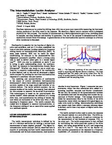
II. Method for Separating the Intermodulation Products: Theory and Simulations The proposed method, called pulse subtraction intermodulation (PSI), consists of a multi-pulse technique that is able to reinforce the intermodulation products. PSI can
521
also remove the second harmonics, one of the 2 transmitted bands and its third harmonic. In particular, 2 composite pulses are generated in the following manner: 1) The first composite pulse, CP1, is constituted by 2 burst signals at different frequencies called, respectively, F1 and F2, which are combined by a sum operation (Fig. 1). 2) The second composite pulse, CP2, is constituted by summing F1 and F2* where F2* is an inverted replica of F2 (F2* is in phase opposition respect with F2) (Fig. 1). For each line of sight, the 2 composite pulses CP1 and CP2 are then subsequently transmitted into a medium and the 2 echo signals (respectively, ECP1 and ECP2) are received from the transducer, digitized, and stored in memory. When an ultrasonic composite pulse passes through a nonlinear medium, the different frequency components interact with each other, and sum and difference frequency products are generated. These intermodulation components are contained in the received echo signals and must be separated from fundamental and harmonics contents. This is achieved by a point-to-point subtraction of the received echo signals. Subtraction of the 2 RF signals generates a new signal that contains only intermodulation products and some residual components (such as one of the transmitted bands and its third harmonics) but is deprived of one transmitted component and of both second harmonics. The PSI scheme can be theoretically analyzed considering a system described by the following nonlinear equation (for simplicity, a static model with terms up to third order is considered):
S o(t) = K ( S i(t) + eS i 2(t) + V S i 3(t) ) |eS i(t) 2| 1,
|V S i(t) 3| 1.
(1)
By considering input signal Si(t) as 2 composite pulses, S1(t) and S2(t), generated according to the proposed method:
S 1(t) = A1 cos(w 1t) + A 2 cos(w 2t)
(2)
S 2(t) = A1 cos(w 1t) - A 2 cos(w 2t),
(3)
the outputs S1o(t) and S2o(t) of the model in (1), present (4) and (5) (see next page). It is worth noting that some of the expression terms in (4) and (5) appear with opposite signs, and hence, the resulting signal D12(t) (difference signal) maintains only a few terms of the basic expressions.
IEEE Transactions on Ultrasonics, Ferroelectrics, and Frequency Control ,
522
S 1o(t) =
vol. 56, no. 3,
March
2009
ö æ ö 3 K eA1 2 K eA 2 2 æç 3 + + ç KA1 + K V A1 3 + K V A1A 2 2 ÷÷÷ cos(w 1t) + çç KA 2 + K V A 2 3 + K V A1 2A 2 ÷÷÷ cos(w 2t) ç ç 2 2 2 2 è ø è ø 1 1 K eA 2 2 K eA1 2 cos(2w 2t) + K V A1 3 cos(3w 1t) + K V A 2 3 cos(3w 2t) cos(2w 1t) + 2 2 2 2 + A1A 2K e cos((w 1 - w 2)t) + A1A 2K e cos((w 2 + w 1)t)
+
3 + K V A1 2A 2 cos((w 2 + 2w 1)t) + 2 3 + K V A1A 2 2 cos((2w 2 + w 1)t) + 2 S 2o(t) =
(4)
3 K V A1 2A 2 cos((w 2 - 2w 1)t) 2 3 K V A1A 2 2 cos((2w 2 - w 1)t) 2
ö æ ö 3 K eA1 2 K eA 2 2 æç 3 + ç KA1 + K V A1 3 + K V A1A 2 2 ÷÷÷ cos(w 1t) - çç KA 2 + K V A 2 3 + K V A1 2A 2 ÷÷÷ cos(w 2t) çè çè 2 2 2 2 ø ø 1 1 K eA 2 2 K eA1 2 cos(2w 2t) + K V A1 3 cos(3w 1t) - K V A 2 3 cos(3w 2t) cos(2w 1t) + 2 2 2 2 - A1A 2K e cos((w 1 - w 2)t) - A1A 2K e cos((w 2 + w 1)t) +
(5)
3 3 - K V A1 2A 2 cos((w 2 + 2w 1)t) - K V A1 2A 2 cos((w 2 - 2w 1)t) 2 2 3 3 2 + K V A1A 2 cos((2w 2 + w 1)t) + K V A1A 2 2 cos((2w 2 - w 1)t) 2 2
D 12(t) = S 1o(t) - S 2o(t) æ ö 3 = K eA 2 2 + 2 çç KA 2 + K V A 2 3 + K V A1 2A 2 ÷÷÷ cos(w 2t) çè 2 ø + K V A 2 3 cos(3w 2t) + 2A1A 2K e cos((w 1 - w 2)t) + 2A1A 2K e cos((w 2 + w 1)t)
5) Three intermodulation products of third order that are at the frequencies f2, (f2 + 2f1) and |f2 − 2f1| (diagonal shading in Fig. 2).
The considered static mathematical model provides useful results explaining the distortion mechanism of intermodulation focused on PSI scheme. In any case, math+ 3K V A1 2A 2 cos((w 2 + 2w 1)t) ematical models, which describe the cumulative process + 3K V A1 2A 2 cos((w 2 - 2w 1)t) of distortion during propagation, lead to a more refined (6) prediction of the behavior of propagating acoustic waves. For this reason, the Khokhlov-Zabolotskaya-Kuznetsov (KZK) nonlinear propagation equation has been used in Frequency components of S1o(t) and S2o(t), as well as the past [17]–[23] to model nonlinear propagation of ulD12, can be reported in the frequency domain (Fig. 2) trasound in a medium when its nonlinear properties are when burst signals are considered instead of pure tone known. The KZK equation was employed by our group for signals. the study of the PSI method and it was numerically solved The signal D12(t) is constituted by: by FEMLAB (Comsol, Inc. Burlington, MA), a powerful partial differential equation (PDE) solver considering the 1) A constant term (independent from the frequency) acoustic parameters of water (ultrasound speed of 1500 that is related to a continuous component (black in m/s, medium density of 998 kg/m3, B/A parameter of 5, Fig. 2). and attenuation of 0.0025 dB/cm/MHz), propagation dis 2) A term at frequency f2 that is related to the compo- tance of 25 cm, and plane wave approximation. Simulated nents with opposite signs in the transmitted pulses results of Fig. 3 are obtained for 2 composite pulses (gen(dark gray in Fig. 2). erated according to the PSI scheme of Fig. 2) constituted 3) A term at frequency 3f2 that is the third harmonics by 2 sinusoidal bursts F1 and F2 with the same amplitude of the transmitted component at frequency f2 (white of 100 kPa and frequencies of, respectively, 4 MHz and 6 in Fig. 2). MHz. 4) Two intermodulation products of second order that Fig. 3(a) is related to the case of 2 composite pulses are located at the difference |f2 − f1| and sum (f2 + CP1 and CP2 resulting from F1 and F2 burst signals, f1) frequencies in the spectrum (dotted shading in which exhibit the same time duration of 6 μs, i.e., the Fig. 2). same bandwidth in the spectral domain for the 2 trans-
Biagi et al. :
multipulse technique exploiting the intermodulation of ultrasound waves
523
Fig. 1. PSI procedure: For each line of sight, the composite pulses CP1 and CP2 are transmitted in the medium and the received echo signals are subtracted. Note: The amplitudes of the waveforms are plotted on different vertical scales.
524
IEEE Transactions on Ultrasonics, Ferroelectrics, and Frequency Control ,
vol. 56, no. 3,
March
2009
Fig. 2. Top: Harmonic and intermodulation components of a burst signal composed of 2 frequency components at f1 and f2, which undergoes a cubic nonlinear distortion. Bottom: Final spectrum after the PSI procedure. The rejection of one fundamental component, its third harmonic, both second harmonics, and a few intermodulation components of the third order can be noted.
mitted bands; the simulated spectra for the 2 distorted transmitted pulses are reported at the top and center. On the bottom of Fig. 3(a), the spectrum of the subtracted echo signals according to the PSI scheme shows that only one of the 2 transmitted bands (the one at 6 MHz) is still present, as well as the intermodulation products of second order at 2 MHz and 10 MHz and of third order at 14 MHz. The transmitted band at 4 MHz and its harmonics (at 8 MHz and 12 MHz) are completely removed, creating a free spectral region and significant separation of intermodulation components. It is worthwhile to note that the intermodulation spectral components have a bandwidth comparable to the transmitted ones. In fact, the intermodulation phenomenon occurs during the entire time duration of the composite pulse leading to the generation of an intermodulation pulse that has the same time duration as the original composite pulse, i.e., the same bandwidth. Fig. 3(b) concerns the simulated result when composite pulse CP1 and CP2 are obtained by F1 and F2 signals with different time durations, i.e., different spectral bandwidths. In this example, CP1 is a sum of the F1 signal (Gaussian shape, 4 MHz central frequency, 1.2 μs time duration, and 200 kPa acoustic pressure) and the F2 signal (Gaussian shape, 6 MHz central frequency, 16 μs time duration, and 50 kPa acoustic pressure). The second composite pulse CP2 is obtained according the PSI method. The simulated spectra of the first and second composite pulses are reported, respectively, on the top and in the center of Fig. 3(b). The presence of narrowband and
broadband harmonics due to nonlinear propagation can be noted. The spectrum of the subtracted signals is reported at the bottom of the figure. It can be observed that one of the 2 transmitted bands (the one at 4 MHz) is removed, as well as its second harmonics, whereas the intermodulation products of second order at 2 MHz and 10 MHz and of third order at 14 MHz are still present. In this last regard, it can be stated that when composite pulses are composed of 2 frequency components F1 and F2 with different time durations, i.e., different spectral bandwidth, the bandwidth of the intermodulation products due to nonlinear propagation is comparable to the bandwidth of the broadband component. In fact, intermodulation occurs only when the 2 frequency components are simultaneously present in the composite pulse and, hence, the time duration of the intermodulation signals is set by the component signal with the shortest time duration. It can be stated that the bandwidth of the intermodulation products is the same as the broadest one between the 2 frequency component signals. The generation of 2 composite pulses starting from F1 and F2 component signals with different time durations appears to be suitable for developing a broadband imaging technique with great benefits in terms of axial resolution. The removal of the broadband transmitted component and its harmonics according to the method allows the creation of a free region in the spectral domain where intermodulation products can be located with a good separation from the remaining frequency contents. Moreover, the
Biagi et al. :
multipulse technique exploiting the intermodulation of ultrasound waves
525
Fig. 3. (a) The spectra of composite pulses CP1 (top) and CP2 (center) obtained by F1 and F2 signals with the same time duration. On the bottom, the spectrum of the subtracted signals according to the PSI scheme is reported; only one of the 2 transmitted bands (6 MHz) is still present, as well as the intermodulation products of second order (2 MHz and 10 MHz) and of third order (14 MHz). (b) Spectra of the second couple of composite pulse CP1 (top) and CP2 (center). Frequency component signals F1 and F2 have different time duration, i.e., different bandwidth in the spectral domain. At the bottom, the spectrum of the final signal resulting from PSI method is reported. Note: simulations were performed at a distance of 25 cm from the transducer surface assuming B/A parameter equal to 5.
bandwidth of the intermodulation products is the same as the broadband component signal, which is removed by the final subtraction. The free spectral region can be additionally increased if the unremoved fundamental component is narrowed as much as possible. To reduce side lobes of the remaining narrowband fundamental (ensuring minimal interference with the intermodulation bands), different kinds of narrowband component signals can be employed instead of uniformly weighted (rectangular windowed) sinusoidal burst signals. A low level for the side lobe amplitude is ensured by a fam-
ily of windowed signals. Windowed signals are signals with an amplitude modulation according to well-known window types such as Hanning, Hamming, or Gaussian, which allow smoothing of the initial and final part of the ultrasonic pulse. This kind of signal is able to reduce the spectral side lobes of the ultrasonic pulse, especially when applied to transducers driven outside their central frequency. In this case, a smoothed excitation signal is able to force the transducer to resonate at the driving frequency and eliminate its free oscillation response located at the initial and final part of the ultrasonic pulse. For windowed ex-
526
IEEE Transactions on Ultrasonics, Ferroelectrics, and Frequency Control ,
vol. 56, no. 3,
March
2009
Fig. 4. Generation of the composite pulses CP1 and CP2 starting from their basic components F1 and F2.
citation signals, considerations concerning the generation of intermodulation products remain unchanged with the advantage of an effective removal of undesirable frequency contents that could overlap intermodulation contents. Further considerations can be made regarding the amplitude of the intermodulation products and hence the signal to noise ratio (SNR). It is worth noting in Fig. 3 that amplitudes of the sum and difference intermodulation contents are not the same: The sum intermodulation product is 25 dB greater than the difference one. For this reason, the employment of the intermodulation products at sum frequency leads to a higher SNR. The amplitude of final intermodulation signal is a consequence of the single amplitude of both frequency component signals F1 and F2. The amplitude of each frequency component can be set separately when the composite pulse is created with advantageous implications in terms of SNR, especially when broadband and narrowband windowed components are present in the same composite
pulse. In this case, time durations and amplitudes of the 2 frequency components F1 and F2 need to be optimized separately as a trade-off between SNR and acoustic intensity limitations in terms of mechanical index (MI) and spatial peak pulse average intensity (ISPPA) [24]) imposed by safety regulations for ultrasound equipment.
III. Method for Separating the Intermodulation Products:
Simulated Results In this section, the procedure to obtain the ultrasonic pulses employed for the experimentation is described and the simulation of the proposed method with the KZK nonlinear model is reported. Fig. 4 reports the composite pulses and their basic components employed in the experimentation. The first composite pulse CP1 (left column), is constituted by the temporal sum of 2 Gaussian-shaped signals with different
Biagi et al. :
multipulse technique exploiting the intermodulation of ultrasound waves
527
Fig. 5. Simulation of propagation of the composite pulse CP1 and CP2 in a nonlinear medium with B/A = 5 at a distance of 10 cm. (a) Propagated signal and spectrum of composite pulse CP1; (b) propagated signal and spectrum of composite pulse CP2; (c) final PSI signal and spectrum obtained according to the proposed method.
time durations and center frequencies: the first one (component F1) is a 5 MHz Gaussian shaped signal with short time duration (1.2 μs). Component F2 is a 7.5 MHz center frequency signal with time duration of 16 μs. The second composite pulse CP2 (right column) is the temporal sum of basic component F1 with F2*, which is the phase inversion signal of the F2 basic component. Simulated signals according to the KZK model whose inputs are CP1 and CP2 are reported, respectively, in Fig. 5(a) and Fig. 5(b). Because the experimentation was carried out in a water tank, simulations were performed with the B/A parameter equal to 5 and a propagation distance of 10 cm. The transmitted components (narrowband and broadband components) are clearly recognizable in these spectra in contrast to nonlinear contents such as harmonics and intermodulation components, which are overlapped and undistinguished.
Results of the subtraction between the distorted pulse CP1 and CP2 are reported in Fig. 5(c): Suppression of the broadband fundamental and the second harmonics can be noted in the spectrum. The presence of the narrowband transmitted frequency (7.5 MHz), the sum (12.5 MHz) and difference (2.5 MHz) intermodulation products according to the theory in Section II are worth noting. Moreover, an intermodulation product of third order is present at 17.5 MHz and a fourth- order intermodulation product is present at 7.5 MHz. In Fig. 6(a), the spectrum of the PSI final signal is reported as a continuous line. A low pass FIR filter—dashed line in Fig. 6(a)—and a pass band FIR filter—dot-dashed line in Fig. 6(a)—are employed to isolate, respectively, the difference and sum frequency intermodulation products from the other spectral contents. Fig. 6(b) and Fig. 6(c) show the 2 intermodulation signals, difference and sum frequencies, respectively, obtained by means of digital FIR filters.
528
IEEE Transactions on Ultrasonics, Ferroelectrics, and Frequency Control ,
vol. 56, no. 3,
March
2009
Fig. 6. (a) Continuous line: spectrum of the PSI final signal. Basic dash line: low pass (LP) filter (Hamming FIR filter with 191 taps). Double dash line: band pass (BP) filter (Hamming FIR filter with 191 taps). (b) Difference frequency intermodulation signal extracted by means of the LP filter. Sum frequency intermodulation signal extracted by means of the BP filter.
Fig. 7. Left: A block diagram of the acquiring system. As transmitting unit, a single-element ultrasonic transducer is connected to a linear power amplifier (ENI 350L) driven by an arbitrary waveform generator (Agilent, 33250A). Right: transducer arrangements for the first 2 setups employed for the experimentation.
Considering the filtered signals in Fig. 6(b) and in Fig. 6(c), it can be affirmed that their temporal width is comparable to the broadband component of CP1 and CP2 reported in Fig. 4, and hence, the final axial resolution of the method is imposed by the broadband component of the transmitted composite signals.
IV. Experimental Setup Three experimental setups were devised to assess the proposed method (Fig. 7). The first one consists of an experimental transmission system based on 2 single-element transducers aligned ac-
Biagi et al. :
multipulse technique exploiting the intermodulation of ultrasound waves
529
Fig. 8. PSI method applied to the transmitted signals acquired by the hydrophone described in setup 1. (a) Acquired composite pulse CP1 and its spectrum; (b) acquired composite pulse CP2 and its spectrum; (c) subtraction signal between composite pulses CP1 and CP2 and its spectrum with the frequency response of the low pass filter (dashed line) and pass band filter (dot-dashed line) superimposed; (d) sum (right) and difference (left) intermodulation signals extracted from the signal shown in Fig. 8(c) by filtering processing.
530
IEEE Transactions on Ultrasonics, Ferroelectrics, and Frequency Control ,
vol. 56, no. 3,
March
2009
Fig. 9. Experimental assessment of the sum and difference intermodulation amplitude versus the acoustic pressures employing setup 1. (a) Definition of negative and positive peak pressure values (PPK-, Ppk+) on an acquired ultrasonic signal; (b) spectrum of the final PSI signals for the case of Fig. 9(a); (c) experimental evaluation of PPK- and Ppk+ values versus the transducer driving voltage; (d) values of sum and difference intermodulation contents evaluated in the spectrum obtained by PSI and normalized by the maximum level of the narrowband fundamental content.
cording to the beam axis. The transmitting transducer is a BDN10 (Gilardoni, Mandello del Lario, Lecco, Italy) with a diameter of 10 mm, a planar face, a center frequency of 5 MHz, and a −6dB relative bandwidth of 50%. The receiving transducer is a calibrated hydrophone (PVDF coplanar shielded membrane hydrophone type No. 699/1/00002/200, GEC-Marconi, Caswell, Towcester, UK). Because the near field distance N for the BDN10 transducer is 8.3 cm at 5 MHz and 12.45 cm at 7.5 MHz, the distance between the 2 transducers is set at 10 cm. An 80 MHz arbitrary waveform generator (AWG 33250A; Agilent Technologies, Palo Alto, CA) generated the composite pulses according to the proposed method and was also the trigger for the whole system. The transmit signals were amplified by a broadband 50 dB RF power amplifier (350LA; ENI, Rochester, NY). For accurate and low-noise acoustic pressure measurements, a 500-MHz analog bandwidth and 1-GS/s sampling frequency oscilloscope (TDS 520C, Tektronix Inc.,
Portland, OR) was employed in connection with the calibrated membrane hydrophone. A set of 24 different driving voltages (i.e., 24 different acoustic intensities) ranging between 24 and 162 Vpp was employed to evaluate the acoustic levels of the sum and difference intermodulation content generated as a consequence of signal distortion. The second setup for this experimentation (setup 2) is a system based on a pulse-echo technique with 2 singleelement transducers positioned confocally at a 20° angle. The distance from the transducer faces and the acoustic beam intersection is 10 cm. The BDN10 transducer was employed as transmitter, and the receiving probe, a V382 (Panametrics, Waltham, MA) with a diameter of 1.3 cm, was a 3.2-MHz center frequency unfocused transducer with a minus 6 dB relative bandwidth of 100%. RF received signals were amplified with a low noise, 40-dB RF amplifier (5052 PR, Panametrics, Waltham, MA) and sampled at 40 MHz using one channel of a fast echographic multiparameters multi-image novel appara-
Biagi et al. :
multipulse technique exploiting the intermodulation of ultrasound waves
531
Fig. 10. Experimental assessment of the PSI method: RF echo signals and related spectra acquired with the setup 2. (a) Two RF echo signals related, respectively, to the CP1 and CP2 composite pulses; (b) superposition of the spectra of the echo signal of Fig. 10(a); (c) spectrum of the RF subtracted signal; (d) final intermodulation signal as a result of band pass filtering.
532
IEEE Transactions on Ultrasonics, Ferroelectrics, and Frequency Control ,
vol. 56, no. 3,
March
2009
Fig. 11. Radiation diagrams calculated in the frequency domain by means of Fourier Transform. (a) Black line: radiation diagram of the difference intermodulation component (2.5 MHz); grey line: radiation diagram of the fundamental (5 MHz). (b) Black line: radiation diagram of the sum intermodulation component (12.5 MHz); grey line: radiation diagram of the fundamental (5 MHz).
tus (FEMMINA platform) [25], [26], which is echographic equipment able to acquire ultrasonic signals in real-time modality. The 2 setups were employed with the same kind of composite pulses to assess the method both in transmission and in echo modalities. The transmitted sequence consists of 2 composite pulses, CP1 and CP2, with a mutual delay time of 150 μs. The repetition frequency of the sequence is 10 Hz. Echo signals were received and processed according to the described method. A tissue-mimicking phantom was employed in the second setup as a nonlinear and scattering water-based medium. The phantom consisted of a latex tube, 4 cm in diameter and 80 μm thick, filled with a water test fluid (Model 707-G, ATS Laboratories, Inc., Bridgeport, CT) and including a calibrated dispersion of 30-μm diameter plastic particles. A third measurement set-up was employed for ultrasound beam profile characterization (set-up 3). The transmitting transducer (BDN10) and the coplanar hydrophone, moved by a micrometric scanning system, were immersed in a water tank filled with degassed water. The beam diagrams of intermodulation components were obtained by using the PSI processing method.
V. Experimental Results A. Setup 1: Transmission Setup Fig. 8 shows the experimental results obtained from setup 1 when the ultrasonic pulses CP1 and CP2 theoretically described in Fig. 4 and simulated in Fig. 5 and in Fig. 6 are transmitted into the water tank. Fig. 8(a) and Fig. 8(b) show the experimental ultrasonic pulses (and their spectra) received by the hydrophone after a 10 cm propagation in water. The intermodulation components are difficult to recognize in the spectra of distorted CP1 and CP2 signals because they are overlapped by the fundamental and second harmonic bands. After the subtraction between CP1 and CP2 signals, second-order intermodulation components appear in the spectrum of Fig. 8(c) and can be separated by low-pass—dashed line of Fig. 8(c)—and pass-band— dot-dashed line of Fig. 8(c)—filters. In Fig. 8(d), the 2 intermodulation pulses extracted from the signal of Fig. 8(c) are shown. It is worth noting how their time duration is comparable to the time duration of the broadband component of CP1 and CP2 (Fig. 4, basic component signal F1) and their similarity with the simulated signals (Fig. 6, on the bottom).
Biagi et al. :
multipulse technique exploiting the intermodulation of ultrasound waves
Fig. 9 reports the results from experimentation performed using setup 1. These results show the behavior of sum and difference intermodulation spectral contents at different increasing acoustic intensities. Fig. 9(a) shows how the measured negative Ppk- and positive Ppk+ peak pressures are defined on a distorted composite pulse. In Fig. 9(b), the spectrum of subtraction signal according to the PSI method is reported: the residual transmitted narrowband at 7.5 MHz, the sum and difference intermodulation contents at, respectively, 12.5 MHz and 2.5 MHz, the residual third-order intermodulation content at 17.5 MHz, and the residual fourth-order intermodulation contents at 7.5 MHz and 22.5 MHz are all worth noting. Fig. 9(c) reports the positive (Ppk+) and negative (Ppk-) acoustic pressure levels versus the peak-to-peak driving voltage for the CP1 and CP2 composite signals. Fig. 9(d) shows the spectral levels of the sum and difference intermodulation content of the PSI final signal versus the peak-to-peak driving voltage applied to the transducer. B. Setup 2: Echographic Setup Fig. 10 reports acquired signals coming from experimental setup 2. Two portions of the RF echo signals related to CP1 and CP2 described in Fig. 4 are shown in Fig. 10(a). The 2 RF signals present a maximum value in the central portion due to spatial overlapping of the transmitting and receiving acoustic beams according to the 20-degree alignment of transducers. Spectra of the RF echo signals are superimposed in Fig. 10(b). Narrowband content at 7.5 MHz is related to the component F2 of the transmitted CP, and broadband spectral contents in the range 1.5 MHz to 5.5 MHz are related to both the transmitted component F1 and the intermodulation difference product. It should be noted that the 2 spectral curves are almost superimposable, except in the region between 1.5 and 3 MHz, where the difference frequency intermodulation content is present as shown in the spectrum of the subtracted signal reported in Fig. 10(c). The broadband transmitted components are removed from the spectral domain, and only the difference intermodulation product and the transmitted narrowband component F2 remain according to the theory. The sum intermodulation product is located outside the receiving transducer bandwidth and hence is overcome by the electronic noise. The difference intermodulation signal of Fig. 10(d) is obtained by the pass-band filtering (91-tap Hanning FIR filter) of the subtraction signal resulting from PSI processing. C. Setup 3: Beam Profile In Fig. 11, beam radiation diagrams at −18 dB, coming from experimental setup 3, are shown for the sum and difference intermodulation components. Fig. 11(a) represents a comparison between the fundamental broadband beam (dark gray line) and the difference intermodulation beam (black line). Fig. 11(b) represents a comparison of the fun-
533
damental broadband beam (dark gray line) with the sum intermodulation beam (black line). VI. Discussion The proposed method allows extraction of the nonlinear intermodulation contents from the RF echo signal, making them available for further processing algorithms for tissue characterization or contrast imaging. For these aims, the intermodulation spectral contents must be separated from the transmitted bands and harmonics to avoid mutual spectral interference. PI technique operating with multifrequency composite pulses represents the gold standard for nonlinear echographic imaging, allowing fundamental band suppression and reinforcement of intermodulation and harmonic contents. Unfortunately, PI technique applied to multifrequency composite pulses leads to a superposition of the harmonic and intermodulation contents and thus limits the effectiveness of this technique. The investigation of the PSI technique, both theoretically and experimentally, helps overcome PI drawbacks by removing harmonic contents (especially second harmonics) in addition to one of the 2 fundamental transmitted bands. An improved version of this technique consists of transmitting for the composite pulses a narrowband component with low spectral side lobes to improve the band separation and free up the frequency region for broadband transmitted components, as appears in Fig. 3 and in Fig. 5. This can be achieved by employing as first frequency component a long sinusoidal burst with an amplitude modulation and a broadband pulse as second frequency component (Fig. 5). In this paper, we have demonstrated, both through numerical simulations and experimentally, that the bandwidth of the intermodulation product is connected to the bandwidth of the broadband frequency component of CP regardless of the time duration of the narrowband component in the composite pulse. The intermodulation frequency bands are the spectral replica of the broadband transmitted ones, as derived from Fig. 5, and hence, the final axial resolution of the method is controlled by the transmitted broadband component in the composite pulse. The narrowband spectral content of the received echo signals can be easily removed by a notch or rejection filter by considering that the smoothness of its shape ensures the lowness of spectral side lobes. Another interesting implication of windowed signals is that they avoid free oscillations, which occur around the transducer central frequency when rectangular windowed burst sinusoidal signals are employed. Moreover it is possible to move the frequency of the narrowband signals to appropriately choose the frequencies of intermodulation components with respect to the frequency response of the employed transducer. Further considerations concern the lateral resolution of the method that is imposed by the lateral dimension of the acoustic beam. As reported in [27], [28], the beam
534
IEEE Transactions on Ultrasonics, Ferroelectrics, and Frequency Control ,
cross section of difference intermodulation component is narrower if compared with that obtained by using the difference frequency as fundamental. Moreover it is possible to assert that beam lateral dimensions of nonlinear contents are lower than the fundamental ones. As experimentally demonstrated in Fig. 11, the acoustic beam dimension associated with the intermodulation products is lower than the beam associated to the fundamental components leading to an improvement of lateral resolution of the final echographic image. Furthermore, the intermodulation acoustic beam shows low side lobes with the benefits of artifact reduction in the final echographic image. The PSI method was tested under different transmitted acoustic intensities and frequencies. The method is flexible and adaptable to different insonifying conditions because the intermodulation products are located in the frequency domain in dependence on the mutual position of the 2 fundamental transmitted components F1 and F2. For instance, the difference frequency intermodulation content can be positioned at lower frequencies than the fundamental bands and hence exploits the low tissue attenuation or, alternatively, the difference frequency intermodulation content can be placed in the center of the transducer bandwidth leading to a high SNR. If the basic components of the CP are appropriately chosen, the sum frequency intermodulation content is well separated after PSI and appears, both from simulations and experimentally, with a higher amplitude level with respect to the difference frequency content. Because the sum frequency intermodulation band occurs generally at higher frequencies, its high amplitude is useful for balancing the greater ultrasound attenuation. Unfortunately, 2 transmissions for each sight line are required; as a consequence, the PSI method suffers from potential motion artifacts as well as any multipulse technique. Moreover, the need for a signal with a long time duration leads to a further reduction of the final frame rate. VII. Conclusion In this work, a method for extracting nonlinear intermodulation contents from an echographic signal backpropagated from a nonlinear medium is proposed. The method is based on the transmission of 2 composite pulses for each line of sight and then on the subtraction of the 2 acquired echographic signals. The subtraction procedure makes it possible to reject harmonics from the resulting signal (especially second ones) and to achieve good isolation of intermodulation sum and difference frequency contents, making them available for further processing algorithms. It has been demonstrated that the intermodulation spectral bandwidth is a replica of the transmitted bandwidth of the broadband component in the composite pulse if sinusoidal burst signals are employed, and hence, PSI can be suitable for high-resolution imaging techniques. We have also demonstrated the feasibility of the PSI technique
vol. 56, no. 3,
March
2009
both theoretically and experimentally. The development of optimal waveforms or coded signals exploitable for a pulse compression method will be the focus of further work. References [1] M. A. Averkiou and M. F. Hamilton, “Nonlinear distortion of short pulses radiated by plane and focused circular pistons,” J. Acoust. Soc. Am., vol. 102, pp. 2539–2548, Nov. 1997. [2] M. A. Averkiou and M. F. Hamilton, “Measurement of harmonic generation in a focused finite amplitude sound beam,” J. Acoust. Soc. Am., vol. 98, no. 6, pp. 3439–3442, Dec. 1995. [3] L. Filipczynsky, T. Kujawska, R. Tymkiewicz, and L. Wojcik, “Non linear and linear propagation of diagnostic ultrasound pulses,” Ultrasound Med. Biol., vol. 25, no. 2, pp. 285–299, 1999. [4] M. A. Averkiou, “Tissue harmonic imaging,” in Proc. IEEE Ultrasonics Symp., 2000, pp. 1563–1572. [5] M. A. Averkiou, D. N. Roundhill, and J. E. Powers, “A new imaging based on the nonlinear properties of tissue,” in Proc. IEEE Ultrasonics Symp., 1997, pp. 1561–1566. [6] B. Ward, A. C. Backer, and V. F. Humprey, “Nonlinear propagation applied to the improvement of resolution in diagnostic medical ultrasound,” J. Acoust. Soc. Am., vol. 101, no. 1, pp. 143–154, Jan. 1997. [7] P. N. Burns, “Harmonic imaging with ultrasound contrast agents,” Clin. Radiol., vol. 51, suppl. 1, pp. 50–55, Feb. 1996. [8] N. de Jong, A. Bouakaz, and P. J. Frinking, “Harmonic imaging for ultrasound contrast agents,” in Proc. IEEE Ultrasonics Symp., 2000, pp. 1869–1876. [9] P. J. Frinking, A. Boukaz, J. Kirkhorn, F. J. Ten Cate, and N. de Jong, “Ultrasound contrast imaging: Current and new potential methods,” Ultrasound Med. Biol., vol. 26, no. 6, pp. 965–975, 2000. [10] D. Hope Simpson, C. T. Chin, and P. N. Burns, “Pulse inversion doppler: A new method for detecting nonlinear echoes from microbubble contrast agents,” IEEE Trans. Ultrason. Ferroelectr. Freq. Control, vol. 46, no. 2, pp. 372–382, 1999. [11] W. L. Konrad and L. F. Carlton, “Design and application of high power parametric sonars,” Oceans, vol. 5, pp. 310–315, Sept. 1973. [12] L. Bjorno, “Forty years of nonlinear ultrasound,” Ultrasonics, vol. 40, pp. 11–17, May 2002. [13] M. B. Moffett and R. H. Mellen, “Model for parametric acoustic sources,” J. Acoust. Soc. Am., vol. 61, no. 2, pp. 325–337, Feb. 1977. [14] A. L. Vyas, V. S. Balaji, and R. G. Gupta, “Design consideration of parametric arrays,” in Proc. Underwater Technology Int. Symp., 1998, pp. 98–102. [15] W. L. Konrad and M. B. Moffett, “Parametric acoustic source beamwidth control,” IEEE J. Oceanic Eng., vol. OE-3, no. 3, pp. 57–59, July 1978. [16] M. Averkiou, “Ultrasonic diagnostic imaging of nonlinearly intermodulated and harmonic frequency components,” U.S. Patent 6 440 075, Aug. 27, 2002. [17] S. Makarov and M. Ochmann, “Nonlinear and thermoviscous phenomena in acoustics, Part II,” Acustica, vol. 83, pp. 197–222, 1997. [18] E. A. Zabolotskaya and R. V. Khokholov, “Quasi-plane waves in the nonlinear acoustics of confined beams,” Sov. Phys. Acoust, vol. 15, pp. 35–40, 1969. [19] V. P. Kutnezov, “Equations of non linear acoustics,” Sov. Phys. Acoust, vol. 16, pp. 467–470, 1971. [20] Y. Li and J. A. Zagzebski, “Computer model for harmonic ultrasound imaging,” IEEE Trans. Ultrason. Ferroelectr. Freq. Control, vol. 47, no. 5, pp. 1259–1272, Sept. 2000. [21] H. Hobaek and B. Ystad, “Experimental and numerical investigation of shock wave propagation in the focal region of a focused sound field,” Acustica, vol. 83, pp. 978–986, 1997. [22] S. Makarov and M. Ochmann, “Nonlinear and thermoviscous phenomena in acoustics, Part I,” Acustica, vol. 82, pp. 579–606, 1996. [23] F. E. Fox and W. A. Wallace, “Absorption of finite amplitude sound waves,” J. Acoust. Soc. Am., vol. 26, pp. 994–1000, 1954. [24] U.S. Department of Health and Human Services, Food and Drug Administration, “Information for manufacturers seeking marketing clearance of diagnostic ultrasound system and transducers,” Center for Devices and Radiology Health, Sep. 1997.
Biagi et al. :
multipulse technique exploiting the intermodulation of ultrasound waves
[25] M. Scabia, E. Biagi, and L. Masotti, “Hardware and software platform for real-time processing and visualization of echographic RF signals,” IEEE Trans. Ultrason. Ferroelectr. Freq. Control, vol. 49, no. 10, pp. 1444–1452, Oct. 2002. [26] L. Masotti, E. Biagi, M. Calzolai, L. Capineri, and S. Granchi, “FEMMINA: A fast echographic multiparametric multi-imaging novel apparatus,” in Proc. IEEE Ultrasonics Symp., 1999, pp. 739– 748. [27] W. Konrad and L. Carlton, “Design and application of high power parametric sonars,” Oceans, vol. 5, pp. 310–315, Sept. 1973. [28] A. H. Quazi and W. L. Konrad, “Underwater acoustic communications,” IEEE Commun. Mag., vol. 20, no. 2, pp. 24–30, Mar. 1982.
Elena Biagi was born in Florence, Italy, in 1956. She received the “Laurea” degree in electronic engineering from the University of Florence, Florence, Italy, in 1982. From 1982 to 1991, her research was mainly devoted to ultrasonic material characterization and medical imaging. Since 1991, she has been an assistant professor in the Electronics and Telecommunications Department of the University of Florence; she became an associate professor for optoelectronics in 2001. Her current interests deal with the design of dedicated ultrasonic transducers, signal processing for biological tissue spectral parameters extraction, design arid realization of fast electronics for radiofrequency real-time processing. Since 1996, her teaching activity has concerned optoelectronics and sensors.
Luca Breschi was born in Prato, Italy, in 1973. He received the “Laurea” degree in electronic engineering from the University of Florence, Florence, Italy, in 1999. In 2002, he received the Ph.D. degree in nondestructive testing from the Electronics and Telecommunications Department of the University of Florence. His main research activities are nonlinear interaction between ultrasound and tissues and signal process-
535
ing for tissue characterization. He developed several signal processing algorithms for biological tissue characterization and diagnostic systems for cancer detection by means of contrast agents. He is also holds several patents and is the author of several national and international scientific papers.
Enrico Vannacci was born in Pistoia, Italy, in 1978. He received the “Laurea” degree in electronic engineering from the University of Florence, Florence, Italy. In 2008, he received the Ph.D. degree in nondestructive testing from the Electronics and Telecommunications Department of the University of Florence. His research activities include acoustic characterization of ultrasound contrast agents, ultrasound nonlinear imaging, and signal processing for tissue characterization. Currently, he is involved in devising new diagnostics systems for the assessment of the acoustic nonlinear properties of tissues.
Leonardo Masotti was born in Faenza, Italy, in 1939. He received the “Laurea” degree in electronic engineering from the University of Engineering in Bologna, Italy, in 1963 and the Italian National Libera Docenza degree in applied electronics in 1970. His present position is full professor of electronics and research director of the Ultrasound and Non Destructive Testing Laboratory at the Department of Electronics and Telecommunications of the University of Florence, Florence, Italy. He has been a member of the Scientific Committee of the Istituto di Elaborazione dell’lnformazione of the Consiglio Nazionale delle Ricerche (CNR) in Pisa since 1983 and the Scientific Committee of Istituto di Fisiologia Clinica of CNR in Pisa since 1989. He has been president of the Consorzio Centro di Eccellenza Optronica (CEO) since 1992. He also takes part in the Technical Direction Committee of the High Tech Network for the Tuscany Region.