BRAIN RESEARCH ELSEVIER
Brain Research 734 (1996) 213-222
Research report
Neural parameters contributing to temperature compensation in the flight CPG of the locust, Locusta migratoria Hongjian Xu 1, R. Meldrum Robertson * Department of Biology, Queen's University, Kingston, Ont. KTL 3N6, Canada
Accepted 15 May 1996
Abstract
Elevated thoracic temperature increases the wingbeat frequency of flying locusts. We investigated the extent to which temperature-induced changes in resting membrane potential and postsynaptic potential amplitude contribute to the effects of increased temperature on the frequency of the central flight rhythm. Flight neurons were hyperpolarized by changing the K + concentration of the superfusing saline from 10 mM to 2 mM. 5 min of low-K + superfusion hyperpolarized flight motoneurons from -42.8 mV to -50.1 mV with a concomitant decrease of the frequency of the central flight rhythm from 11.6 Hz to 10.5 Hz. The amplitude of postsynaptic potentials was halved after 10 rain of zero Ca2+/high Mg 2+ superfusion, but the frequency of the central rhythm did not change significantly. GABAergic inhibitory connections were reduced in amplitude using picrotoxin. This treatment increased the frequency of the central rhythm from 11.6 Hz to 12.9 Hz, and increased the thermosensitivity of the rhythm frequency. We conclude that the excitatory effect of increased temperature on rhythm frequency is not mediated by temperature effects on membrane potential and/or synaptic potential amplitude. We propose that the inhibitory effect of temperature-induced hyperpolarization of the membrane potential compensates for the excitatory effect of temperature on rhythm frequency (e.g. via increased conduction velocity). We further suggest that some measure of temperature compensation is afforded by equal effects on the amplitudes of excitatory and inhibitory postsynaptic potentials, such that the net effect on the level of excitation is zero. Keywords: Insect; Locust; Flight; Motor pattern; Temperature; Membrane potential; PSP amplitude
1. Introduction
Ambient temperature can have considerable impact on the operation of neuronal circuits and the form and existence of subsequent rhythmic behaviors, particularly in poikilotherms (e.g. heartbeat frequency in leeches [2], teleost and ascidian swimming [3], pyloric contractions in lobsters [16,17]). Therefore, a complete understanding of the adaptive operation of neuronal circuits is not possible without also considering the impact of the thermal regime of the organism under investigation. The natural habitat of the African migratory locust, Locusta migratoria, is characterized by variable and high temperatures [45]. Although air density decreases at higher temperatures, many of the aerodynamic requirements of a
* Corresponding author. Fax: + 1 (613) 545-6617; E-mail:
[email protected] I present address: Department of Biochemical Pharmacology, 321 Hochestetter Hall, SUNY at Buffalo, Buffalo, NY 14260-0001, USA.
flight system remain the same at different temperatures. A compelling question is therefore how the neuronal circuits controlling kinematic parameters of the wingbeat maintain appropriate output in spite of a wide range of operating temperatures. In the laboratory it has been demonstrated that the locust flight system is capable of operating in the range from 24°C to 42°C [24,46]. One parameter of the wingbeat, wingbeat frequency, changes minimally with ambient temperature in locusts [46] (0.27 H z / ° C , [9]) and one implication of this finding is that compensatory mechanisms exist within the flight system of locusts to stabilize the output frequency. Flight motor patterns of locusts are generated by neuronal circuits that include peripheral as well as central components [25,31,47]. A number of flight interneurons have been identified (reviewed in [31,32]) and some have been shown to support plateau potentials which may contribute to rhythm generation in the system [27-29]. A simple circuit of flight interneurons which could account for several main features of the central motor pattern has been described [37], and recently a computer simulation
0006-8993/96/$15.00 Copyright © 1996 Elsevier Science B.V. All rights reserved. PII S0006-8993(96)00635-X
214
H. Xu, R.M. Robertson / Brain Research 734 (1996) 213-222
has demonstrated that this circuit is capable of generating robust flight rhythms [11]. Temperature has profound effects on neuronal properties in the locust nervous system [1,5,14,21,22,26,40]. The effects of temperature on some cellular, synaptic and circuit properties of locust flight neurons have also been described [33,48]. Amongst other things, it has been proposed that: (1) hyperpolarization of the resting membrane potential is involved in automatic temperature compensation at the cellular level [48], and (2) the frequency of the rhythm is unaffected by the effects of temperature on the amplitude of postsynaptic potentials within permissive limits [33]. The main goal of the experiments described here was to test these ideas by mimicking particular changes in neural properties wrought by temperature, and assessing the effect on flight rhythm generation. Also, Miles [22] has suggested that temperature compensation in the afferent pathway from wind-sensitive head hairs of the grasshopper may be a result of equal effects of temperature on concurrent excitatory and inhibitory inputs. Effects of temperature on the different inputs may thus cancel each other out. We tested this idea in the flight circuitry by pharmacologically changing the relative strengths of excitatory and inhibitory synaptic connections. The specific objectives of this study were: (1) to determine the influence of resting membrane potential (RMP) on the central flight rhythm by manipulating membrane potentials with reduced extracellular K + (e.g. [42]); (2) to study the effect of postsynaptic potential (PSP) amplitude on the central flight rhythm by manipulating PSP amplitude with reduced extracellular Ca 2+ (e.g. [4]); and (3) to examine the response of the central flight rhythm to changes in the relative strength of excitatory and inhibitory postsynaptic potentials (EPSPs and IPSPs) in the central circuitry by manipulating IPSP amplitude with picrotoxin (e.g. [37]).
2. Materials and methods
Adult male locusts (Locusta migratoria) were obtained from a colony maintained in the Department of Biology, Queen's University. Only mature animals ( > 10 days after the imaginal moult) were used. Colony conditions were 31 4- I°C, with a 16:8 h light/dark cycle.
2.1. Preparations We used a standard preparation of the deafferented locust flight system [34]. The wings and legs were cut off and the locust was pinned to a cork board. The thoracic nervous system was exposed by a dorsal dissection that removed the gut and overlying tissue. Nerves 3, 4, and 5 of the mesothoracic and metathoracic ganglia [7] were severed. These two ganglia were supported on a stainless steel platform. The preparation was initially superfused with
locust saline which could be replaced with ion-substitution or drug solutions. The temperature of the solutions was controlled with a heating coil around a glass pipette that directed the saline into the thoracic cavity. A copper/constantan thermocouple was placed adjacent to the mesothoracic ganglion to monitor thoracic temperature and the preparation was grounded through an Agar/electrolyte bridge. Most experiments were performed at room temperature (22-25°C). The experiments using picrotoxin also examined the thermosensitivity of the rhythm frequency in the 20-35°C range.
2.2. Intracellular and extracellular recordings Short flight sequences were obtained by blowing air over the head of the animal. The time of depression in the flight rhythm was monitored using an electromyographic (EMG) electrode (50 ~m copper wire, insulated except at the tip) inserted into one of the dorsal longitudinal (DL) muscles. Spontaneous action potentials of the forewing stretch receptor or hindwing tegulae were recorded from nerve 1 of the mesothoracic or metathoracic ganglion with a monopolar silver wire electrode insulated from the saline with vaseline. Standard criteria for presumed monosynaptic connections were used: a constant latency between presynaptic spikes and postsynaptic potentials, and synaptic latency 1 ms or less after subtracting a conduction delay of 1 ms. This conduction delay was confirmed numerous times from recordings of afferent activity simultaneously with a peripheral extracellular electrode and a central intracellular electrode. Intracellular recordings were made using 1 M KAc filled glass microelectrodes (resistance around 40 Mf~) inserted in the neuropile processes of flight motoneurons and interneurons in the mesothoracic and metathoracic ganglia. Recordings were maintained for at least 10 rain, allowing sufficient time to change the superfusing solutions. Electrical signals were amplified conventionally and all recordings were stored on VHS videotape using a Neurodata neurocorder (DR-886) for subsequent analysis.
2.3. Solutions Locust saline contained (in retool/l) NaC1, 147; KCI, 10; CaC12, 4; NaOH, 3; HEPES, 10 (pH 7.2). To reduce extracellular K + to 2 mM, 8 mM mannose replaced the KC1. The zero Ca 2+ solution contained 20 mM MgCI 2 instead of CaC12 and 10 mM EDTA was added. For pharmacological experiments, 5 ! 10 _5 M picrotoxin (Sigma Chemical Co., St. Louis, MO) was added to locust saline. Stock solutions of picrotoxin were maintained in a refrigerator for up to a week while being used in experiments. The pH for all ion-substitution or drug solutions was adjusted to 7.2.
H. Xu, R.M. Robertson / Brain Research 734 (1996) 213-222
215
RMP normal saline
-46 mV
low K+ 5 rain
-54 mV
low K+ 10 min
'
%
'
-
-
-
-
'
-
"
J
~
recovery
-66 mV
-42
m.V
i140 mV 100 ms
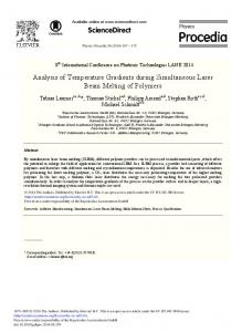
Fig. 1. Effects of low K + treatment on resting membrane potential and central flight rhythm. Intracellular recordings from a depressor motoneuron during expression of the flight rhythm before and after low K + treatment. Flight-initiating stimuli were delivered at the time of the brief depolarizing potential seen at the beginning of each trace (giving rise to action potentials in the top and bottom traces), Note the decrease in rhythm frequency after low K + treatment. Note also the gradual increase of the delay for initiation of bursting activity.
The concentrations of the ion-substitution and drug salines indicated above are higher than the concentrations that will actually be experienced by the neurons in the flight circuitry. The nature of saline substitution in a semi-intact preparation that contains crevices difficult to wash out, and tissues able to act as reservoirs is such that complete substitution of the superfusing saline and the extracellular fluid is impossible within the duration of these experiments. Also, these global treatments could have non-specific effects on neural parameters other than the ones we were targeting, especially at high concentrations. For these reasons we used only the minimum concentrations necessary to mimic the effects of temperature described previously [33,48] and did not examine an extended range of concentrations.
from four flight sequences. EPSP amplitudes were measured from on-line averages of approximately 20 EPSPs. To test for differences, the data were tested for normality and equal variance and the appropriate statistical tests were performed using the statistical software package SigmaStat (Jandel Scientific, Corte Madera, CA). Significance was assessed at P < 0.05.
3. Results
3.1. Influence of resting membrane potential on rhythm frequency Reduced extracellular K + gradually decreased the rhythm frequency although there was no obvious effect on the amplitude of action potentials or on the amplitude of the membrane potential oscillations (Fig. 1). After 5 min of superfusion with low K + saline, the resting membrane potential of flight motoneurons hyperpolarized by 7.3 mV, from -42.8_+ 1.6 mV to - 5 0 . 1 _+2.0 mV (mean_+ S.E.M.). Simultaneously, the mean frequency of the cen-
2.4. Data analysis Data presented here were collected from experiments performed with at least seven animals for each treatment. Rhythm frequency was recorded as the mean instantaneous frequency (reciprocal of period) of five consecutive cycles A
B
1.5
1.5
c-
~ n o 1.0 z-~
z~,
0.5 0.0
CONT
lOW K +
0.5 0.0
CONT
Low K +
Fig. 2. Superfusion with low K + saline hyperpolarizes the membrane potential (A) and decreases the rhythm frequency (B) of flight motoneurons. Measurements of membrane potential and rhythm frequency were normalized to control values (CONT). Bars indicate means and standard errors before, and 5 min after, low K + treatment. Asterisks indicate significant differences.
H. Xu, R.M. Robertson/Brain Research 734 (1996) 213-222
216
Table 1 Absolute values of deafferented flight rhythm frequency before and after 5 min or less of different ion substitution and drug treatments Treatments
LowK + (n=9) Zero Ca2+/High Mg 2+ ( n = 9 ) Picrotoxin (n = 11 )
normal saline
Rhythm frequency (Hz) Control
After treatment
11.6_+1.6 11.2±1.3 11.6 _+ 1.2
10.5±1.7 * 11.0_+ 1.5 12.9 ± 0.8
zero Ca++ 5 min
Values represent the mean ± S.E.M. * Significant difference from control values (paired t-tests; P < 0.05).
recovery 5 min recovery 10 min
tral flight rhythm decreased from 11.6 + 1.6 Hz to 10.5 + 1.7 Hz (Table 1). To accommodate variability in the control (pre-treatment) values for membrane potential and rhythm frequency, post-treatment values were normalized by division with control values. Five min of superfusion with reduced extracellular K + significantly hyperpolarized the membrane to 1.2_+ 0.02 of the control membrane potential (paired t-test: t = - 8 . 8 l , d f = 8, P < 0.0001) (Fig. 2A), and significantly decreased the rhythm frequency to 0.9 _+ 0.01 of the control frequency (paired t-test: t = 9.06, df = 8, P < 0.0001) (Fig. 2B).
NlC
10 ms
Fig. 3. Effects of zero Ca2+/high Mg 2+ treatment on EPSP amplitude recorded in an unidentified flight interneuron. Each of the five top traces is an average of 20 EPSPs triggered from the extracellular recorded action potentials of the hindwing tegulae (NIC, only one sweep shown). Note that EPSP amplitude decreased after zero Ca~+/high Mg 2+ treatment and that the effect was reversible.
3.2. hTfluence of PSP amplitude on rhythm frequency
hindwing tegula (e.g. Fig. 3) were reduced by superfusion with zero Ca2+/high Mg 2+ saline. However, there was little effect of the same treatment on the frequency of flight rhythm generation (Fig. 4). Ten recordings of affer-
The amplitudes of EPSPs generated by action potentials from a forewing stretch receptor (not shown) or from a
1!t I L
j'JJ ,
L 4my
zero Ca++ 10 min
L,, L,
IUvilr
"
j I
i;
%
i
ii
I
z e r o Ca + + 10 min
20 zero C o + + 15 rnin
recovery 15 min
/
~y
L / ~ , i,
U U ~.
-
L
~
_
.
--
~ / .
U
~ .
~
~
_
~
_
~
~
.
z
mV
~
~ 1 O0
"-~ ms
Fig. 4. Effects of zero Ca2+/high Mg 2+ treatment on the rhythmic bursting, activity of a tergosternal elevator motoneuron. Although the waveform amplitude was attenuated, zero Ca2+/high Mg 2+ treatment had minimal effect on the rhythm frequency. Note the reduction in the intraburst firing frequency, particularly at the start of flight sequences.
217
H. Xu, R.M. Robertson/Brain Research 734 (1996)213-222
A
B
1.5-
1.5 t-
1.o
.
=~ 1.o
-~E E