0163-769X/07/$20.00/0 Printed in U.S.A.
Endocrine Reviews 28(3):339 –363 Copyright © 2007 by The Endocrine Society doi: 10.1210/er.2006-0046
Notch Signaling in Development and Cancer Victoria Bolo´s, Joaquı´n Grego-Bessa, and Jose´ Luis de la Pompa Departmento de Inmunologı´a y Oncologı´a, Centro Nacional de Biotecnologı´a/Consejo Superior de Investigaciones Cientı´ficas, E-28049 Madrid, Spain Notch is an evolutionarily conserved local cell signaling mechanism that participates in a variety of cellular processes: cell fate specification, differentiation, proliferation, apoptosis, adhesion, epithelial-mesenchymal transition, migration, and angiogenesis. These processes can be subverted in Notch-mediated pathological situations. In the first part of this review, we will discuss the role of Notch in vertebrate central nervous system development, somitogenesis, cardiovascular and endocrine development, with attention to the mechanisms by which Notch regulates cell fate specification and patterning in these tissues.
In the second part, we will review the molecular aspects of Notch-mediated neoplasias, where Notch can act as an oncogene or as a tumor suppressor. From all these studies, it becomes evident that the outcome of Notch signaling is strictly contextdependent and differences in the strength, timing, cell type, and context of the signal may affect the final outcome. It is essential to understand how Notch integrates inputs from other signaling pathways and how specificity is achieved, because this knowledge may be relevant for future therapeutic applications. (Endocrine Reviews 28: 339 –363, 2007)
I. Introduction: Elements of the Notch Signaling Pathway II. Notch in Vertebrate CNS Development A. Notch promotes progenitor diversification and inhibits neuronal differentiation B. Notch in gliogenesis III. Notch in Somitogenesis IV. Notch in Cardiovascular Development and Homeostasis V. Notch in Endocrine Development: Pancreas, Gut, and Bone Endocrine Cells A. Pancreatic development B. Gut development C. Bone development VI. Notch in Cancer A. Notch in hematological tumors B. Notch as an oncogene in solid tumors: breast and gut cancer C. Differential roles of NOTCH in two types of skin cancer: keratinocyte-derived carcinoma and melanomas D. Notch in EMT and tumor progression VII. Concluding Remarks
Drosophila, based on its dominant wing-notching phenotype (1). The study of the embryonic lethal phenotype caused by complete lack of Notch function (2) and its complex allelic series and genetic interactions (3) brought Notch to the forefront, so that in the mid-1980s the Drosophila Notch gene product was identified (4, 5). Notch is a local signaling mechanism that is evolutionarily conserved throughout the animal kingdom. Mammals have four Notch proteins (Notch 1– 4; Refs. 6 –10 and Fig. 1A) that are membrane-bound type I receptors (with a single-pass transmembrane domain), harboring a large extracellular domain involved in ligand binding, and a cytoplasmatic domain involved in signal transduction. The extracellular domain contains a variable number of epidermal growth factor (EGF)-like repeats that are critical for binding interactions (11, 12). The EGF-like repeats are followed by three cysteinerich LIN12/Notch repeats (LNR) that prevent signaling in the absence of the ligand. The Notch intracellular domain (NICD) contains a RAM23 domain (13), six ankyrin/cdc10 repeats involved in protein-protein interactions (14), two nuclear localization signals (N1 and N2), a transcriptional activation domain (TAD) that differs among the four receptors, and a PEST sequence [rich in proline (P), glutamic acid (E), serine (S) and threonine (T)] that negatively regulates protein stability (15). The Notch receptors are synthesized as single precursor proteins that are cleaved by a furin-convertase activity (16) at site 1 or S1 (Fig. 1B) during transport to the cell surface, where they are expressed as heterodimers (17). The mammalian Notch ligands Delta1 (18), Delta3 (19), Delta4 (20), Jagged1 (21), and Jagged2 (22) are named after the Drosophila homologs Delta and Serrate, respectively, and are also membrane-bound. They have an amino-terminal domain termed DSL (for Delta, Serrate and LAG-2 domain), followed by a variable number of EGF-like repeats. In addition, Jagged1 and Jagged2 harbor a cysteine-rich domain (CR; Fig. 1A). Notch signaling is regulated by posttranslational modification events, such as glycosylation, and by other modifications involving the extracellular domains of both receptors and ligands, such as the extension of sugar residues by the
I. Introduction: Elements of the Notch Signaling Pathway
N
OTCH IS ONE of the fundamental signaling pathways that regulate metazoan development and adult tissue homeostasis. The Notch mutant was initially described in
First Published Online April 4, 2007 Abbreviations: A-P, Anterior-posterior; bHLH, basic helix-loop-helix; CNS, central nervous system; CoR, corepressor; CR, cysteine-rich domain; DSL, Delta, Serrate and LAG-2 domain; E, embryonic day; EGF, epidermal growth factor; EMT, epithelial-mesenchyme transition; FGF10, fibroblast growth factor 10; HNSCC, head and neck SCC; HSC, hematopoietic stem cell; Lfng, lunatic fringe; LNR, LIN12/Notch repeats; MAN, Mastermind-Like 1; MMTV, mouse mammary tumor virus; NICD, Notch intracellular domain; PAE, porcine aortic endothelial; PEST, sequence rich in proline (P), glutamic acid (E), serine (S) and threonine (T); PKA, protein kinase A; PPR, PTH/PTHrP receptor; PSM, presomitic mesoderm; SCC, squamous cell carcinoma; Shh, sonic hedgehog; TACE, TNF-␣-converting-enzyme; TAD, transcriptional activation domain; T-ALL, T cell acute lymphoblastic leukemia; WAP, whey acidic protein. Endocrine Reviews is published by The Endocrine Society (http://www.endo-society.org), the foremost professional society serving the endocrine community.
339
340
Endocrine Reviews, May 2007, 28(3):339 –363
A
Bolo´s et al. • Notch in Development and Cancer
Regulation and dimerization Destruction Signaling Ligand binding
Jagged1 & 2
Notch1
Delta1 & 4 Notch2 Delta3 Notch3
Notch4 pm
pm
Cell 1
Cell 2
PEST domain
RAM23
TAD
HD
NLS
LNR
Cdc10/ ankyrin repeats
EGF-like repeats
B
EGF-like repeats
C
S1 cleavage and secretion of the receptor to the cell membrane
DSL domain
CR
Endocytosis
Cell 1 pm
Ligand-Receptor interaction, extracellular domain endocytosis and S2 cleavage
Mib/Neu
U U
cytoplasm pm
Cell 2
Jagged 1 & 2
S1 cleavage
S2 cleavage
Cell 1
Fringe
pm
Furin TACE
D
S3 (γγ-secretase) cleavage and N1ICD release to the nucleus
pm
cytoplasm
cytoplasm
Golgi
a leav S3 c
E
CoR CSL Transcriptional regulation
ge
Aph-1
Cell 1
Nct Pen-2
PS1/2
NICD CoR CSL
deltex
γ-secretase
NICD
nm
cytoplasm
MAM
Nucleus
Numb NICD
Target genes
MAM
CSL Nucleus
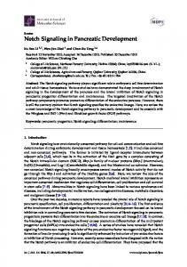
FIG. 1. Notch receptors and ligands and schematic representation of Notch signaling. A, Vertebrates have four Notch receptors (Notch1– 4) expressed in the signal-receiving cell (Cell 1). The extracellular domain of Notch has between 29 and 36 EGF-like repeats (36 in Notch1 and Notch2, 34 in Notch3, 29 in Notch4) involved in ligand binding, followed by three cysteine-rich LNRs. The LNR domain prevents ligand-independent activation of the receptor and is followed by the heterodimerization domain (HD). The cytoplasmic part of the receptor contains the RAM23 domain, six cdc10/ankyrin repeats, two nuclear localization signals (NLS), a transcriptional transactivation domain (TAD), and a PEST sequence. Vertebrate receptors can be activated by at least five ligands (Jagged1 and 2 and Delta1, 3, and 4), expressed in the signaling cell (Cell 2). The ligands share an N-terminal DSL structure. Both Delta and Jagged ligands have EGF-like repeats in the extracellular domain, but only Jagged1 and Jagged2 harbor an additional cysteine-rich (CR) sequence downstream of the EGF-like repeats. B, The Notch receptor is secreted to the cell membrane in a furin convertase-dependent step (site 1 or S1 cleavage) that takes place in the Golgi. In this cell compartment, the glycosyltransferase fringe elongates previously attached fucose residues. Notch is expressed in the cell membrane as a heterodimer. C, Binding to Delta or Jagged ligands initiates two consecutive proteolytic cleavage events; the first is mediated by the ADAM protease TACE and occurs on the extracellular side of Notch, near the transmembrane domain (site 2 cleavage). D, The second cleavage (S3) occurs within the transmembrane domain and is mediated by ␥-secretase activity, a complex composed of four different integral membrane proteins: presenilin, nicastrin (Nct), Aph-1, and Pen-2 (286). NICD is released and translocates to the nucleus. In the cytoplasm, the Numb protein negatively regulates Notch signaling, possibly by promoting receptor turnover. Deltex proteins may transduce Notch signals independently of CSL. E, In the nucleus NICD binds to the CSL transcription factor, converting it from a transcriptional repressor into a transcriptional activator by displacing a CoR complex and recruiting coactivators such as MAML1 (MAM). This leads to transcriptional activation of downstream target genes. pm, Plasma membrane; nm, nuclear membrane.
Bolo´s et al. • Notch in Development and Cancer
glycosyl transferase fringe (23, 24) (Fig. 1B). This prevents Notch activation by Jagged but not by Delta ligands (Refs. 25 and 26; reviewed in Ref. 27). Notch signaling initiates through ligand-receptor interactions between neighboring cells, leading to two consecutive proteolytic cleavages of the receptor, which ultimately liberate NICD (Fig. 1, C and D). Thus, after ligand binding, the ubiquitin ligases mind bomb (28, 29) or neuralized (30 –32) interact with the ligand intracellular domain to promote its ubiquitination and internalization (28) (Fig. 1C). Ligand endocytosis leads to a conformational change in Notch that allows ADAM (metalloprotease and disintegrin) protease TACE (TNF-␣converting-enzyme), to cleave the receptor at a second site (S2) on the extracellular side, near the transmembrane domain (33) (Fig. 1C); the released extracellular portion of the receptor is then transendocytosed to the ligand-expressing cell (34) (Fig. 1C). The third cleavage (S3) occurs within the transmembrane domain and is mediated by a ␥-secretase activity whose key components are presenilin and nicastrin (35) (Fig. 1D). This final cleavage liberates NICD, which subsequently translocates to the nucleus where it binds via its RAM23 domain to the transcription factor CSL (CBF1 in humans, Suppressor of Hairless in Drosophila, LAG in Caenorhabditis elegans), also called RBPJK in mice. In the absence of Notch activity, CSL proteins bind to promoters of its target genes and recruit histone deacetylases (36) and corepressors (CoR; Fig. 1E) that inhibit transcription. The corepressor molecules include SMRT/NcoR (37) and SHARP/MINT/SPEN (38). The NICD/CSL interaction converts CSL from a transcriptional repressor into a transcriptional activator by displacing the corepressor complex and recruiting coactivators (Fig. 1E) such as Mastermind-Like 1 (MAM; Fig. 1E) (39) and histone acetyltransferase (40). A number of additional proteins modulate Notch signaling, including the RING-domain E3 ubiquitin ligase deltex (41, 42) and the phosphotyrosinebinding domain (PTB)-containing proteins numb and numblike (43), which act as context-dependent negative or positive Notch regulators (Fig. 1D). To date, only a few target genes have been identified; some are Notch-dependent in various tissues, whereas others are tissue-specific. The best-known Notch target genes are members of the basic helix-loop-helix (bHLH) hairy/enhancer of split (Hes) family, the related HRT/Herp (Hes-related repressor protein) transcription factor family (44), the cell cycle regulator p21 (45), the Notch pathway element Notch-regulated ankyrin repeat protein (Nrarp) (46), deltex1, and the pre-T cell receptor-␣ gene (47). For an in-depth study of the molecular intricacies of Notch signaling elements see Refs. 48 –51. Here we will examine the role of Notch in specific developmental processes such as central nervous system (CNS) development, somitogenesis, cardiovascular development, and endocrine development, with attention to the distinct mechanisms by which Notch regulates cell specification and patterning in these tissues. We will then analyze the role of Notch in cancer, both in leukemia and in solid tumors, and describe studies that suggest possible means of therapeutic intervention. As a rule, when we refer to humans, the elements of the pathway are named in capital letters (i.e., NOTCH1 receptor or NOTCH1 gene), and when we refer to
Endocrine Reviews, May 2007, 28(3):339 –363 341
experimental models, the elements of the pathway are named in lowercase (Notch1 protein or Notch1 gene). II. Notch in Vertebrate CNS Development
During vertebrate CNS development, the primitive neuroepithelium gives rise to two main lineages, neurons and glia. Neurons are generated in embryonic life from multipotent progenitors close to the ventricle and, after their final mitotic division, migrate away from their birthplace to their ultimate destinations, where they terminally differentiate and integrate into the brain circuitry. Glial cells, in contrast, are generated in the proliferating subventricular zone at late embryonic and early postnatal stages. During the past 10 yr, the role of Notch in the differentiation, morphogenesis, and function of the CNS has become increasingly valued. The phenotypic analyses of Notch-targeted mutants in mice and functional manipulation in other vertebrates have greatly benefited from knowledge generated by research in Drosophila neurogenesis (52). Poulson (2) was the first to associate lack of Notch function with an embryonic lethal phenotype in Drosophila; it is caused by failure of the early neurogenic ectoderm to segregate neural and epidermal cell lineages. In homozygous Notch mutant embryos, all cells become neuroblasts, which leads to hypertrophy of the neural tissue at the expense of the epidermis, giving rise to the so-called neurogenic phenotype (2). In vertebrates, Notch is required when the epidermal and neural lineages have already segregated; its inactivation results in a “neurogenic phenotype” represented by premature differentiation of neuronal progenitors, leading to the interpretation that Notch maintains a progenitor state and inhibits differentiation. A. Notch promotes progenitor diversification and inhibits neuronal differentiation
Notch1 was the first Notch pathway gene to be disrupted by homologous recombination (53, 54). Mutant embryos die at midgestation [embryonic day 11 (E11)] with defective somitogenesis and placentation, although little attention was given to a possible neural phenotype. Subsequently, a detailed analysis of neural development in Notch1 targeted mutants was reported (55). This study examined the expression of pathway components such as Hes1, Hes5, and Delta1 and of early differentiation markers such as NeuroD (or Neurod2), Math4A (or Neurog2), and NSCL-1 (or Nhlh1). Consistent with the view that Notch activity is needed for progenitor maintenance, expression of these markers was increased in mutants. In addition, Hes5 expression was reduced in Notch1 mutants, although Hes1 expression appeared to be unaffected. This result was unexpected in light of the Hes1 mutant phenotype (56) and the extensive literature supporting the idea that Hes1 is a primary Notch/CSL target (reviewed in Ref. 57). Similar results in RBPJk targeted mutants (55) suggested that whereas Hes1 may well be a true Notch target, it is also likely to be activated by other signaling pathways. After the original Notch1 deletion studies, targeted alleles of Notch2 (58, 59), Notch3 (60), and Notch4 (61) were also generated, as well as conditional (floxed) alleles of
342
Endocrine Reviews, May 2007, 28(3):339 –363
Notch1 (62). Although Notch3 and Notch4 do not have detectable phenotypes when deleted, Notch2 mutants, similarly to Notch1 mutants, die around E11 (58). In contrast to Notch1 mutants, however, Notch2 mutants do not show alterations in Hes5 expression in the CNS. Notch2 mutants undergo widespread cell death in the CNS starting around E9 (58), but it is not clear whether this phenotype reflects a role for Notch2 in the developing CNS or whether it occurs as a consequence of other embryonic perturbations. To circumvent the early lethality of Notch1 deletion, several studies have addressed the effect of deleting this receptor in specific brain structures. In one case, conditional CreloxP-mediated recombination was used to delete Notch1 from the medial cerebellar primordium (63). Consistent with the traditional model of Notch function in the nervous system, the authors found that Notch1 deletion resulted in upregulation of the proneural genes Mash1 and Math1 and precocious neuronal differentiation. More recently, conditional deletion of Notch1 in the neural progenitor pool using a nestin-Cre promoter also resulted in precocious neuronal differentiation (64). Deletion of Notch1 in the telencephalon, using the foxg1-Cre line, led to reduced neuron numbers later in development, most likely resulting from precocious neuron differentiation and earlier progenitor pool depletion (65). In support of this finding, the telencephalic deletion of Notch1 led to a reduction in progenitor frequency (assayed as neurospheres) in vitro (66). This result is consistent with the reduced neurosphere frequencies observed after standard deletion of Notch1, RBPJk, PS1 and PS2 (67), or Hes1 and Hes5 (68). In summary, the conditional deletions of Notch1 support the view that Notch signaling inhibits neuron differentiation and maintains the neural progenitor pool. In addition to the receptor mutations, many Notch ligand mutations have been examined in mice. A few studies have analyzed the effect of deleting Delta1 on neural development (66, 69). One of these studies found that Delta1 mutants had decreased Hes5 expression, consistent with the predicted reduction in Notch activation (66). This finding was previously documented in a comparative study of the role of Notch in somitogenesis (70). In addition, the study by Yun et al. (66) found that Delta1 targeted mutants showed a decrease in radial progenitor markers and an increase in neuronal markers. These findings support the view that Notch signaling inhibits neuron differentiation in the developing CNS. Based on comparisons of the Delta1, Mash1, and other mutants, however, the authors suggest that Notch signaling might also regulate the diversification of the progenitor pool into distinct progenitor subtypes. This function would precede the role of Notch in inhibiting the differentiation of mature cell types (neurons and oligodendrocytes) and could convert a homogeneous proliferative pool into a heterogeneous mixture of stem cells, neuroblasts, and glioblasts. Further evidence that Notch signaling may generate progenitor diversity was obtained by in vitro analysis of Delta1 targeted mutants (69). This report suggested that Notch signaling first specifies glial progenitors and then functions in those cells to promote astrocyte vs. oligodendrocyte fate. Both this study and the work described above indicate that in mice, Notch influences multiple choice points in the neural progenitor lineage.
Bolo´s et al. • Notch in Development and Cancer
As discussed previously, the Notch signaling cascade is transduced primarily through the transcriptional regulator CSL (RBPJK in mice), when nuclear translocation of NICD converts CSL from a repressor to an activator. Consistent with the Notch1 mutant phenotype, RBPJk targeted mutants show altered gene expression, suggestive of widespread precocious neuron differentiation (such as decreased Hes5 and increased Delta1 and NeuroD) (55). As in the case of the other Notch pathway genes, conditional deletion of RBPJk in the CNS is likely to be highly informative. The most widely accepted Notch/CSL targets are the Hes (71) and the HRT gene families (72). Although there are seven Hes genes, not all are clear Notch targets, and studies in the mammalian CNS have focused on Hes1 and Hes5. Hes1 mutant embryos show severe defects in neural development, including lack of cranial neural tube closure and anencephaly (56). Because these animals die perinatally, it is possible to examine alterations in gene expression at late developmental stages. Consistent with the canonical model, precocious neurogenesis in Hes1 mutants was suggested by early expression of Mash1, NSCL1, and neurofilament markers. Studies with double mutant combinations reveal that Hes1 and Hes5 have redundant functions in the CNS, regulating the differentiation of the neural progenitor pool. Hes1; Hes5 double mutants thus have a more severe phenotype than single Hes1 and Hes5 mutant phenotypes combined (68). Hes1 mutants also show increased Hes5 expression, suggesting the existence of a compensatory mechanism between these Notch targets (56). Lastly, although Hes5 mutants showed some precocious neuronal differentiation, these animals were largely normal, suggesting that Hes1 is able to compensate for lack of Hes5 almost completely. Inactivation of recently identified Notch signaling elements also leads to altered neurogenesis. Targeted mutagenesis of mind bomb1 causes a phenotype relatively similar to that of Notch1 or RBPJk mutants, which result in embryonic lethality at E10.5. There is also a strong neurogenic phenotype in the CNS, with premature neurons undergoing apoptosis soon after differentiation. Aberrant neurogenesis is a direct consequence of lowered Hes1 and Hes5 expression resulting from the inability to generate N1ICD (73). B. Notch in gliogenesis
In contrast to its “permissive” role in neuronal differentiation, Notch appears to have an instructive role in gliogenesis, directly promoting the differentiation of many glial subtypes. The results of in vivo studies involving mouse (74, 75), zebrafish (76), chick (77, 78), and Xenopus (79) are consistent with an instructive role for Notch signals in gliogenesis, through their conventional bHLH targets. Activation of Notch signaling favors the generation of Mu¨ller glia cells at the expense of neurons, whereas reduced Notch signaling induces production of ganglion cells, causing a reduction in the number of Mu¨ller glia. In vertebrates (55, 80) as in Drosophila (81) early neurogenesis, Notch signaling operates among cells belonging to an equivalence group— because they express the same set of molecules and are functionally equivalent—and controls their commitment to differentiate via a mechanism termed
Bolo´s et al. • Notch in Development and Cancer
Endocrine Reviews, May 2007, 28(3):339 –363 343
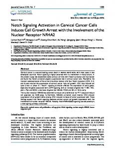
lateral inhibition (82). How a given cell within an equivalence group adopts the neuronal fate or not depends on the Notch ligand expression level. If a progenitor cell expresses more Delta ligand than its neighbors, it will become a neuron and will transmit an inhibitory signal to the Notch-expressing progenitors in contact with it, preventing these progenitors from differentiating prematurely into neurons and from expressing Delta. As a consequence, the cell that produces more ligand forces its neighbors to produce less; ultimately, individual neighboring cells are driven into different developmental pathways (83). Molecular genetic studies show that in this situation, ligand production is regulated by a negative feedback loop (84). This interpretation of the gene expression patterns has been documented extensively by experiments in Xenopus (79, 80), chick (78, 85), zebrafish (86 – 88), and mouse (55). Another example of lateral inhibition is the formation of sensory hair cells in the vertebrate inner ear (89), where a single cell within an equivalence domain expresses high Delta levels. More detailed analyses of Notch function in the CNS has revealed that Notch is likely to regulate progenitor pool diversification and neuronal maturation (90). Emerging data also suggest that Notch signaling has a role in neuronal function in the adult brain (91). It will be of great interest to determine which components of the Notch signaling cascade function in each of these processes. Figure 2 summarizes the roles of Notch in embryonic and adult CNS. III. Notch in Somitogenesis
The somites are blocks of paraxial mesoderm cells lying at either side of the neural tube. They give rise to the vertebrae of the axial skeleton and their associated muscles and tendons, which retain a segmental or metameric pattern. In all vertebrates, somites are generated sequentially from the preNotch
Notch Notch
somitic mesoderm (PSM), the unsegmented paraxial mesoderm at the caudal end of the embryo (92). At the rostral end of the PSM, clefts appear, and successive blocks of somite tissue split off; meanwhile, the embryo grows caudally, with a relatively constant amount of PSM tissue. Once formed, somitic cells differentiate progressively to give rise to five major cell types: the bone, cartilage and tendons of the trunk, skeletal muscles of the body, and the dermis of the back (93, 94). Each somite is also subdivided into anterior and posterior compartments (A-P polarity), a subdivision that is already established in the anterior PSM (95). In addition, the different morphological specification of somites is established early in the PSM and relies mostly on the activity of Hox genes (96). The connection between somitogenesis and the nested expression domains of Hox genes in the paraxial mesoderm has been established (97), although the coordination of these two patterning processes is poorly understood. Notch involvement in somitogenesis was first suggested by the defects in somite morphology observed in mice with targeted mutations in the Notch1 (54) and RBPJk (98) genes. In Notch1 mutants, the PSM generates irregular somites in which the positioning of segmental boundaries is abnormal (54). Similarly, Delta1 targeted mutants show abnormal somitogenesis with loss of A-P polarity (99). This phenotype is more severe in RBPJk mutants, which have fewer somites and a largely unsegmented paraxial mesoderm (70, 98) (Fig. 3, A–D). Studies using dominant-negative or constitutively activated Notch showed that perturbing Notch signaling in Xenopus produces similar phenotypes; somitic cells differentiated normally into myocytes, but the segmental organization of these cells is lost (100). The similarity of these phenotypes suggested that Notch is critical in the patterning process leading to somite boundary formation (92) and the establishment of the A-P polarity of somites (99, 101).
Notch
Neuroblast
Notch
Early neuronal differentiation
Notch Notch
Notch Neuronal maturation and function
Stem cell
Adult neural stem cell
Adult neurogenesis
Notch
Oligodendrocytes
Notch Glioblast
Notch
Astrocytes
FIG. 2. Notch signaling in the developing and adult CNS. Processes that require Notch activity are labeled with green arrows, and those that require Notch inhibition are labeled with red truncated arrows. The green semicircular arrows indicate the requirement of Notch for progenitor pool maintenance. See text for details. [Adapted from K. Yoon and N. Gaiano: Nature Neuroscience 8:709 –715, 2005 (287) with permission from Macmillan Publishers Ltd.]
344
Endocrine Reviews, May 2007, 28(3):339 –363
Bolo´s et al. • Notch in Development and Cancer
E
F Delta
Notch Lfng
2 sp Me
Hes1/Hes7
Lfng CSL
CSL Hes1/7 Hes1/7
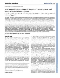
FIG. 3. Notch signaling is required for somite boundary formation. A–D, Dorsal view of the somitic region of E8.5 embryos. A, Wild-type embryo. B, Delta1 targeted embryo. Note irregular somites (arrowhead) and unsegmented paraxial mesoderm. C, Notch1 mutant embryo. Note irregular and fused somites (arrowhead). D, RBPJk mutant embryo. Note a few irregular somites (arrowhead) and a large stretch of unsegmented paraxial mesoderm. Scale bar, 50 m. E, Oscillatory expression in the PSM (black). chairy1 mRNA sweeps repeatedly across the PSM in a posterior-to-anterior direction, and each cycle is synchronous with the formation of a new somite. Blackened area represents the moving front of chairy1 expression. F, Notch and the segmentation clock. In the posterior PSM, Delta-Notch signaling activates Lfng, Hes1, and Hes7. Lfng potentiates Delta-Notch interaction (green arrow), whereas Hes1 and Hes7 binding to E boxes (brown) repress Lfng and their own transcription, via a negative feedback loop. After Hes1/Hes7 proteins disappear, their transcription is initiated de novo by activators such as Notch. Thus, Lfng expression oscillates in the same phase as Hes1/7 expression. In the anterior PSM, Mesp2 binds to N boxes (blue) in the Lfng promoter and activates its expression. Lfng in this situation inhibits Delta-Notch1 interaction (red truncated arrow). As a result, both waves of Notch1 activity and Lfng are arrested, and a boundary between Notch1 activity domain and the Mesp2 expression domain is generated, which leads to a new segmental boundary. [Modified from Refs. 105, 108, 109, and 113.]
A great advance in the molecular analysis of segmentation was the discovery of cHairy1, a chick gene whose expression oscillates within the PSM, with a periodicity corresponding to a segmental cycle (102) (Fig. 3E). The cHairy1 expression pattern provided the first molecular insight into a potential segmental clock. In addition, cHairy1 belongs to a large family of bHLH transcriptional repressors, including proteins encoded by genes that are known to be direct Notch targets in other species, such as Hes1 (103) in the mouse and the Enhancer of Split [E(spl)] genes in Drosophila (104). These observations, together with the defective somitogenesis of Notch targeted mutants described above, raised the possibility that the dynamic changes in cHairy1 expression within the PSM could be driven by changes in Notch signaling, as part of a segmentation clock. Oscillatory expression of other bHLH repressor genes in the PSM was later observed in other vertebrate embryos, although the gene with dynamic expression is not always the same. Hes7 (105) and Her1/7 (106, 107) are thus rhythmically expressed in the mouse and zebrafish embryo, respectively. In Xenopus, homologs of the oscillating zebrafish Her1 gene are expressed in the PSM, but their expression does not oscillate (101). The emerging picture is that Notch signaling undergoes a dramatic spatial and temporal change within the PSM during each segmental cycle. Segmentation appears to rely on two major components; an oscillator, the segmentation clock, sets the periodicity of somite formation, and a dynamic wave-front defines the level at which PSM cells respond to the clock, providing a mechanism that spaces the segment boundaries (108). How does the segmentation clock work? A negative feedback loop has been proposed as the major mechanism. A study by Hirata et al. (109) showed that the segmentation clock can be mimicked in cultured cells. After serum stimulation, expression of both Hes1 mRNA and protein oscillates with a 2-h cycle. This coincides with the cycle of the segmentation clock in mouse (Fig. 3E), suggesting that the oscillators in cultured cells and the PSM share the same mechanism. Hes1 protein oscillation is delayed by approximately 15 min relative to Hes1 mRNA oscillation. Both Hes1 mRNA and Hes1 protein have very short half-lives, which enable 2-h cycle oscillatory gene expression. Hes1 protein is degraded by the ubiquitin-proteasome pathway (109). When stabilized by proteasome inhibitors, Hes1 protein constitutively represses its own transcription by binding directly to the Hes1 promoter. Conversely, in the absence of functional Hes1 protein, Hes1 transcription is constitutively up-regulated. The negative feedback loop in which Hes1 protein periodically represses its own transcription is thus the central mechanism for Hes1 oscillation (Fig. 3F). It is likely that Hes1 protein represses Hes1 mRNA synthesis, but is rapidly degraded, allowing the next cycle of mRNA synthesis (109). Because Hes1 transcription is constitutively up-regulated in the absence of functional Hes1 protein, there appears to be a steady level of transcriptional activators in cultured cells after serum stimulation. Because a simple negative feedback loop would be insufficient to maintain stable oscillation, another cycling factor might be involved. Such a cycling factor is not yet known for cultured cells, but for the segmentation clock, lunatic fringe
Bolo´s et al. • Notch in Development and Cancer
(Lfng) in mouse (Fig. 3F) and chick and deltaC in zebrafish might be implicated in maintaining stable oscillation in the PSM. Dale et al. (110) found that Lfng also establishes a negative feedback loop in chick, and that, in addition to Lfng mRNA, Lfng protein exhibits oscillatory expression in the PSM. Activation of Notch signaling induces Lfng transcription, but Lfng protein inhibits Notch signaling and thereby represses its own transcription. Lfng thus also periodically represses its own expression, like Hes1 and Hes7. Analysis of the regulatory region of Lfng in mouse confirmed that it includes a site that mediates direct stimulation by activated Notch (111) but showed that it contains sites that mediate inhibition by bHLH proteins such as the Hes family members (112). This led to a model in which the inhibitory action of Hes genes plays a key role, and Lfng is assumed to potentiate Delta-Notch signaling (Fig. 3F), as in Drosophila (25). In contrast to this proposition, Morimoto et al. (113) have shown that Notch1 activity in mice oscillates in the posterior PSM. Somite boundaries would form at the interface between Notch1-activated and Notch1-repressed domains. This interface would be generated by suppression of Notch activity by the bHLH transcription factor Mesp2 through induction of Lfng that, in contrast with its role in the wing margin (25), would inhibit Notch activation. It thus becomes clear that the function of Lfng could vary, depending on cell context and biological system. Another important finding is that Wnt signaling is also involved in the segmentation clock (114). Lfng oscillation is lost in vestigial tail mice (hypomorphic Wnt3a mutants), indicating that the Wnt pathway interacts with the Notch pathway, although the precise mechanism remains to be determined. Although great advances have been made in the past decade in understanding aspects of the somitogenesis process, many aspects, such as the molecular machinery that underlies the segmentation clock, remain to be explained. For a more detailed review on the molecular aspects of somitogenesis see Refs. 108, 115, and 116.
IV. Notch in Cardiovascular Development and Homeostasis
The cardiovascular organ system is the first to form during vertebrate embryogenesis, when the heart develops from the cardiogenic mesoderm to form the double-walled primary heart tube. This tube consists of two cell types: an inner layer of endocardial endothelial cells, and an outer myocardial layer which is separated from the inner layer by an extracellular matrix (the cardiac jelly). Endocardial endothelium and cardiomyocytes together constitute the primitive heart tube. The first rhythmic cardiac contractions are initiated at this double-walled stage of heart development (117). Interaction between endocardial endothelium and myocardium leads to formation of ventricular trabeculae, highly organized sheets of cardiomyocytes forming muscular ridges lined by endocardial cells (118). The formation of this spongy, trabeculated pattern substantially increases the endocardial endothelial surface area. Tissue interactions between the myocardium and endocardium in the atrioventricular canal and outflow tract regions lead to epithelial-mesenchyme transition (EMT) of en-
Endocrine Reviews, May 2007, 28(3):339 –363 345
docardial cells, to participate in cardiac valves and membranous septa formation (119). Cushion formation is followed by the development of more compact myocardium in the periphery of the developing cardiac ventricles. Vascular development initiates with the differentiation of endothelial precursors, or angioblasts, into endothelial cells (120). These cells assemble to form a primitive vascular plexus of uniformly sized vessels composed entirely of endothelial cells by the process of vasculogenesis. This primitive vascular plexus is then remodeled to form the veins, arteries, and capillaries through the process of angiogenesis. Different Notch ligands (20, 61) and receptors (6, 10, 121, 122), as well as their downstream effectors and target genes (72), are expressed in the vascular system. Functional studies in zebrafish (123) and mice (61) showed that Notch is a critical regulator of cardiovascular development. Evidence for a crucial function of Notch in vascular development and homeostasis is the finding that the human disease CADASIL (cerebral autosomal dominant arteriopathy with subcortical infarction and leukoencephalopathy), which involves the NOTCH3 gene, causes stroke and vascular dementia (124). In addition, JAGGED1 haploinsufficiency leads to the dominant inherited Alagille syndrome, which among other features is characterized by vascular anomalies (125, 126). Analysis of mice with targeted mutations in Notch1 or RBPJk revealed that these mutants have severe pericardial distension, indicative of a circulatory defect (53, 54, 98). Likewise, mutations of the Delta1 (99) and Jag1 (127) genes lead to vascular abnormalities and hemorrhage, indicative of an underlying vascular defect. Analysis of the Notch1; Notch4 double mutants provided an insight into Notch function in the vasculature. Whereas Notch4 mutants are viable and fertile, the Notch1; Notch4 double mutants show normal vasculogenesis, but the process of angiogenesis is affected in the embryo proper, the yolk sac, and the placenta (61). This phenotype is more severe than that of Notch1 mutants alone, suggesting that Notch1 and Notch4 have partially redundant roles during embryonic vascular development. From this study, it became clear that Delta4 was the key ligand for Notch1 and Notch4 receptors in the vasculature. In fact, Delta4-targeted mutants and endothelial-specific RBPJk-targeted mutants show a loss of arterial identity and arteriovenous malformations (128, 129), indicating that Notch signaling is required for arterial specification and patterning during development. These studies also suggested a relationship between Notch and EphrinB2/EphB4, another local cell signaling pathway involved in arterial-venous specification (130). Targeted mutagenesis of potential Notch target genes in the cardiovascular system has helped to delineate the Notch signaling pathway that leads to arterial specification. HRT1; HRT2 double mutants die after E9.5 and display severe angiogenic remodeling defects and massive hemorrhage, probably due to impaired arterial fate determination and maturation. This phenotype is very similar to that of zebrafish gridlock (grl) mutants, that show impaired aorta maturation (131). Grl encodes for the zebrafish homolog of mammalian HRT2. These report identified HRT/CHF1/Hesr/Herp genes as essential Notch effectors in vascular development. The view of Notch as a key regulator of arterial fate is consistent with
346
Endocrine Reviews, May 2007, 28(3):339 –363
Bolo´s et al. • Notch in Development and Cancer
data indicating that ectopic activation of Notch signaling leads to repression of venous fate (132) and to severe vascular patterning defects (133). More recently, our understanding of how arterial-venous specification occurs has been refined by the study of an endothelial-specific mutant of the COUP-TFII transcription factor. Suppression of Notch signaling activity by COUP-TFII regulates vein identity, which may thus be genetically controlled and not derived by a default pathway (134, 135) (Fig. 4). Specific Notch ligands and receptors are expressed in the heart from early developmental stages. Delta4 (61), Notch1 (6), and Notch4 (10) are transcribed in the endocardial lineage from gastrulation onward, whereas other ligands and receptors show restricted expression in the myocardium from midgestation (136, 137). The Notch targets HRT1 and HRT2 are expressed in the endocardium and/or myocardium at different stages of cardiogenesis (72). Studies in Xenopus (138) and in mouse embryonic stem cells (139) indicate that cardiomyogenic commitment and differentiation require Notch signaling inhibition. In vivo studies in mouse and zebrafish nevertheless indicate that abrogation of Notch signaling does not affect primary cardiac cell fate determination and differentiation (139). In RBPJk-targeted mutants, early cardiogenic differentiation and chamber identity is unaffected, but valve development is severely disrupted, presumably because of defective endocardial maturation and signaling (140). During valvulogenesis in the E9.5 mouse embryo, endocardial cells undergo EMT and form the cardiac cushions that later remodel to give rise to the thin, finely sculpted mature valve leaflets of the E14.5 embryo (141). RBPJk and Notch1 mutants have a collapsed endocardium and lack mesenchymal cushion cells, indicating that endocardial EMT is defective in Notch VEGF COUP-TFII NP-1, Flk1 NP-1, Flk1 Delta4/Notch1,3,4 HRT/ Hey/grl
Delta4/Notch1,3,4 HRT/ Hey/grl
EphB4 Flt4
EphrinB2
Artery
EphB4 Flt4
EphrinB2
Vein
FIG. 4. Models of arterial-venous differentiation. In presumptive arterial endothelium, VEGF binds to its receptors neuropilin-1 (NP-1) and Flk1 that would in turn activate endothelial specific Notch signaling activity, leading to HRT2/gridlock-mediated repression of EphB4 and Flt4 receptor genes and activation of EphrinB2 expression, triggering the arterial differentiation program. In presumptive vein endothelium, COUP-TFII suppresses NP-1 and Flk1 to downregulate Notch activity and therefore release repression of EphB4 and Flt4 expression and down-regulate EphrinB2 expression, leading to vein differentiation. [Adapted from L.R. You et al.: Nature 435:98 – 104, 2005 (134) with permission from Macmillan Publishers Ltd.]
pathway mutants. Ultrastructural analysis of E9.5 mutant atrioventricular canal endocardia reveals that cells remain in close association, abnormally maintaining adherens junctions, and do not invade the cardiac jelly, despite showing features of activated premigratory endocardial cells. These observations correlate with a specific reduction in the transcription of the snail repressor (snai1) (142) in the atrioventricular canal region. Concomitantly, VE-cadherin expression remains abnormally stabilized in the atrioventricular canal and outflow tract endocardium of the mutants, suggesting that the lack of snail expression prevents down-regulation of VE-cadherin in this tissue, blocking endocardial EMT. These findings demonstrate that Notch activity is required for endocardial EMT (140). Explant assays with Notch1 and RBPJk mutants demonstrate an impaired endocardial EMT, a phenotype reproduced in wild-type explants cultured with the ␥-secretase inhibitor N-[N-(3,5-difluorophenacetyl)-l-alanyl]-S-phenylglycine t-butyl ester (DAPT). This finding is supported by Notch inhibition experiments in zebrafish that impair valve development and by gain-of-function experiments in which transient overexpression of N1ICD in the heart leads to formation of hypertrophic atrioventricular valves (140). This result contrasts with recent zebrafish data reporting that restricted N1ICD overexpression in the endocardium of atrioventricular canal inhibits EMT (143). Conditional activation of Notch1 in the cardiac lineage of the mouse using a MesP1-CRE driver impairs atrioventricular myocardial differentiation and ventricular myocardium maturation of MesP1-CRE; N1ICD transgenic mice, but EMT occurs normally (144). This discrepancy may be explained by the fact that N1ICD RNA was ectopically expressed in both endocardium and myocardium (140), which may generate additional myocardial-derived EMT-inductive signals. In the case of the experiments by Beis et al. (143), N1ICD was overexpressed exclusively in the endocardium. Our own data infecting wild-type atrioventricular canal explants with a retrovirus expressing N1ICD are consistent with this possibility, because N1ICDtransduced explants transform more than controls (J. GregoBessa, L. Luna-Zurita, and J. L. de la Pompa, unpublished data). The relevance of Notch in human cardiac valve development and homeostasis was demonstrated by the recent finding that NOTCH1 mutations cause aortic valve disease in humans (145). Studies in mice in the same report show that similarly to Notch1, its target genes HRT1/Hey1 and HRT2/Hey2 are expressed in the aortic valve leaflets at E17.5 where they repress Runx2, a regulator of osteoblast cell fate. These results suggest that NOTCH1 mutations cause an early developmental defect in the aortic valve and a later derepression of calcium deposition that causes progressive aortic valve disease (145). Jag1 and Notch2 are also linked to cardiac development. Mice doubly heterozygous for Jag1 and Notch2 mutations exhibit developmental abnormalities characteristic of Alagille syndrome, such as jaundice, growth retardation, defective bile duct differentiation, and abnormal kidney, eye, and heart development. The cardiac defects include right ventricular hypoplasia, pulmonic valve stenosis, atrial and ventricular septal defect, and dextropositioning of the aorta (59). The Notch targets HRT1/Hey1 and HRT2/Hey2 have also been linked to cardiac development. HRT2-targeted mutant mice have several cardiac anomalies. Almost all HRT2
Bolo´s et al. • Notch in Development and Cancer
mutants show growth retardation and die within 10 d of birth. Surviving HRT2 mutants have enlarged atria and ventricles, and echocardiographic analysis revealed abnormal cardiac hemodynamics, including stenosis and regurgitation of the tricuspid valve, mitral valve regurgitation, membranous ventricular septal defect, and secundum atrial septal defect (146). The phenotypic variation reported in HRT2 mutants probably results from the use of different genetic backgrounds and/or functional redundancy between HRT2 and other HRT family members (147). Analysis of HRT2 mutant mice suggests that HRT2 is required for formation of the atrioventricular valves. In addition, mice lacking both HRT1 and HRT2 die during embryogenesis due to severe cardiovascular malformations, including those in the development of the atrioventricular cushions. Few cells undergo EMT in the HRT1; HRT2 mice (148), suggesting that HRT1 and HRT2 function synergistically in the EMT process. In addition, HRT1; HRT2 mice show defects in trabecular myocytes (148). Although the ventricular chamber containing both the compact and trabecular zones forms initially, subsequent apoptosis of trabecular zone myocytes leads to poor trabecular formation. It is thus clear that HRT2 is important for correct cardiomyocyte development. The interaction between HRT1-expressing endocardial and epicardial cells and HRT2-expressing cells in the compact layer of the ventricle is also considered necessary to produce and/or maintain trabecular myocytes (149). Our own data (295) support a role for Notch in the development of ventricular myocardium, as the trabeculation-defective phenotype of standard and endocardial-specific Notch 1 and RBPJk mutants indicates. We propose that Notch mediates an endocardium-myocardium interaction critical for trabeculation and ventricular chamber morphogenesis and identify two distinct Notch-dependent processes: 1) transition of primitive myocardial epithelium to trabecular and compact myocardium (EphrinB2- and NRG1dependent); and 2) maintenance of a proliferating trabecular cardiomyocyte population (BMP10-dependent) during this transition. The defective ventricular phenotype of Notch mutants is reminiscent of a congenital disorder termed isolated ventricular noncompaction (IVNC) (296), which is characterized by altered myocardial structure. Thus, Notch signaling may be altered in infants with conditions including malseptation, abnormal valve development or conduction system defects, all of which are related to abnormal trabeculation. Notch activity in the cardiovascular system functions via a mechanism termed lateral induction (83), by which Notch generates contiguous domains of cells that share the same fate, an embryonic field. This signaling mechanism also occurs in flies during wing margin boundary formation (25). Other examples of Notch-mediated lateral induction in vertebrates include induction of proneural domains in the ear (150), formation of the limb bud margin (151), and somite boundary formation (83). In these cases, Notch activation promotes ligand production via a positive feedback loop, so that signaling occurs simultaneously in a developmental field. Loss of Notch signaling leads to down-regulation of ligand expression throughout the embryonic field, indicating the existence of a positive feedback loop. Figure 5 summarizes the processes in which Notch is in-
Endocrine Reviews, May 2007, 28(3):339 –363 347
volved during cardiogenesis. In a search for novel therapeutic approaches for cardiac disease, and in view of the role of Notch in the maintenance of an uncommitted state (152) and in inhibition of the cardiogenic fate in vitro (139), it will be of great interest to explore the role of Notch in adult cardiac homeostasis. V. Notch in Endocrine Development: Pancreas, Gut, and Bone Endocrine Cells A. Pancreatic development
The pancreas is an endoderm-derived organ formed by three main cell types, the exocrine acinar cells that produce digestive enzymes (i.e., carboxypeptidase), the ␣-and -endocrine cells (islets of Langerhans) that produce the hormones regulating nutrient homeostasis (insulin and glucagon), and the elaborate branched ductal tree that connects with the intestine (153, 154). Pancreas formation begins early in development (around E9.0 in the mouse) with the formation of two cell buds, ventral and dorsal. These buds express the Pdx1 transcription factor (155, 156) and arise from a specialized endodermal epithelium located in the foregut region that will give rise to the duodenum (157). During pancreatic organogenesis, the two buds undergo a series of morphogenetic, proliferative, and lineage specification events, grouped in three “developmental transitions,” to give rise to the mature and functional pancreas (for a review, see Refs. 158 and 159). At E13.5, the developing pancreas is formed by a branched Pdx1-positive epithelial tree, without morphological signs of exocrine differentiation. Endocrine and exocrine precursors are marked by two members of the bHLH transcription factors family, Ngn3 and Ptf1-P48, respectively (160). Specific Notch pathway elements and downstream effectors are expressed in the developing pancreas, suggesting a role for Notch in pancreatic development (161–163). Lossof-function of various Notch pathway genes (RBPJk, Delta1, and Hes1) leads to up-regulation of the proendocrine gene Ngn3 (164), a direct Hes1 target (165), and consequent accelerated and increased differentiation of pancreatic endocrine cells (161, 162). Conversely, forced Notch activity in the embryo blocks both endocrine and exocrine pancreas development (166, 167), and in pathological situations it leads to dedifferentiation of exocrine cell types in pancreatic epithelium (168). The physiological relevance of Notch in exocrine pancreas has been shown in the mouse, where Notch is active in committed exocrine progenitors cells and whose ectopic activation in pancreatic bud explants represses acinar cell differentiation (169). The available data indicate that Notch regulates the progressive recruitment of endocrine and exocrine cell types from a common precursor pool in developing pancreas. The inhibitory effect of Notch signaling in exocrine differentiation has been well characterized in zebrafish, where endocrine and exocrine cells arise independently (170). Thus, accelerated exocrine differentiation is observed in zebrafish mind bomb mutants or upon expression of a dominant negative Su (H) version in wild-type embryos (169). Similarly to the CNS, Notch operates in the developing pancreas by lateral inhibition. This would explain the ini-
348
Endocrine Reviews, May 2007, 28(3):339 –363
Bolo´s et al. • Notch in Development and Cancer
Ventricle
Atrio-ventricular canal Notch1, 4
Endocardium
Notch1, 2, 4 Delta4
Delta4
NICD/RBPJK NICD/RBPJK NICD/RBPJK
Snai1
Jag1
Snai1
Snai1
VE-C
VE-C
Snai1
HR T 2
Notch 1, 2, 4
? HRT2
EMT
EMT
?
?
Ventricular development: Trabecular and compact zone Valve formation+Remodeling (HRT2?) TGFß2 (+ Other signaling pathways)
Myocardium
HRT2 (+Other signaling pathways)
FIG. 5. Notch activity and cardiac development. The endocardial endothelium expresses specific Notch ligands and receptors simultaneously, behaving as an embryonic field. In the atrioventricular canal and outflow tract regions (data not shown), Delta4/Notch1 receptor interactions lead to snai1 expression, which represses vascular endothelial cadherin (VE-C) expression, allowing endocardial cells to down-regulate adhesion, undergo EMT, and acquire a mesenchymal phenotype. Notch activity in the endocardium is also required for the production of a soluble signal that activates TGF2 expression in the myocardium. TGF2 binding to its endocardial receptors would also lead to snail expression and EMT induction. In the ventricular endocardium, Notch signaling mediated by HRT2 (among other factors) is required for ventricular chamber development and trabeculation. Other signaling pathways active in the myocardium may cooperate with Notch in this process. In addition, myocardial HRT2 would also be involved.
tially dispersed distribution of endocrine cells within the pancreatic epithelium. Consistent with this idea, Hes1 mutants show increased expression of the Delta 1 and Delta 3 ligands (162). Nevertheless, additional observations suggest that Notch signaling in the pancreas does not function only via lateral inhibition and perhaps respond to additional signals, emanating from the mesenchyme. Fibroblast growth factor 10 (FGF10) produced in pancreatic mesenchyme has been shown to be essential for precursor pool maintenance (171). Transgenic mice overexpressing FGF10 in pancreatic epithelia show pancreatic hyperplasia and a block in exocrine, ductal, and endocrine differentiation (172, 173). In these mice and in contrast to the wild-type situation (161, 162), Notch1 and Notch2 as well as Hes1 are ubiquitously expressed in pancreatic precursor cells, whereas ngn3 expression is abrogated (172, 173). Thus, ectopic FGF10 signaling is capable of maintaining Notch signaling activity in the pancreas. These observations have led to the suggestion that another mechanism different from lateral inhibition and termed “suppressive maintenance” sustains Notch signaling activity in the pancreas and suppress its differentiation (159, 172). The mechanism underlying suppressive maintenance is not well understood, but it may involve FGF10-dependent activation of the Jag 1 and Jag2 Notch ligands in pancreatic
epithelium (172). It remains to be seen what is the effect of Notch signaling abrogation on the expression of its ligands. A further refinement of our view of Notch in pancreatic development has come from the analysis of Hes1-targeted mutant mice. Ptf1 is misexpressed in discrete regions of the primitive stomach and duodenum and throughout the bile duct of Hes1 mutants. Ptf1-expressing cells are reprogrammed to multipotent pancreatic progenitors that differentiate into mature pancreatic exocrine, endocrine, and duct cells. These data demonstrate that Notch is required for proper regional specification of pancreas in developing foregut endoderm through Ptf1 regulation (174). Because Notch is involved in the development and homeostasis of a variety of self-renewing tissues (175, 176), understanding Notch function in pancreatic development will help to design protocols to control -cell development in vitro and thus treat diseases such as diabetes, in which the mass of insulin-producing -cells is reduced. B. Gut development
The gastrointestinal tract comprises the small intestine and colon. Small intestine epithelium is organized into finger-like villi and adjacent invaginations, the crypts of Lieberku¨hn; the
Bolo´s et al. • Notch in Development and Cancer
colon has a flat epithelial surface with no villi (177). In small intestine, the crypt compartment contains stem cells and progenitors, whereas the villus compartment is made up entirely of differentiated cells. Pluripotent stem cells, which reside at the crypt bottom, give rise to transit amplifying cells, which divide robustly before terminal differentiation. Four lineages can be distinguished in the gastrointestinal tract, i.e., absorptive enterocytes, mucus-secreting goblet cells, hormone-secreting enteroendocrine cells, and lysozyme- and cryptidin-producing Paneth cells (reviewed in 175). Enteroendocrine cells can be further subdivided on the basis of the hormones they secrete (178). Intestine homeostasis depends on a small number of evolutionarily conserved pathways, including bone morphogenetic protein (BMP)/TGF, sonic hedgehog (Shh), wingless (Wnt) and Notch (179). The analysis of mutant zebrafish or targeted mice deficient for different Notch pathway elements or target genes has implicated Notch in the regulation of the earliest intestinal cell fate decisions. Thus, a recent study describes increases in secretory cells at the cost of absorptive cells in the intestines of zebrafish that are mutant for DeltaD and mind bomb (180). Different Notch pathway elements are expressed in murine crypts (181). The Notch target Hes1 is expressed in crypts (162). Analysis of the developing fetal intestine of Hes1 mutant mice revealed an increase in mucosecreting and enteroendocrine cells at the expense of absorptive enterocytes (162). The crypt progenitor pool in the small intestine seemed unaffected, as judged by an analysis of proliferative activity. Math1 is a target gene of Hes1-mediated repression in several organs, including the intestine (182). Math1 mutant mice die neonatally. Although the crypt-villus architecture was essentially undisturbed in the mutant mice, commitment toward the secretory lineage had entirely halted (182). These results have been interpreted to indicate that Hes1 and Math1 are required to skew the fate of differentiating cells leaving the transit amplifying compartment toward an enterocyte or a secretory phenotype, respectively (162, 182). Conditional deletion of the RBPJK effector in the epithelium of the small intestine and colon using different CREdriver lines led to a decrease in Hes1 expression, and Math1 was up-regulated throughout the crypt compartment, whereas it is normally expressed only in secretory cells (183). In addition, the number of goblet cells was increased. In the small intestine, Paneth cells appeared in near-normal numbers at the bottom of the crypts. However, the rapidly dividing transit amplifying compartment, which normally occupies the remainder of the crypt, was entirely replaced by postmitotic goblet cells that expressed Math1 protein but not Hes1. Examination of proliferation by different techniques indicated that basically all epithelial cell division had halted. Essentially identical observations were made in the colon (183). As an explanation for the different severity of the phenotypes observed upon Hes1 deletion and conditional RBPJk deletion in colon, it has been argued that other Hes genes such as Hes6 are affected by conditional RBPJk deletion (183). This phenotype was confirmed using a highly efficient ␥-secretase inhibitor. Cell proliferation had entirely halted and histological markers revealed that the tissue changes were indistinguishable from those observed after RBPJk deletion (183).
Endocrine Reviews, May 2007, 28(3):339 –363 349
A reciprocal phenotype to that of RBPJk deletion in colon was obtained in transgenic mice overexpressing N1ICD in all cells of the intestinal epithelium, by virtue of the villin-CRE driver (184). N1ICD; villin-CRE transgenic mice have a complete lack of goblet cells in all intestinal tracts. In addition, enteroendocrine cells are markedly reduced, as well as Paneth cells (184). Microscopic examination of earlier developmental stages revealed that already at E18.5, N1ICD expression affects the architecture of the villi, and thus the differentiation of secretory cell lineages along the duodenalileal axis and the cranial-to-caudal wave of intestinal differentiation was inhibited (184). Transcriptional analysis revealed a direct correlation between N1ICD expression and elevated levels of Hes1 transcription in the intestinal epithelium of N1ICD transgenic mice, although no other Hes genes were affected. Expression of the Hes1 targets Math-1 (185) and ngn3 (165), involved in secretory cell lineage specification, was reduced in N1ICD; villin-CRE mice. This finding was consistent with the idea that the expression of Math-1 and ngn3 is repressed by Notch activation (184). This intestinal phenotype is reminiscent of Math-1 (lack of goblet and enteroendocrine cells) and opposite to that of Hes1 mutant mice (an excess of secretory cells at the expense of enterocytes). Thus, the gain-of-function phenotype of N1ICD; villin-CRE mice provides direct evidence that Notch signals target Hes1 in the intestine, explaining mechanistically the differentiation defects observed. Overall, these genetic data indicate that Notch-mediated Hes1 expression regulates a binary cell fate decision between adsorptive and secretory cell fates. However, Hes1-deficient mice do not show a change in the proliferative status of the intestinal precursor pool (162), whereas Notch activation profoundly affects the proliferation potential of intestinal progenitors (184), suggesting that other Notch targets may be responsible for the increased in proliferation. The effect of deregulated Notch signaling in colon homeostasis and cancer will be discussed in Section VI.B. C. Bone development
Bone is a dynamic tissue that is constantly renewed and degraded. Two main types of bone cells are responsible for these processes: bone-forming osteoblasts of mesenchymal origin, and bone-resorbing osteoclasts of hematopoietic origin. Bone formation and resorption are coordinated so that bone remodeling is balanced. When this equilibrium is altered in a way that bone resorption exceeds bone formation, osteoporosis occurs. This disease is prevalent in old age and is characterized by bone loss and a high risk of fractures. Therefore, knowledge about molecular events involved in osteoblast differentiation is crucial. The in vitro potential of Notch pathway in osteoclastogenesis and osteoblastogenesis has been investigated in several reports. Evidence indicates that Notch down-regulates osteoclastogenesis activation, reduces the surface expression of c-Fms, which is a receptor for macrophage colony-stimulating factor, in osteoclast precursor cells and enhances the expression of osteoprotegerin in stromal cells, which results in the down-regulation of osteoclastogenesis (186). However, controversial results have been obtained with respect to os-
350
Endocrine Reviews, May 2007, 28(3):339 –363
teoblastic differentiation. Continuous NICD expression inhibits bone morphogenetic protein 2-induced osteoblast differentiation in osteoblast precursor cells (187). In contrast, transient expression of N1ICD in osteoblast precursor cells leads to an enhanced bone mineral deposition (188). Notch1 is expressed in the mesenchymal condensation area and subsequently in the hypertrophic chondrocytes during chondrogenesis (189). Another study shows that Notch1, Delta1, and Jagged1 are expressed in cultured osteoblast precursor cells as well as in differentiating osteoblasts during bone regeneration, and that Notch1 is activated in these cells (190). These results suggest that Notch signaling plays an important role in the commitment of mesenchymal cells to the osteoblastic cell lineage (190). Concomitant expression of Delta1 and Jagged1 during in vivo bone regeneration suggests that there is a functional redundancy between Delta1 and Jagged1 and that these ligands direct osteoprogenitor cells to the differentiated status through identical signaling pathways (190). These data suggest a therapeutic potential for Notch in bone regeneration as well as osteoporosis. Mammalian bone marrow architecture involves hematopoietic stem cells (HSCs) in close proximity to the endosteal surfaces of bone (191), with more differentiated cells arranged in a graduated fashion as the central longitudinal axis of the bone is approached (192), suggesting a relationship between HSCs and osteoblasts. Osteoblasts produce hematopoietic growth factors (193) and are activated by PTH or the locally produced PTHrP through the PTH/PTHrP receptor (PPR). Experiments involving transgenic mice expressing in osteoblastic cells a constitutively active PPR under the control of the alpha1(I) collagen promoter showed increased numbers of trabeculae and trabecular osteoblastic cells in the long bones of transgenic mice (194). In addition, the number of HSCs was also increased in these mice, but mature cell number was not changed (194). Because this phenotype was reminiscent of the mode of Notch action in hematopoiesis (195) and Jag1 is expressed by marrow stromal cells (196) and by murine osteoblasts (197), the levels of Jag1 protein were analyzed in the bone marrow of transgenic mice. col1-caPPR transgenic mice showed a marked increase in Jag1 expression in osteoblastic cells. The response of hematopoietic stem cells to increased Jag1 expression was measured by N1ICD staining, which was increased in transgenic mice. These data support a model in which PPR activation in the osteoblastic population increases cell number and the overall production of Jag1. This, in turn, may activate Notch on primitive hematopoietic cells, resulting in expansion of the stem cell compartment (194). In another study, these authors demonstrated that PTH treatment led to increased levels of Jagged1 in osteoblastic cells both in vivo and in vitro, in a protein kinase A (PKA)-dependent manner (198). Because Jagged1 is very important in stromal-HSC interactions and PTH regulates HSC expansion through osteoblastic activation, these studies suggested that stimulation of osteoblastic PKA activation may alter the HSC niche. This is of great therapeutic importance because this study suggests that alternative means to stimulate osteoblastic PKA activation may alter the HSC niche. In addition, Jagged1/Notch signaling modulates osteoblastic differentiation (189), and Jagged1 may play a critical role in mediating the effects of PTH on osteoblasts.
Bolo´s et al. • Notch in Development and Cancer
VI. Notch in Cancer
Experimental evidence supports the idea that signaling pathways essential for embryonic development also have a role in regulating self-renewing tissues (199, 200). Mutations in these pathways (such as TGFB, Wnt, and ErbB) often lead to tumorigenesis, as is also true for Notch (reviewed in Ref. 200). An interesting aspect of Notch is its apparently opposite functions in tumor development, because it can act as an oncogene or as a tumor suppressor. Although the mechanism underlying this dual Notch action is being explored, the outcome of Notch signaling activity depends on signal strength, timing, cell type, and context (reviewed in Ref. 201). The result of altered Notch signaling depends on its normal function in a given tissue. Notch thus acts as an oncogene if its normal function is as a gatekeeper of stem cells or as a regulator of precursor cell fate; its tumor suppressor activity is detected in tissues in which Notch signaling initiates terminal differentiation events (202). Table 1 summarizes the involvement of abnormal Notch signaling in cancer. The oncogenic function of Notch is shown by the finding that truncated forms of all four Notch isoforms (Notch1Notch 4), resulting in constitutively active Notch signaling, have transforming activity in vitro (203) and in various animal models (204 –208). Furthermore, deregulated expression of wild-type Notch receptors, ligands, and targets is found in many human solid tumors (209, 210) and hematological malignancies (176, 210). Notch inhibition by antisense retrovirus or by pharmacological ␥-secretase blockade has antineoplastic effects in Notch-expressing transformed cells in vitro and in xenograft models in vivo (Refs. 211–214; reviewed in Refs. 176 and 209). Notch alone may not be a very efficient oncogene, however, and it must associate with another oncoprotein to cause transformation. Although such partners have not yet been identified in naturally occurring tumors, transformation can be induced in vitro in various cell types by expressing NICD with certain oncoproteins (215, 216). The Notch tumor suppressor function may be a peculiarity of the mouse skin system, or it may also apply in man and include human keratinocytes as well as other human epithelial cell types (217). Available evidence thus suggests that, with the possible exception of some human epidermal malignancies, Notch signaling inhibition is a viable strategy for treatment of certain solid and hematopoietic tumors (176, 209). JAG1 expression in head and neck squamous cell carcinoma (HNSCC) cells triggers Notch activation in neighboring endothelial cells and promotes network formation (218). In xenograft models, HNSCC cells overexpressing JAG1 formed larger tumors with increased vascularization, and JAG1 protein levels were notably higher in HNSCC samples than in normal samples (218). These results offer a causal link between Notch signaling and tumor angiogenesis and describe a novel juxtacrine signaling mechanism from tumors to surrounding vasculature. As we saw in Section IV, Notch is essential for angiogenic remodeling in the embryo and for vascular homeostasis in the adult. This study shows that Notch is also involved in pathological angiogenesis (218), suggesting a possible direction for therapeutic intervention in tumors. In this regard, a recent report has shown that VEGF-induced Delta4 acts as a negative regulator of tumor
Bolo´s et al. • Notch in Development and Cancer
Endocrine Reviews, May 2007, 28(3):339 –363 351
TABLE 1. Abnormal Notch signaling in tumorigenesis and EMT Tumor type/process
Hematological tumors T cell malignancies (T-ALL)
B cell malignancies Solid tumors Breast cancer
Gut cancer Skin cancer Keratinocytederived carcinoma Melanocytederived carcinoma Cervical cancer
EMT
Function
Human models
Oncogenic Notch signaling
Constitutively active NOTCH1: t(7; 9) (q34:q34.3) (223), activating mutations (224, 289)
Oncogenic Notch signaling Tumor suppressive Notch signaling
Notch1, Notch2, and Jagged1 (229 – 231) Intracellular Notch1– 4, Jagged 1–2 (232)
Oncogenic Notch signaling
Notch1 and Jagged-1 (252, 253)
Tumor suppressive Notch signaling Oncogenic Notch signaling
Notch2 (252)
Tumor suppressive Notch signaling
Notch signaling in basal cell carcinoma (260)
Oncogenic Notch signaling
Notch1 in primary melanoma (265, 266) NOTCH2 and HEY1 (269)
Oncogenic Notch signaling Tumor suppressive Notch signaling EMT promoter
Notch1 in early disease stage (293)
Animal models
Activating mutations in Notch1 in mouse models of T-ALL (290). Constitutively active Notch2 (291). Constitutively active Notch3. Transgenic mice; lck promoter-driven intracellular Notch3 (205). DLL4 overexpressing mice (292). Intracellular Notch1– 4, Jagged 1–2 (232) Constitutively active Notch4 in transgenic mice: MMTv insertion (239), MMTv-intracellular Notch4 (237); WAP- intracellular Notch4 (240). Constitutively activated Notch1 in transgenic mice: MMTv insertion (242), MMTv-intracellular Notch1 (207, 208). Constitutively activated Notch3 in transgenic mice: MMTvintracellular Notch3 (208). Notch signaling in APC min mice (183)
Notch1 in late disease stage (293, 294) JAGGED1 in prostate cancer metastasis (284). MCF 10A cell line (212). Human keratinocytes (283).
angiogenesis (219). Thus, tumor-derived VEGF induces Delta4 expression in angiogenic endothelial cells to negatively regulate vascular growth, acting to restrain excessive vascular sprouting and branching and allowing angiogenesis to proceed at a productive rate. Thus, increasing Delta4/Notch activity resulted in decreased vascular density associated with reduced sprouting and branching of the vascular network. On the contrary, the blockade of Delta4/Notch signaling produced enhanced angiogenic sprouting and branching, resulting in a marked increase in tumor vessel density but decreased tumor vessel function (219). These data suggest that an alternative treatment of tumors may be based on the promotion of “nonfunctional” tumor angiogenesis. A. Notch in hematological tumors
Notch signaling mediates hematopoietic cell fate determination in the embryo (220) and in the adult (221) and is also a critical factor in the maintenance of a pool of self-renewing HSCs (222). Deregulated expression of Notch pathway ele-
Conditional ablation of Notch1 in murine epidermis (257). Conditional transgenic mice with epidermal inhibition of Notch signaling (SM22Cre⫹/DNMAML1⫹ mice) (259).
PAE cell line (140).
ments can thus lead to development of hematological malignancies. The prototypical Notch-associated cancer is human acute T cell acute lymphoblastic leukemia/lymphoma (T-ALL), which constitutes approximately 15–20% of ALL in children and adults. The NOTCH1 gene was discovered due to its involvement in a chromosome translocation [t (7; 9)] seen in some human T-ALL; this leads to expression of N1ICD in a T cell receptor--regulated manner (Fig. 6A) (223). Notch1 was later shown to be essential for normal T cell progenitor development (62). Although t (7; 9) is rare (less that 1% of T-ALL; Fig. 6A), the majority of human T-ALL have gain-of-function mutations in NOTCH1, leading to aberrant increases in downstream signaling (Fig. 6A) (224), placing the NOTCH pathway at the center of T-ALL pathogenesis. Weng et al. (224) found that more than 50% of human T-ALL without specific (7; 9) chromosome translocation, including tumors from all major molecular oncogenic subtypes, have activating mutations that involve the NOTCH1 extracellular heterodimerization domain and/or the C-ter-
352
Endocrine Reviews, May 2007, 28(3):339 –363
A
Bolo´s et al. • Notch in Development and Cancer
Hematological tumors: human T-cell acute lymphoblastic leukemia (T-ALL) 50% T-ALL
Activating mutations
Mutations in extracellular HD Notch1
Mutations and/or deletions in intracellular PEST domain
Ligand-independent activation Delayed proteosomal degradation
B
Solid tumors: mouse mammary tumors Viral insertion in Notch1/4 locus
LTR
MMTV
LTR
Mouse mammary tumors
Truncated Notch1/4 Ligand-independent constitutive Notch activation
FIG. 6. Oncogenic Notch signaling in hematological malignancies and solid tumors. A, Chromosome translocations and activating mutations within the human NOTCH1 gene cause human T-ALL. The t (7; 9) translocation in T-ALL patients is characterized by juxtaposition of the 3⬘ portion of the human NOTCH1 gene with the T cell receptor  (TCR) locus. This leads to expression of truncated NOTCH1 transcripts and consequent production of dominant active, ligand-independent forms of the NOTCH1 receptor, causing T-ALL. This rare event occurs in less than 1% of all T-ALL patients. Schematic diagram of the full-length human NOTCH1 protein, showing “hot spots” of mutations found in more than 50% of T-ALL patients. B, Integration of the MMTV between the LNRs and the transmembrane domain of the Notch1 or Notch4 gene cause mammary tumors in the mouse.
minal PEST destruction box (Fig. 6A). The detection of mutations in multiple molecular subtypes of T-ALL is the basis for the conclusion that NOTCH1 appears to collaborate with various other proteins that are also deregulated in T-ALL (176). A recent study also identified c-MYC as an important direct target of NOTCH1 in T-ALL and in a critical stage of normal pre-T cell development, showing that c-MYC inhibitors interfere with progrowth effects of activated NOTCH1 and that forced c-MYC expression rescues NOTCH1-dependent T-ALL cell lines from Notch withdrawal (225). Based on these findings, a phase I/II clinical trial was recently begun using a NOTCH pathway inhibitor to treat patients with refractory T-ALL. If this trial is successful, NOTCH pathway inhibitors may soon be considered therapeutic agents that target cancer-specific molecular lesions (reviewed in Ref. 176). Although the Notch receptor is expressed throughout the hematolymphoid compartment, its transforming potential appears to be restricted to developing T cells. Several studies have explored this issue in malignant B cells, with conflicting results; three reports suggest that constitutive Notch signaling in malignant B cells leads to growth inhibition and/or apoptosis (226 –228), whereas three groups found that Notch signaling promotes malignant B cell proliferation (229 –231). A recent study of the effect of constitutive Notch signaling in malignant murine and human B cells showed generalized Notch-mediated growth inhibition and apoptosis in immature and mature human and murine B cell malignancies, including therapy-resistant subtypes (232). This suggests that Notch signaling may be of therapeutic use for certain B cell tumors, although more research is still required.
B. Notch as an oncogene in solid tumors: breast and gut cancer
1. Breast cancer. Stem cells thought to reside in the mammary gland are thought to renew mammary gland cells through cycles of pregnancy, lactation, and involution during a woman’s lifetime (233). There is increasing evidence that stem cells might be targets of transformation during mammary carcinogenesis. The Notch signaling pathway is implicated in the self-renewal of normal mammary stem cells (234, 235), and recent work suggests a role for the Notch pathway in breast cancer (236). Notch4 was identified as a mouse mammary tumor virus (MMTV) insertion site in mammary tumors (Fig. 6B); provirus was inserted within the Notch4 gene (originally known as the int-3 locus) (237). This MMTV model system has proven useful for identification and characterization of genes involved in malignant transformation of normal mammary epithelium (238). In the case of the Notch4 gene, provirus insertion leads to expression of a truncated Notch that lacks most of the extracellular portion of the protein but contains Notch4 transmembrane and intracellular domains (N4ICD) (239). Transgenic mice harboring this constitutively active N4ICD under the regulation of the MMTV promoter show arrested mammary gland development and eventually develop poorly differentiated adenocarcinomas (237). Additional evidence confirms the Notch4/ int-3 gene effect in mammary epithelial differentiation and mammary tumorigenesis; this is derived from studies in which N4ICD was expressed from the whey acidic protein (WAP) promoter in transgenic mice, which restricts its activity to secretory mammary epithelial cells of pregnant mice (240). As predicted, secretory lobule growth and differenti-
Bolo´s et al. • Notch in Development and Cancer
ation were inhibited, and mammary tumors were histologically identical to MMTV-N4ICD tumors. Other workers subsequently identified a 1.8-kb human NOTCH4/Int3 RNA species (designated h-Int3sh). h-Int3sh RNA encodes a protein lacking the CBF1-binding region (RAM23) of the NOTCH4/Int3 intracellular domain. Although WAP-Int3sh transgenic mouse lines develop mammary tumors, the latency period is long (241); the authors thus speculate that the N4ICD-induced block of mammary gland development and tumorigenesis is the result of an increasing CBF1-dependent NOTCH4/Int3 signaling gradient. Notch1 involvement in mammary tumorigenesis is being studied extensively. The first evidence that aberrant Notch1 signaling has a role in mammary tumorigenesis came from studies in the MMTV model, which attempted to identify genes that collaborate with Neu/erbB2 in mammary tumorigenesis (Fig. 6B). An MMTV insertion in the Notch1 locus in MMTV-Neu mammary tumors causes N1ICD expression (242). HC11 mouse mammary epithelial cells expressing N1ICD-encoding cDNA are transformed, form colonies in agar, but are unable to form tumors in nude mice, indicating that acquisition of malignant characteristics requires additional genetic events (242). Other studies showed that transgenic activation of N1ICD in mammary glands leads to development of lactation-dependent tumors that regress at weaning (207, 208); with time, these regressing neoplasms apparently become nonregressing adenocarcinomas (207). Recent evidence shows that c-myc is a direct transcriptional target of aberrant Notch1 signaling, with a fundamental role in MMTV-N1ICD-induced murine mammary tumorigenesis (243). The study of NUMB expression in human breast cancer also supports NOTCH signaling pathway involvement in breast cancer. Pece et al. (244) showed that NUMB-mediated negative regulation of NOTCH signaling is lost in 50% of human mammary carcinomas. This is due to specific NUMB ubiquitination and proteasomal degradation (244) and indicates that enhanced NOTCH signaling activity occurs in these mammary carcinomas. NOTCH1 is also implicated as a downstream effector of oncogenic Ras in human mammary tumorigenesis (245). H-Ras is thought to have a central role early in human mammary carcinogenesis (246). The Ras signaling network and its mediators in human neoplastic cells thus have considerable importance for the development of antineoplastic agents for breast cancer (247). Weijzen et al. (245) suggested that N1ICD mediates the tumorigenic effects of oncogenic Ras. Analysis of several human breast cancer cases showed high NOTCH1 expression in all Ras-positive tumors. The authors also show that NOTCH1 down-regulation in Ras-transformed human cells led to a marked decrease in cell proliferation, and inhibition of Ras signaling blocked N1ICD up-regulation, indicating that Ras acts upstream of NOTCH1. Nonetheless, they showed that N1ICD overexpression in the absence of oncogenic Ras was consistently unstable in human cell lines that do not bear Ras mutations. In addition, expression of oncogenic Ras leads to higher DELTA1 and PRESENILIN1 protein levels, which may increase NOTCH1 processing (245). Another study showed that Notch4 also requires Ras signaling to exert its oncogenic effect (248), whereas Notch
Endocrine Reviews, May 2007, 28(3):339 –363 353
antagonism by transgenic expression of Deltex affects H-Rasinduced mammary tumorigenesis (207). Crossing MMTV/ v-H-Ras and MMTV-DTX1 mice and monitoring the bitransgenic progeny for tumor development showed that Deltex expression greatly inhibited the oncogenic effects of H-Ras expression in mammary glands (207). The authors also showed that in MMTV-H-Ras mice (which eventually will develop mammary tumors), H-Ras induced cyclin D1 upregulation, even in a precancerous state. In contrast, the H-Ras/hDTX1 bitransgenic mice showed a marked reduction in the amount of cyclin D1 compared with MMTV-H-Ras mice. These results would be consistent with the finding that N1ICD expression activates cyclin D1 gene transcription in cultured human cells (249) and that mice lacking cyclin D1 were resistant to development of H-Ras- or Neu/Erb2-induced mammary tumors (250). Recent work also suggests a relationship between WNT and NOTCH pathways in breast tumorigenesis. Ectopic WNT-1 expression in human mammary epithelial cells increases WNT signaling and produces a tumorigenic state via a NOTCH-dependent mechanism (251). This suggests that deregulation of WNT signaling is an early event in NOTCH-dependent mammary epithelial transformation. Other groups are using gene expression profile analysis to test the correlation of NOTCH pathway components, clinical outcome, and tumor clinicopathological parameters in human breast cancer. In tissue samples from breast cancer patients, Parr et al. (252) quantified NOTCH1 and NOTCH2 expression in association with clinical outcome and showed aberrant NOTCH1 and NOTCH2 levels in breast cancer tissues compared with normal breast tissue. Examination of the clinicopathological parameters for breast cancer patients indicated that high NOTCH1 levels may be associated with poor prognosis, whereas increased NOTCH2 levels correlated with greater probability of survival (252). NOTCH1 may thus have tumor-promoting functions, whereas NOTCH2 could have a tumor-suppressive role in human breast cancer, supporting the suppression of NOTCH-1 activity as a therapeutic strategy. Tissue microarray studies showed high JAGGED1 and/or NOTCH1 expression levels in human breast cancer, associated with poor overall survival compared with patients with low levels of these genes (253). Because high-level JAGGED1 and NOTCH1 coexpression showed a synergistic effect on overall survival, this type of breast tumor could also benefit from ␥-secretase inhibitorbased therapy. Recent evidence suggests an antiapoptotic role for the NOTCH pathway in human breast cancer. An increase in RBP-JK-dependent NOTCH signaling in the normal human breast cell line MCF 10A thus protects them from druginduced apoptosis by abolishing the p53-mediated response (212). Other authors show that NOTCH1 signaling confers chemoresistance by inhibiting p53 activation of transcription and thus, apoptosis (254). 2. Gut cancer. As we have seen in Section V.B, Notch is essential for gut development and homeostasis. The Wnt cascade is also implicated in crypt progenitor cell maintenance; it is considered a major force behind the proliferative potential of intestinal adenomas and adenocarcinomas (179). In
354
Endocrine Reviews, May 2007, 28(3):339 –363
their study of the role of Notch signaling in intestine development and cancer, van Es et al. (183) showed that the Wnt signaling pathway remained active in RBPJkfloxed/floxed; P450Cre mutant mice. Their data indicated that Notch might function downstream of Wnt in the intestine and that both pathways may synergize as gatekeepers in the intestinal epithelium. This study also showed that spontaneous adenomas in ApcMin (multiple intestine neoplasia) mutant mice have high Hes-1 expression levels, similar to the intestinal crypts. The Notch pathway is thus activated in intestinal adenomas in these mice, as is the Wnt cascade (175). Notch pathway inhibition by ␥-secretase inhibitors in ApcMin mice induces goblet cell differentiation and reduces adenoma proliferation (183). Taken together, these data indicate that maintenance of undifferentiated, proliferative cells in crypts and adenomas requires activation of the Notch pathway in concert with the Wnt cascade. They also suggest that Notch signaling could provide an alternative target-drug strategy for intestinal neoplastic disease therapy (183). Thus, NOTCH and WNT inhibitors could be combined in an approach for colorectal neoplasia treatment (211). C. Differential roles of NOTCH in two types of skin cancer: keratinocyte-derived carcinoma and melanomas
In human primary keratinocytes, increased NOTCH1 activity promotes commitment of self-renewing stem cells to transit-amplifying populations that continue to proliferate, although only for a limited time (255). In primary mouse keratinocytes, Notch acts as a tumor suppressor gene, promoting exit from the cell cycle and entry into differentiation (256, 257). Conditional ablation of Notch1 in murine epidermis results in epidermal hyperplasia, skin carcinoma (basal cell carcinoma-like tumors), and facilitation of chemical-induced skin carcinogenesis. This is explained in part by reduced p21Waf1/Cip1 protein levels (257), because p21Waf1/Cip1⫺/⫺ mice are also more sensitive to chemical-induced carcinogenesis (258). Indeed, Notch1 mutant keratinocytes are highly susceptible to ras oncogene malignant transformation; Notch1 mutant cells infected with a retrovirus transducing the ras oncogene and injected sc into nude mice form aggressive squamous cell carcinoma (SCC), whereas wild-type cells do not (257). Loss of Notch1 activity may thus cooperate with ras oncogene transformation in keratinocyte tumor development. Accordingly, conditional transgenic mice with epidermal-restricted inhibition of Notch signaling (SM22Cre⫹; DNMAML1⫹ mice) have a hyperplastic epidermis and develop both spontaneous SCC and dysplastic precursor lesions (259); these mice also show enhanced accumulation of nuclear -catenin and cyclin D1 in suprabasilar keratinocytes and in lesional cells from SCC, as also observed in human SCC (259). The antioncogenic effect of Notch1 in murine skin appears to be mediated by p21Waf1/Cip1 induction and by repression of Shh and Wnt signaling (Fig. 7) (45, 257, 260). Notch1 induces p21Waf1/Cip1 expression directly by targeting NICD/ RBPJK to the p21Waf1/Cip1 promoter (256) and indirectly through the calcineurin/NFAT (nuclear factor of activated T cells) pathway (Fig. 7) (261). Mice with skin-specific Notch1
Bolo´s et al. • Notch in Development and Cancer
Notch1
Skin
Hes-1
p21
NFAT E2F
1:p 21
Wnt4
Wnt/β β-catenin signaling
Shh
Proliferation
FIG. 7. Tumor suppressive Notch signaling pathway in the skin. Activation of Notch modulates signaling pathways that inhibit proliferation, preventing neoplastic transformation. The diagram shows functional and/or biochemical interactions (discussed in the text). NFAT, Nuclear factor of activated T cells.
deletion develop spontaneous basal cell carcinoma-like tumors; human basal cell carcinomas also show reduced NOTCH1, NOTCH2, and JAGGED1 expression (260). In mice and in man, this tumor type is frequently associated with aberrant Shh signaling. Consistent with this, Notch1 deletion in the mouse epidermis leads to aberrant expression of Gli2, a downstream component of the Shh pathway. Loss of NOTCH in human epidermis could also lead to aberrant SHH signaling, thus contributing to the development of basal cell carcinomas (Fig. 7). Wnt signaling is suppressed by Notch1 activation and is elevated in keratinocytes and tumors as a consequence of loss of Notch1 function (257). Increased Wnt/-catenin signaling is biologically important because it is associated with maintenance of keratinocytes in their stem cell compartment (262) and with keratinocyte-derived malignancies (263). Devgan et al. (45) recently established the mechanism for Wnt signaling suppression by Notch1 activation in keratinocytes, showing that Notch1 activation down-regulates this pathway by suppressing Wnt-4 expression. p21Waf1/Cip1 mediates this negative regulation; Notch1 activation increases levels of p21Waf1/Cip1 protein, which subsequently associates with E2F1 transcription factors at the Wnt4 promoter, down-regulating Wnt4 expression (Fig. 7) (45). The function of Notch signaling in epidermis and in keratinocytes is thus to induce terminal differentiation processes, as well as to withdraw proliferating cells from the cell cycle. A long-term consequence of Notch1 deletion in murine skin is the development of basal cell carcinoma-like tumors, suggesting that Notch exerts tumor-suppressive functions in this tissue. Melanoma is a skin cancer that originates from melanocytes. In human skin, melanocytes are positioned at the epidermal-dermal junction and are interspersed among the basal keratinocytes (264). Melanocytes are tightly controlled by keratinocytes and maintain a nonproliferative status. Transformation to melanoma is the pathological consequence of disruption of cell control, which is environmentally initiated and is linked to specific genetic aberrations.
Bolo´s et al. • Notch in Development and Cancer
Recent data suggest that NOTCH signaling does not inhibit melanoma as it does in keratinocyte-derived carcinoma (260). In vivo and in vitro experiments show that NOTCH1 activation is insufficient to transform melanocytes but enables primary melanoma cells to gain metastatic capability (265). This oncogenic role of NOTCH is stage-specific. Thus, NOTCH signaling advances primary melanoma but has little effect on metastatic melanoma (265). A novel mechanism for NOTCH signaling in melanoma progression has been defined in which the oncogenic effect of NOTCH1 on primary melanoma cells is mediated by -CATENIN, which is upregulated after NOTCH1 activation. Inhibiting -CATENIN expression reverses NOTCH1-enhanced tumor growth and metastasis. In primary melanoma cells, -CATENIN thus acts as a downstream target of NOTCH1 signaling (265). Concurring with these findings, another report showed that NOTCH1 promotes primary melanoma progression by activating MAPK/PI3K-AKT survival pathways and up-regulating N-CADHERIN expression (266). NOTCH1 also down-regulates microtubule-associated protein 2 expression in primary melanoma cell lines (267). Abundant microtubule-associated protein 2 is often associated with longer disease-free survival rate in melanoma patients (268). Although previous work showed that NOTCH1 has a critical role in melanoma progression, the involvement of other NOTCH family members is not excluded. NICD are expressed in primary lesions of human malignant melanoma (reviewed in Ref. 209). NOTCH2 and HEY1 genes are also up-regulated in melanomas compared with melanocytes (269). These data indicate that NOTCH signaling is implicated in primary melanoma progression and suggest that certain members of the NOTCH pathway are potential therapeutic targets in the development of new melanoma treatments. Qin et al. (270) showed that ␥-secretase inhibitors can induce apoptosis in melanoma cell lines with markedly enhanced levels of activated NOTCH1 receptor. ␥-Secretase inhibitor treatment triggers apoptosis in melanoma cells but only causes G2/M growth arrest in melanocytes, without subsequent cell death (270); this treatment also induced the proapoptotic BH3-only protein NOXA in melanoma cells, but not in normal melanocytes (Ref. 270; reviewed in Ref. 271). D. Notch in EMT and tumor progression
EMT is a fundamental process that implies loss of cell polarity and intercellular adhesion, as well as acquisition of a migratory phenotype, leading to mesenchymal cell formation from primitive epithelium. EMT takes place during critical phases of embryonic development such as gastrulation or the formation of the cardiac valve primordium (see Section IV). EMT also occurs during tumor progression when cells from a primary epithelial tumor change phenotype, become mesenchymal, and disseminate as single carcinoma cells, invading other organs and leading to tumor metastasis. EMT might also be involved in the dedifferentiation program that leads to malignant carcinoma (272, 273). The mechanisms underlying EMT are under intense study, and it is clear that developmental and metastatic EMT are governed by the same signaling pathways (272, 273). Loss of expression of the epithelial adhesion protein E-
Endocrine Reviews, May 2007, 28(3):339 –363 355
CADHERIN indicates progression from an in situ to an invasive carcinoma (274). In most differentiated tumors, ECADHERIN production is maintained, and there is an inverse correlation between E-CADHERIN levels and cancer grade or patient survival (274, 275). In accordance, a causal role has been established for E-cadherin loss in the transition from adenoma to carcinoma in mouse models (276). Transcription factors detected at EMT sites during embryonic development were recently identified as key transcriptional repressors of E-cadherin expression during tumor progression (277). One of these molecules is the zinc finger protein SNAIL (SNAI1), an E-CADHERIN repressor; carcinoma cell lines lacking E-CADHERIN produce considerable amounts of SNAI1, and E-CADHERIN-positive cell lines transfected with SNAI1 undergo EMT (278, 279). We previously showed that Notch is critical for promotion of EMT during cardiac valve development via Snai1 induction (see Ref. 140 and Section IV), and that N1ICD overexpression in immortalized porcine aortic endothelial (PAE) cells induces EMT, with induction of Snail and repression of VE-cadherin (140). The data indicated that Notch is upstream of Snai1 in the EMT process that generates the valve primordia and in the PAE system. Other authors showed that JAGGED1 activation of endogenous NOTCH receptors in human endothelial cells also promoted EMT as endothelial cells expressing activated NOTCH1 or NOTCH4 repressed VE-CADHERIN (280). NOTCH might thus be implicated in EMT during tumor progression, potentially via SNAI1 induction. After stable expression of N1ICD or an RBP-JK/VP16 fusion protein, the normal human breast cell line MCF 10A showed marked changes in cell shape associated with a reduction in E-CADHERIN protein levels (212). N1ICD induction in the human adenocarcinoma cell line MCF7 promotes migratory behavior associated with E-CADHERIN loss (our unpublished observations). TGF is another well-known inducer of EMT during embryonic development and the later stages of tumor progression. During early tumor development, however, TGF inhibits the growth of most epithelial cell types, acting as a tumor suppressor (272). Another mechanism of NOTCHinduced tumor development and progression may involve modulation of the TGF signaling pathway. N1ICD was recently shown to suppress the growth-inhibitory effects of TGF by sequestering the transcriptional coactivator p300 from SMAD3, a downstream effector of TGF (281). Moreover, N4ICD binds directly to and inhibits SMAD2, SMAD3, and SMAD4 signaling activity, resulting in attenuated TGF signaling in MCF-7 breast cancer cells (282). Cells with activated NOTCH might thus be resistant to the growth inhibitory effects of TGF, promoting tumor development. Another recent study shows that RNA silencing of HEY-1 or JAGGED-1 or the chemical inactivation of NOTCH inhibits TGF-induced EMT in human keratinocytes (283). The ability of TGF to induce EMT may thus be Notch signalingdependent. The authors also found that TGF induces HEY1 and JAGGED1 expression at the onset of EMT in epithelial cells from mammary gland, kidney tubules, and epidermis. The HEY1 expression profile is biphasic, consisting of immediate-early SMAD3-dependent, NOTCH-independent activation, followed by delayed JAGGED1/NOTCH-depen-
356
Endocrine Reviews, May 2007, 28(3):339 –363
dent activation (283). In our PAE system, N1ICD did not appear to affect TGF signaling, suggesting that Notch signaling may be TGF-independent in this setting (140). Distinct pathways and/or transcription factors might thus participate in EMT via E-cadherin repression in distinct cell lines and tumor types. A study based on immunohistochemical analysis of human prostate tumors suggests a role for the NOTCH pathway in prostate cancer metastasis (284). JAGGED1 expression is notably higher in metastatic prostate cancer than in localized prostate cancer or benign prostate tissues. This finding supports a model in which dysregulation of JAGGED1 protein levels plays a role in prostate cancer progression and metastasis and suggests that JAGGED1 may be a useful marker to distinguish slow-progressing from aggressive prostate cancers (284). Although genomic studies show that expression of NOTCH pathway components is increased in a variety of solid tumors (285), a systematic search comparing the expression status of NOTCH pathway elements and potential target genes in metastatic and nonmetastatic tumors remains to be carried out.
VII. Concluding Remarks
Notch is a key regulator of many developmental processes during embryonic and adult life. In the embryo, Notch activity via lateral inhibition or induction mechanisms generates molecular differences between adjacent cells. In the vertebrate CNS, Notch maintains the neural progenitor state and inhibits differentiation. In contrast, during gliogenesis Notch seems to have an instructive role, directly promoting the differentiation of different glial subtypes. More detailed analyses have also revealed that Notch regulates progenitor pool diversification and neuronal maturation. Emerging data suggest that Notch signaling has a role in neuronal function in the adult brain. During somitogenesis, spatial and temporal changes in Notch signaling in the PSM are linked to segmental clock activity and are critical for segmental patterning. Oscillatory gene expression in the PSM is likely to involve Notch activation of Hes genes that negatively feed back on their own transcription, as well as the expression of ligands and/or Lfng. A major question is how the transition occurs between oscillatory Notch activity in the caudal PSM to fixed Notch activity in the anterior PSM. During vascular development, Notch is dispensable for vasculogenesis but is essential to establish the arterial endothelial fate and for angiogenesis, suggesting that arterial/ venous specification may be important for proper vascular remodeling. In addition, the vein phenotype is not achieved by default but rather requires the inhibition of Notch activity in presumptive vein territory. Studies in humans with vascular pathologies involving NOTCH suggest that this pathway may be required to maintain or stabilize the cells that contribute to blood vessels. In the heart, Notch is not necessary to establish the primary cardiac fates in vivo, but its activity regulates crucial cell communication events between endocardium and myocardium during both the formation of the valve primordial and
Bolo´s et al. • Notch in Development and Cancer
ventricular development and differentiation. Human studies indicate that NOTCH is critical for cardiac valve homeostasis, and more information about the implication of NOTCH in other human disorders involving the cardiovascular system is likely to come soon. During pancreas development, Notch is required for timely cell lineage specification of both endocrine and exocrine pancreas. In the gut, Notch and Wnt have a gatekeeper function for crypt progenitor cells, and Notch appears to influence binary fate decisions of cells that must choose between the secretory and adsorptive lineages. Deregulation of the Wnt pathway is central to the development of colorectal cancers in man. It remains to be shown whether Notch deregulation also follows the Wnt cascade in this respect. In bone development, Notch may expand the HSC compartment and participate in commitment to the osteoblastic lineage, suggesting a potential therapeutic role for Notch in bone regeneration and osteoporosis. Aberrant Notch activity is involved in hematological and solid tumor formation. The best-established role of Notch within the hematopoietic system is the ability to influence and/or specify cell fates of lymphoid progenitors. It has become clear that aberrant Notch signaling in humans due to activating mutations in the NOTCH1 receptor promotes T-ALL. Notch1 is thus an established oncogene in the hematopoietic system. The evidence suggests that Notch signaling activity has an oncogenic role in human breast cancer, which may be facilitated by cooperation with other signaling pathways such as Neu/ErbB2 and WNT. Deregulated NUMB activity also supports NOTCH involvement in breast cancer, where it acts as a downstream effector of Ras. This contrasts dramatically with Notch1 function in the skin, where Notch1 appears to induce terminal differentiation and also acts as a tumor suppressor. The observation that Notch can have such opposite functions in different selfrenewing organs indicates that the outcome of Notch activation depends to a great extent on the cell context and factors such as the cell type in which Notch is activated, the specific growth factors in the microenvironment, and the level of Notch activity. During embryonic development, the question is basically the same: How does Notch integrate its activity with other cell inputs to control specific developmental events? Another important matter is to understand how Notch activates distinct target genes according to cell type and time. Genomic studies will undoubtedly increase the spectrum of target genes and allow development of a systematic approach for understanding these different responses. Deregulated Notch signaling activity is involved in disease, but it remains to be determined whether modulation of such a pleiotropic pathway can be a therapeutic target for cancer and in regenerative medicine. Acknowledgments We thank C. Mark for editorial assistance. Address requests for reprints to: Jose´ Luis de la Pompa, Departmento de Inmunologı´a y Oncologı´a, Centro Nacional de Biotecnologı´a/Con-
Bolo´s et al. • Notch in Development and Cancer sejo Superior de Investigaciones Cientı´ficas, Darwin 3, Campus de Cantoblanco, E-28049 Madrid, Spain. E-mail:
[email protected] This work was supported by Grants SAF2004-05204 (Spanish Ministry of Education and Science), GR/SAL/0851/2004 and 200520M072 (Regional Government of Madrid), and LSHM-CT-2005-018630 (European Union 6th FP). The Department of Immunology and Oncology was founded and is supported by the Spanish National Research Council (CSIC) and by Pfizer. J.G.-B. was partially supported by a basic and clinical research fellowship from the Spanish Society for Cardiology. The authors declare no conflict of economic interests with regard to this publication.
Endocrine Reviews, May 2007, 28(3):339 –363 357
20. 21. 22. 23.
References 1. Morgan TH 1917 The theory of the gene. Am Nat 51:513–544 2. Poulson DF 1937 Chromosomal deficiencies and the embryonic development of Drosophila melanogaster. Proc Natl Acad Sci USA 23:133–137 3. Welshons WJ, Von Halle ES 1962 Pseudoallelism at the notch locus in Drosophila. Genetics 47:743–759 4. Wharton KA, Johansen KM, Xu T, Artavanis-Tsakonas S 1985 Nucleotide sequence from the neurogenic locus notch implies a gene product that shares homology with proteins containing EGFlike repeats. Cell 43:567–581 5. Kidd S, Kelley MR, Young MW 1986 Sequence of the notch locus of Drosophila melanogaster: relationship of the encoded protein to mammalian clotting and growth factors. Mol Cell Biol 6:3094 –3108 6. Del Amo FF, Smith DE, Swiatek PJ, Gendron-Maguire M, Greenspan RJ, McMahon AP, Gridley T 1992 Expression pattern of Motch, a mouse homolog of Drosophila Notch, suggests an important role in early postimplantation mouse development. Development 115:737–744 7. Weinmaster G, Roberts VJ, Lemke G 1992 Notch2: a second mammalian Notch gene. Development 116:931–941 8. Lardelli M, Lendahl U 1993 Motch A and motch B–two mouse Notch homologues coexpressed in a wide variety of tissues. Exp Cell Res 204:364 –372 9. Lardelli M, Dahlstrand J, Lendahl U 1994 The novel Notch homologue mouse Notch 3 lacks specific epidermal growth factorrepeats and is expressed in proliferating neuroepithelium. Mech Dev 46:123–136 10. Uyttendaele H, Marazzi G, Wu G, Yan Q, Sassoon D, Kitajewski J 1996 Notch4/int-3, a mammary proto-oncogene, is an endothelial cell-specific mammalian Notch gene. Development 122:2251–2259 11. Fehon RG, Kooh PJ, Rebay I, Regan CL, Xu T, Muskavitch MA, Artavanis-Tsakonas S 1990 Molecular interactions between the protein products of the neurogenic loci Notch and Delta, two EGFhomologous genes in Drosophila. Cell 61:523–534 12. Rebay I, Fleming RJ, Fehon RG, Cherbas L, Cherbas P, ArtavanisTsakonas S 1991 Specific EGF repeats of Notch mediate interactions with Delta and Serrate: implications for Notch as a multifunctional receptor. Cell 67:687– 699 13. Tamura K, Taniguchi Y, Minoguchi S, Sakai T, Tun T, Furukawa T, Honjo T 1995 Physical interaction between a novel domain of the receptor Notch and the transcription factor RBP-J /Su(H). Curr Biol 5:1416 –1423 14. Blank V, Kourilsky P, Israel A 1992 NF-B and related proteins: Rel/dorsal homologies meet ankyrin-like repeats. Trends Biochem Sci 17:135–140 15. Rechsteiner M 1988 Regulation of enzyme levels by proteolysis: the role of pest regions. Adv Enzyme Regul 27:135–151 16. Logeat F, Bessia C, Brou C, LeBail O, Jarriault S, Seidah NG, Israel A 1998 The Notch1 receptor is cleaved constitutively by a furin-like convertase. Proc Natl Acad Sci USA 95:8108 – 8112 17. Blaumueller CM, Qi H, Zagouras P, Artavanis-Tsakonas S 1997 Intracellular cleavage of Notch leads to a heterodimeric receptor on the plasma membrane. Cell 90:281–291 18. Bettenhausen B, Hrabe de Angelis M, Simon D, Guenet JL, Gossler A 1995 Transient and restricted expression during mouse embryogenesis of Dll1, a murine gene closely related to Drosophila Delta. Development 121:2407–2418 19. Dunwoodie SL, Henrique D, Harrison SM, Beddington RS 1997
24. 25. 26.
27. 28.
29. 30. 31. 32.
33.
34. 35. 36.
37.
38.
39.
40. 41.
Mouse Dll3: a novel divergent Delta gene which may complement the function of other Delta homologues during early pattern formation in the mouse embryo. Development 124:3065–3076 Shutter JR, Scully S, Fan W, Richards WG, Kitajewski J, Deblandre GA, Kintner CR, Stark KL 2000 Dll4, a novel Notch ligand expressed in arterial endothelium. Genes Dev 14:1313–1318 Lindsell CE, Shawber CJ, Boulter J, Weinmaster G 1995 Jagged: a mammalian ligand that activates Notch1. Cell 80:909 –917 Shawber C, Boulter J, Lindsell CE, Weinmaster G 1996 Jagged2: a serrate-like gene expressed during rat embryogenesis. Dev Biol 180:370 –376 Moloney DJ, Panin VM, Johnston SH, Chen J, Shao L, Wilson R, Wang Y, Stanley P, Irvine KD, Haltiwanger RS, Vogt TF 2000 Fringe is a glycosyltransferase that modifies Notch. Nature 406: 369 –375 Bruckner K, Perez L, Clausen H, Cohen S 2000 Glycosyltransferase activity of Fringe modulates Notch-Delta interactions. Nature 406: 411– 415 Panin VM, Papayannopoulos V, Wilson R, Irvine KD 1997 Fringe modulates Notch-ligand interactions. Nature 387:908 –912 Shao L, Moloney DJ, Haltiwanger R 2003 Fringe modifies Ofucose on mouse Notch1 at epidermal growth factor-like repeats within the ligand-binding site and the Abruptex region. J Biol Chem 278:7775–7782 Haltiwanger RS 2002 Regulation of signal transduction pathways in development by glycosylation. Curr Opin Struct Biol 12:593–598 Itoh M, Kim CH, Palardy G, Oda T, Jiang YJ, Maust D, Yeo SY, Lorick K, Wright GJ, Ariza-McNaughton L, Weissman AM, Lewis J, Chandrasekharappa SC, Chitnis AB 2003 Mind bomb is a ubiquitin ligase that is essential for efficient activation of Notch signaling by Delta. Dev Cell 4:67– 82 Le Borgne R, Schweisguth F 2003 Notch signaling: endocytosis makes delta signal better. Curr Biol 13:R273–R275 Lai EC, Deblandre GA, Kintner C, Rubin GM 2001 Drosophila neuralized is a ubiquitin ligase that promotes the internalization and degradation of delta. Dev Cell 1:783–794 Deblandre GA, Lai EC, Kintner C 2001 Xenopus neuralized is a ubiquitin ligase that interacts with XDelta1 and regulates Notch signaling. Dev Cell 1:795– 806 Pavlopoulos E, Pitsouli C, Klueg KM, Muskavitch MA, Moschonas NK, Delidakis C 2001 Neuralized encodes a peripheral membrane protein involved in delta signaling and endocytosis. Dev Cell 1:807– 816 Mumm JS, Schroeter EH, Saxena MT, Griesemer A, Tian X, Pan DJ, Ray WJ, Kopan R 2000 A ligand-induced extracellular cleavage regulates ␥-secretase-like proteolytic activation of Notch1. Mol Cell 5:197–206 Parks AL, Klueg KM, Stout JR, Muskavitch MA 2000 Ligand endocytosis drives receptor dissociation and activation in the Notch pathway. Development 127:1373–1385 Kopan R, Goate A 2002 Aph-2/Nicastrin: an essential component of ␥-secretase and regulator of Notch signaling and presenilin localization. Neuron 33:321–324 Morel V, Lecourtois M, Massiani O, Maier D, Preiss A, Schweisguth F 2001 Transcriptional repression by suppressor of hairless involves the binding of a hairless-dCtBP complex in Drosophila. Curr Biol 11:789 –792 Kao HY, Ordentlich P, Koyano-Nakagawa N, Tang Z, Downes M, Kintner CR, Evans RM, Kadesch T 1998 A histone deacetylase corepressor complex regulates the Notch signal transduction pathway. Genes Dev 12:2269 –2277 Oswald F, Winkler M, Cao Y, Astrahantseff K, Bourteele S, Knochel W, Borggrefe T 2005 RBP-J/SHARP recruits CtIP/CtBP corepressors to silence Notch target genes. Mol Cell Biol 25:10379 – 10390 Wu L, Aster JC, Blacklow SC, Lake R, Artavanis-Tsakonas S, Griffin JD 2000 MAML1, a human homologue of Drosophila mastermind, is a transcriptional co-activator for NOTCH receptors. Nat Genet 26:484 – 489 Kurooka H, Honjo T 2000 Functional interaction between the mouse notch1 intracellular region and histone acetyltransferases PCAF and GCN5. J Biol Chem 275:17211–17220 Takeyama K, Aguiar RC, Gu L, He C, Freeman GJ, Kutok JL, Aster
358
42.
43. 44. 45.
46. 47. 48. 49. 50. 51. 52. 53. 54. 55.
56.
57. 58.
59.
60.
61.
62. 63.
Endocrine Reviews, May 2007, 28(3):339 –363 JC, Shipp MA 2003 The BAL-binding protein BBAP and related Deltex family members exhibit ubiquitin-protein isopeptide ligase activity. J Biol Chem 278:21930 –21937 Hori K, Fostier M, Ito M, Fuwa TJ, Go MJ, Okano H, Baron M, Matsuno K 2004 Drosophila deltex mediates suppressor of hairlessindependent and late-endosomal activation of Notch signaling. Development 131:5527–5537 Cayouette M, Raff M 2002 Asymmetric segregation of Numb: a mechanism for neural specification from Drosophila to mammals. Nat Neurosci 5:1265–1269 Iso T, Kedes L, Hamamori Y 2003 HES and HERP families: multiple effectors of the Notch signaling pathway. J Cell Physiol 194: 237–255 Devgan V, Mammucari C, Millar SE, Brisken C, Dotto GP 2005 p21WAF1/Cip1 is a negative transcriptional regulator of Wnt4 expression downstream of Notch1 activation. Genes Dev 19:1485– 1495 Krebs LT, Deftos ML, Bevan MJ, Gridley T 2001 The Nrarp gene encodes an ankyrin-repeat protein that is transcriptionally regulated by the Notch signaling pathway. Dev Biol 238:110 –119 Radtke F, Wilson A, Mancini SJ, MacDonald HR 2004 Notch regulation of lymphocyte development and function. Nat Immunol 5:247–253 Mumm JS, Kopan R 2000 Notch signaling: from the outside in. Dev Biol 228:151–165 Fortini ME 2002 ␥-Secretase-mediated proteolysis in cell-surfacereceptor signalling. Nat Rev Mol Cell Biol 3:673– 684 Baron M 2003 An overview of the Notch signalling pathway. Semin Cell Dev Biol 14:113–119 Bray SJ 2006 Notch signalling: a simple pathway becomes complex. Nat Rev Mol Cell Biol 7:678 – 689 Baker NE 2000 Notch signaling in the nervous system. Pieces still missing from the puzzle. Bioessays 22:264 –273 Swiatek PJ, Lindsell CE, del Amo FF, Weinmaster G, Gridley T 1994 Notch1 is essential for postimplantation development in mice. Genes Dev 8:707–719 Conlon RA, Reaume AG, Rossant J 1995 Notch1 is required for the coordinate segmentation of somites. Development 121:1533–1545 de la Pompa JL, Wakeham A, Correia KM, Samper E, Brown S, Aguilera RJ, Nakano T, Honjo T, Mak TW, Rossant J, Conlon RA 1997 Conservation of the Notch signalling pathway in mammalian neurogenesis. Development 124:1139 –1148 Ishibashi M, Ang SL, Shiota K, Nakanishi S, Kageyama R, Guillemot F 1995 Targeted disruption of mammalian hairy and Enhancer of split homolog-1 (HES-1) leads to up-regulation of neural helix-loop-helix factors, premature neurogenesis, and severe neural tube defects. Genes Dev 9:3136 –3148 Kageyama R, Nakanishi S 1997 Helix-loop-helix factors in growth and differentiation of the vertebrate nervous system. Curr Opin Genet Dev 7:659 – 665 Hamada Y, Kadokawa Y, Okabe M, Ikawa M, Coleman JR, Tsujimoto Y 1999 Mutation in ankyrin repeats of the mouse Notch2 gene induces early embryonic lethality. Development 126:3415– 3424 McCright B, Gao X, Shen L, Lozier J, Lan Y, Maguire M, Herzlinger D, Weinmaster G, Jiang R, Gridley T 2001 Defects in development of the kidney, heart and eye vasculature in mice homozygous for a hypomorphic Notch2 mutation. Development 128:491–502 Krebs LT, Xue Y, Norton CR, Sundberg JP, Beatus P, Lendahl U, Joutel A, Gridley T 2003 Characterization of Notch3-deficient mice: normal embryonic development and absence of genetic interactions with a Notch1 mutation. Genesis 37:139 –143 Krebs LT, Xue Y, Norton CR, Shutter JR, Maguire M, Sundberg JP, Gallahan D, Closson V, Kitajewski J, Callahan R, Smith GH, Stark KL, Gridley T 2000 Notch signaling is essential for vascular morphogenesis in mice. Genes Dev 14:1343–1352 Radtke F, Wilson A, Stark G, Bauer M, van Meerwijk J, MacDonald HR, Aguet M 1999 Deficient T cell fate specification in mice with an induced inactivation of Notch1. Immunity 10:547–558 Lutolf S, Radtke F, Aguet M, Suter U, Taylor V 2002 Notch1 is required for neuronal and glial differentiation in the cerebellum. Development 129:373–385
Bolo´s et al. • Notch in Development and Cancer 64. Yang X, Klein R, Tian X, Cheng HT, Kopan R, Shen J 2004 Notch activation induces apoptosis in neural progenitor cells through a p53-dependent pathway. Dev Biol 269:81–94 65. Yoon K, Nery S, Rutlin ML, Radtke F, Fishell G, Gaiano N 2004 Fibroblast growth factor receptor signaling promotes radial glial identity and interacts with Notch1 signaling in telencephalic progenitors. J Neurosci 24:9497–9506 66. Yun K, Fischman S, Johnson J, Hrabe de Angelis M, Weinmaster G, Rubenstein JL 2002 Modulation of the notch signaling by Mash1 and Dlx1/2 regulates sequential specification and differentiation of progenitor cell types in the subcortical telencephalon. Development 129:5029 –5040 67. Hitoshi S, Alexson T, Tropepe V, Donoviel D, Elia AJ, Nye JS, Conlon RA, Mak TW, Bernstein A, van der Kooy D 2002 Notch pathway molecules are essential for the maintenance, but not the generation, of mammalian neural stem cells. Genes Dev 16:846 – 858 68. Ohtsuka T, Ishibashi M, Gradwohl G, Nakanishi S, Guillemot F, Kageyama R 1999 Hes1 and Hes5 as notch effectors in mammalian neuronal differentiation. EMBO J 18:2196 –2207 69. Grandbarbe L, Bouissac J, Rand M, Hrabe de Angelis M, Artavanis-Tsakonas S, Mohier E 2003 Delta-Notch signaling controls the generation of neurons/glia from neural stem cells in a stepwise process. Development 130:1391–1402 70. Barrantes IB, Elia AJ, Wunsch K, De Angelis MH, Mak TW, Rossant J, Conlon RA, Gossler A, de la Pompa JL 1999 Interaction between Notch signalling and Lunatic fringe during somite boundary formation in the mouse. Curr Biol 9:470 – 480 71. Kageyama R, Ohtsuka T 1999 The Notch-Hes pathway in mammalian neural development. Cell Res 9:179 –188 72. Nakagawa O, Nakagawa M, Richardson JA, Olson EN, Srivastava D 1999 HRT1, HRT2, and HRT3: a new subclass of bHLH transcription factors marking specific cardiac, somitic, and pharyngeal arch segments. Dev Biol 216:72– 84 73. Barsi JC, Rajendra R, Wu JI, Artzt K 2005 Mind bomb1 is a ubiquitin ligase essential for mouse embryonic development and Notch signaling. Mech Dev 122:1106 –1117 74. Furukawa T, Mukherjee S, Bao ZZ, Morrow EM, Cepko CL 2000 Rax, Hes1, and Notch1 promote the formation of Muller glia by postnatal retinal progenitor cells. Neuron 26:383–394 75. Hojo M, Ohtsuka T, Hashimoto N, Gradwohl G, Guillemot F, Kageyama R 2000 Glial cell fate specification modulated by the bHLH gene Hes5 in mouse retina. Development 127:2515–2522 76. Scheer N, Groth A, Hans S, Campos-Ortega JA 2001 An instructive function for Notch in promoting gliogenesis in the zebrafish retina. Development 128:1099 –1107 77. Henrique D, Hirsinger E, Adam J, Le Roux I, Pourquie O, IshHorowicz D, Lewis J 1997 Maintenance of neuroepithelial progenitor cells by Delta-Notch signalling in the embryonic chick retina. Curr Biol 7:661– 670 78. Austin CP, Feldman DE, Ida Jr JA, Cepko CL 1995 Vertebrate retinal ganglion cells are selected from competent progenitors by the action of Notch. Development 121:3637–3650 79. Dorsky RI, Chang WS, Rapaport DH, Harris WA 1997 Regulation of neuronal diversity in the Xenopus retina by Delta signalling. Nature 385:67–70 80. Chitnis A, Henrique D, Lewis J, Ish-Horowicz D, Kintner C 1995 Primary neurogenesis in Xenopus embryos regulated by a homologue of the Drosophila neurogenic gene Delta [see comments]. Nature 375:761–766 81. Skeath JB, Thor S 2003 Genetic control of Drosophila nerve cord development. Curr Opin Neurobiol 13:8 –15 82. Cabrera CV 1990 Lateral inhibition and cell fate during neurogenesis in Drosophila: the interactions between scute, Notch and Delta. Development 110:733–742 83. Lewis J 1998 Notch signalling and the control of cell fate choices in vertebrates. Semin Cell Dev Biol 9:583–589 84. Heitzler P, Bourouis M, Ruel L, Carteret C, Simpson P 1996 Genes of the Enhancer of split and achaete-scute complexes are required for a regulatory loop between Notch and Delta during lateral signalling in Drosophila. Development 122:161–171 85. Henrique D, Adam J, Myat A, Chitnis A, Lewis J, Ish-Horowicz D 1995 Expression of a Delta homologue in prospective neurons in the chick. Nature 375:787–790
Bolo´s et al. • Notch in Development and Cancer 86. Dornseifer P, Takke C, Campos-Ortega JA 1997 Overexpression of a zebrafish homologue of the Drosophila neurogenic gene Delta perturbs differentiation of primary neurons and somite development. Mech Dev 63:159 –171 87. Haddon C, Smithers L, Schneider-Maunoury S, Coche T, Henrique D, Lewis J 1998 Multiple delta genes and lateral inhibition in zebrafish primary neurogenesis. Development 125:359 –370 88. Appel B, Eisen JS 1998 Regulation of neuronal specification in the zebrafish spinal cord by Delta function. Development 125:371–380 89. Eddison M, Le Roux I, Lewis J 2000 Notch signaling in the development of the inner ear: lessons from Drosophila. Proc Natl Acad Sci USA 97:11692–11699 90. Saura CA, Choi SY, Beglopoulos V, Malkani S, Zhang D, Shankaranarayana Rao BS, Chattarji S, Kelleher 3rd RJ, Kandel ER, Duff K, Kirkwood A, Shen J 2004 Loss of presenilin function causes impairments of memory and synaptic plasticity followed by age-dependent neurodegeneration. Neuron 42:23–36 91. Costa RM, Honjo T, Silva AJ 2003 Learning and memory deficits in Notch mutant mice. Curr Biol 13:1348 –1354 92. Pourquie O 2001 Vertebrate somitogenesis. Annu Rev Cell Dev Biol 17:311–350 93. Saga Y, Takeda H 2001 The making of the somite: molecular events in vertebrate segmentation. Nat Rev Genet 2:835– 845 94. Ordahl CP, Le Douarin NM 1992 Two myogenic lineages within the developing somite. Development 114:339 –353 95. Stern CD, Keynes RJ 1987 Interactions between somite cells: the formation and maintenance of segment boundaries in the chick embryo. Development 99:261–272 96. Nowicki JL, Burke AC 2000 Hox genes and morphological identity: axial versus lateral patterning in the vertebrate mesoderm. Development 127:4265– 4275 97. Zakany J, Kmita M, Alarcon P, de la Pompa JL, Duboule D 2001 Localized and transient transcription of Hox genes suggests a link between patterning and the segmentation clock. Cell 106:207–217 98. Oka C, Nakano T, Wakeham A, de la Pompa JL, Mori C, Sakai T, Okazaki S, Kawaichi M, Shiota K, Mak TW, Honjo T 1995 Disruption of the mouse RBP-J gene results in early embryonic death. Development 121:3291–3301 99. Hrabe de Angelis M, McIntyre JN, Gossler A 1997 Maintenance of somite borders in mice requires the Delta homologue DII1. Nature 386:717–721 100. Jen WC, Wettstein D, Turner D, Chitnis A, Kintner C 1997 The Notch ligand, X-Delta-2, mediates segmentation of the paraxial mesoderm in Xenopus embryos. Development 124:1169 –1178 101. Jen WC, Gawantka V, Pollet N, Niehrs C, Kintner C 1999 Periodic repression of Notch pathway genes governs the segmentation of Xenopus embryos. Genes Dev 13:1486 –1499 102. Palmeirim I, Henrique D, Ish-Horowicz D, Pourquie O 1997 Avian hairy gene expression identifies a molecular clock linked to vertebrate segmentation and somitogenesis. Cell 91:639 – 648 103. Jarriault S, Le Bail O, Hirsinger E, Pourquie O, Logeat F, Strong CF, Brou C, Seidah NG, Isra l A 1998 Delta-1 activation of notch-1 signaling results in HES-1 transactivation. Mol Cell Biol 18:7423– 7431. 104. Bray S, Furriols M 2001 Notch pathway: making sense of suppressor of hairless. Curr Biol 11:R217–21 105. Bessho Y, Sakata R, Komatsu S, Shiota K, Yamada S, Kageyama R 2001 Dynamic expression and essential functions of Hes7 in somite segmentation. Genes Dev 15:2642–2647 106. Henry CA, Urban MK, Dill KK, Merlie JP, Page MF, Kimmel CB, Amacher SL 2002 Two linked hairy/Enhancer of split-related zebrafish genes, her1 and her7, function together to refine alternating somite boundaries. Development 129:3693–3704 107. Takke C, Campos-Ortega JA 1999 her1, a zebrafish pair-rule like gene, acts downstream of notch signalling to control somite development. Development 126:3005–3014 108. Giudicelli F, Lewis J 2004 The vertebrate segmentation clock. Curr Opin Genet Dev 14:407– 414 109. Hirata H, Yoshiura S, Ohtsuka T, Bessho Y, Harada T, Yoshikawa K, Kageyama R 2002 Oscillatory expression of the bHLH factor Hes1 regulated by a negative feedback loop. Science 298:840 – 843 110. Dale JK, Maroto M, Dequeant ML, Malapert P, McGrew M,
Endocrine Reviews, May 2007, 28(3):339 –363 359
111.
112. 113. 114. 115. 116. 117. 118.
119. 120. 121. 122.
123. 124.
125.
126.
127.
128.
129.
130.
Pourquie O 2003 Periodic notch inhibition by lunatic fringe underlies the chick segmentation clock. Nature 421:275–278 Morales AV, Yasuda Y, Ish-Horowicz D 2002 Periodic lunatic fringe expression is controlled during segmentation by a cyclic transcriptional enhancer responsive to notch signaling. Dev Cell 3:63–74 Cole SE, Levorse JM, Tilghman SM, Vogt TF 2002 Clock regulatory elements control cyclic expression of lunatic fringe during somitogenesis. Dev Cell 3:75– 84 Morimoto M, Takahashi Y, Endo M, Saga Y 2005 The Mesp2 transcription factor establishes segmental borders by suppressing Notch activity. Nature 435:354 –359 Aulehla A, Wehrle C, Brand-Saberi B, Kemler R, Gossler A, Kanzler B, Herrmann BG 2003 Wnt3a plays a major role in the segmentation clock controlling somitogenesis. Dev Cell 4:395– 406 Weinmaster G, Kintner C 2003 Modulation of notch signaling during somitogenesis. Annu Rev Cell Dev Biol 19:367–395 Aulehla A, Herrmann BG 2004 Segmentation in vertebrates: clock and gradient finally joined. Genes Dev 18:2060 –2067 Manasek FJ 1968 Embryonic development of the heart. I. A light and electron microscopic study of myocardial development in the early chick embryo. J Morphol 125:329 –365 Ben-Shachar G, Arcilla RA, Lucas RV, Manasek FJ 1985 Ventricular trabeculations in the chick embryo heart and their contribution to ventricular and muscular septal development. Circ Res 57:759 – 766 Runyan RB, Markwald RR 1983 Invasion of mesenchyme into three-dimensional collagen gels: a regional and temporal analysis of interaction in embryonic heart tissue. Dev Biol 95:108 –114 Drake CJ, Fleming PA 2000 Vasculogenesis in the day 6.5 to 9.5 mouse embryo. Blood 95:1671–1679 Reaume AG, Conlon RA, Zirngibl R, Yamaguchi TP, Rossant J 1992 Expression analysis of a Notch homologue in the mouse embryo. Dev Biol 154:377–387 Villa N, Walker L, Lindsell CE, Gasson J, Iruela-Arispe ML, Weinmaster G 2001 Vascular expression of Notch pathway receptors and ligands is restricted to arterial vessels. Mech Dev 108:161– 164 Lawson ND, Vogel AM, Weinstein BM 2002 Sonic hedgehog and vascular endothelial growth factor act upstream of the Notch pathway during arterial endothelial differentiation. Dev Cell 3:127–136 Joutel A, Corpechot C, Ducros A, Vahedi K, Chabriat H, Mouton P, Alamowitch S, Domenga V, Cecillion M, Marechal E, Maciazek J, Vayssiere C, Cruaud C, Cabanis EA, Ruchoux MM, Weissenbach J, Bach JF, Bousser MG, Tournier-Lasserve E 1996 Notch3 mutations in CADASIL, a hereditary adult-onset condition causing stroke and dementia. Nature 383:707–710 Li L, Krantz ID, Deng Y, Genin A, Banta AB, Collins CC, Qi M, Trask BJ, Kuo WL, Cochran J, Costa T, Pierpont ME, Rand EB, Piccoli DA, Hood L, Spinner NB 1997 Alagille syndrome is caused by mutations in human Jagged1, which encodes a ligand for Notch1. Nat Genet 16:243–251 Oda T, Elkahloun AG, Pike BL, Okajima K, Krantz ID, Genin A, Piccoli DA, Meltzer PS, Spinner NB, Collins FS, Chandrasekharappa SC, Washburn T, Schweighoffer E, Gridley T, Chang D, Fowlkes BJ, Cado D, Robey E 1997 Mutations in the human Jagged1 gene are responsible for Alagille syndrome. Nat Genet 16:235–242 Xue Y, Gao X, Lindsell CE, Norton CR, Chang B, Hicks C, Gendron-Maguire M, Rand EB, Weinmaster G, Gridley T 1999 Embryonic lethality and vascular defects in mice lacking the Notch ligand Jagged1. Hum Mol Genet 8:723–730 Krebs LT, Shutter JR, Tanigaki K, Honjo T, Stark KL, Gridley T 2004 Haploinsufficient lethality and formation of arteriovenous malformations in Notch pathway mutants. Genes Dev 18:2469 – 2473 Duarte A, Hirashima M, Benedito R, Trindade A, Diniz P, Bekman E, Costa L, Henrique D, Rossant J 2004 Dosage-sensitive requirement for mouse Dll4 in artery development. Genes Dev 18:2474 –2478 Wang HU, Chen ZF, Anderson DJ 1998 Molecular distinction and angiogenic interaction between embryonic arteries and veins re-
360
131. 132.
133. 134. 135. 136.
137. 138. 139. 140.
141. 142. 143.
144.
145. 146.
147. 148.
149. 150.
Endocrine Reviews, May 2007, 28(3):339 –363 vealed by ephrin-B2 and its receptor Eph-B4 [see comments]. Cell 93:741–753 Zhong TP, Childs S, Leu JP, Fishman MC 2001 Gridlock signalling pathway fashions the first embryonic artery. Nature 414:216 –220 Lawson ND, Scheer N, Pham VN, Kim CH, Chitnis AB, CamposOrtega JA, Weinstein BM 2001 Notch signaling is required for arterial-venous differentiation during embryonic vascular development. Development 128:3675–3683 Uyttendaele H, Ho J, Rossant J, Kitajewski J 2001 Vascular patterning defects associated with expression of activated Notch4 in embryonic endothelium. Proc Natl Acad Sci USA 98:5643–5648 You LR, Lin FJ, Lee CT, DeMayo FJ, Tsai MJ, Tsai SY 2005 Suppression of Notch signalling by the COUP-TFII transcription factor regulates vein identity. Nature 435:98 –104 Hirashima M, Suda T 2006 Differentiation of arterial and venous endothelial cells and vascular morphogenesis. Endothelium 13: 137–145 Loomes KM, Underkoffler LA, Morabito J, Gottlieb S, Piccoli DA, Spinner NB, Baldwin HS, Oakey RJ 1999 The expression of Jagged1 in the developing mammalian heart correlates with cardiovascular disease in Alagille syndrome. Hum Mol Genet 8:2443– 2449 McCright B, Lozier J, Gridley T 2002 A mouse model of Alagille syndrome: Notch2 as a genetic modifier of Jag1 haploinsufficiency. Development 129:1075–1082 Rones MS, McLaughlin KA, Raffin M, Mercola M 2000 Serrate and Notch specify cell fates in the heart field by suppressing cardiomyogenesis. Development 127:3865–3876 Nemir M, Croquelois A, Pedrazzini T, Radtke F 2006 Induction of cardiogenesis in embryonic stem cells via downregulation of Notch1 signaling. Circ Res 98:1471–1478 Timmerman LA, Grego-Bessa J, Raya A, Bertran E, Perez-Pomares JM, Diez J, Aranda S, Palomo S, McCormick F, IzpisuaBelmonte JC, de la Pompa JL 2004 Notch promotes epithelialmesenchymal transition during cardiac development and oncogenic transformation. Genes Dev 18:99 –115 Lincoln J, Alfieri CM, Yutzey KE 2004 Development of heart valve leaflets and supporting apparatus in chicken and mouse embryos. Dev Dyn 230:239 –250 Barrallo-Gimeno A, Nieto MA 2005 The Snail genes as inducers of cell movement and survival: implications in development and cancer. Development 132:3151–3161 Beis D, Bartman T, Jin SW, Scott IC, D’Amico LA, Ober EA, Verkade H, Frantsve J, Field HA, Wehman A, Baier H, Tallafuss A, Bally-Cuif L, Chen JN, Stainier DY, Jungblut B 2005 Genetic and cellular analyses of zebrafish atrioventricular cushion and valve development. Development 132:4193– 4204 Watanabe Y, Kokubo H, Miyagawa-Tomita S, Endo M, Igarashi K, Aisaki KI, Kanno J, Saga Y 2006 Activation of Notch1 signaling in cardiogenic mesoderm induces abnormal heart morphogenesis in mouse. Development 133:1625–1634 Garg V, Muth AN, Ransom JF, Schluterman MK, Barnes R, King IN, Grossfeld PD, Srivastava Daa 2005 Mutations in NOTCH1 cause aortic valve disease. Nature 437:270 –274 Kokubo H, Miyagawa-Tomita S, Tomimatsu H, Nakashima Y, Nakazawa M, Saga Y, Johnson RL 2004 Targeted disruption of hesr2 results in atrioventricular valve anomalies that lead to heart dysfunction. Circ Res 95:540 –547 Fischer A, Schumacher N, Maier M, Sendtner M, Gessler M 2004 The Notch target genes Hey1 and Hey2 are required for embryonic vascular development. Genes Dev 18:901–911 Kokubo H, Miyagawa-Tomita S, Nakazawa M, Saga Y, Johnson RL 2005 Mouse hesr1 and hesr2 genes are redundantly required to mediate Notch signaling in the developing cardiovascular system. Dev Biol 278:301–309 Kokubo H, Miyagawa-Tomita S, Johnson RL 2005 Hesr, a mediator of the Notch signaling, functions in heart and vessel development. Trends Cardiovasc Med 15:190 –194 Daudet N, Lewis J 2005 Two contrasting roles for Notch activity in chick inner ear development: specification of prosensory patches and lateral inhibition of hair-cell differentiation. Development 132: 541–551
Bolo´s et al. • Notch in Development and Cancer 151. Irvine KD, Vogt TF 1997 Dorsal-ventral signaling in limb development. Curr Opin Cell Biol 9:867– 876 152. Artavanis-Tsakonas S, Rand MD, Lake RJ 1999 Notch signaling: cell fate control and signal integration in development. Science 284:770 –776 153. Slack JM 1995 Developmental biology of the pancreas. Development 121:1569 –1580 154. Edlund H 2001 Developmental biology of the pancreas. Diabetes 50(Suppl 1):S5–S9 155. Jonsson J, Carlsson L, Edlund T, Edlund H 1994 Insulin-promoterfactor 1 is required for pancreas development in mice. Nature 371:606 – 609 156. Herrera PL 2000 Adult insulin- and glucagon-producing cells differentiate from two independent cell lineages. Development 127: 2317–2322 157. Wessells NK, Evans J 1968 Ultrastructural studies of early morphogenesis and cytodifferentiation in the embryonic mammalian pancreas. Dev Biol 17:413– 446 158. Murtaugh LC, Melton DA 2003 Genes, signals, and lineages in pancreas development. Annu Rev Cell Dev Biol 19:71– 89 159. Jensen J 2004 Gene regulatory factors in pancreatic development. Dev Dyn 229:176 –200 160. Chiang MK, Melton DA 2003 Single-cell transcript analysis of pancreas development. Dev Cell 4:383–393 161. Apelqvist A, Li H, Sommer L, Beatus P, Anderson DJ, Honjo T, Hrabe de Angelis M, Lendahl U, Edlund H 1999 Notch signalling controls pancreatic cell differentiation. Nature 400:877– 881 162. Jensen J, Pedersen EE, Galante P, Hald J, Heller RS, Ishibashi M, Kageyama R, Guillemot F, Serup P, Madsen OD 2000 Control of endodermal endocrine development by Hes-1. Nat Genet 24:36 – 44 163. Lammert E, Brown J, Melton DA 2000 Notch gene expression during pancreatic organogenesis. Mech Dev 94:199 –203 164. Gradwohl G, Dierich A, LeMeur M, Guillemot F 2000 Neurogenin3 is required for the development of the four endocrine cell lineages of the pancreas. Proc Natl Acad Sci USA 97:1607–1611 165. Lee JC, Smith SB, Watada H, Lin J, Scheel D, Wang J, Mirmira RG, German MS 2001 Regulation of the pancreatic pro-endocrine gene neurogenin3. Diabetes 50:928 –936 166. Murtaugh LC, Stanger BZ, Kwan KM, Melton DA 2003 Notch signaling controls multiple steps of pancreatic differentiation. Proc Natl Acad Sci USA 100:14920 –14925 167. Hald J, Hjorth JP, German MS, Madsen OD, Serup P, Jensen J 2003 Activated Notch1 prevents differentiation of pancreatic acinar cells and attenuate endocrine development. Dev Biol 260:426 – 437 168. Miyamoto Y, Maitra A, Ghosh B, Zechner U, Argani P, IacobuzioDonahue CA, Sriuranpong V, Iso T, Meszoely IM, Wolfe MS, Hruban RH, Ball DW, Schmid RM, Leach SD 2003 Notch mediates TGF ␣-induced changes in epithelial differentiation during pancreatic tumorigenesis. Cancer Cell 3:565–576 169. Esni F, Ghosh B, Biankin AV, Lin JW, Albert MA, Yu X, MacDonald RJ, Civin CI, Real FX, Pack MA, Ball DW, Leach SD 2004 Notch inhibits Ptf1 function and acinar cell differentiation in developing mouse and zebrafish pancreas. Development 131:4213– 4224 170. Field HA, Dong PD, Beis D, Stainier DY 2003 Formation of the digestive system in zebrafish. II. Pancreas morphogenesis. Dev Biol 261:197–208 171. Bhushan A, Itoh N, Kato S, Thiery JP, Czernichow P, Bellusci S, Scharfmann R 2001 Fgf10 is essential for maintaining the proliferative capacity of epithelial progenitor cells during early pancreatic organogenesis. Development 128:5109 –5117 172. Norgaard GA, Jensen JN, Jensen J 2003 FGF10 signaling maintains the pancreatic progenitor cell state revealing a novel role of Notch in organ development. Dev Biol 264:323–338 173. Hart A, Papadopoulou S, Edlund H 2003 Fgf10 maintains notch activation, stimulates proliferation, and blocks differentiation of pancreatic epithelial cells. Dev Dyn 228:185–193 174. Fukuda A, Kawaguchi Y, Furuyama K, Kodama S, Horiguchi M, Kuhara T, Koizumi M, Boyer DF, Fujimoto K, Doi R, Kageyama R, Wright CV, Chiba T 2006 Ectopic pancreas formation in Hes1knockout mice reveals plasticity of endodermal progenitors of the gut, bile duct, and pancreas. J Clin Invest 116:1484 –1493
Bolo´s et al. • Notch in Development and Cancer 175. Radtke F, Clevers H 2005 Self-renewal and cancer of the gut: two sides of a coin. Science 307:1904 –1909 176. Aster JC 2005 Deregulated NOTCH signaling in acute T-cell lymphoblastic leukemia/lymphoma: new insights, questions, and opportunities. Int J Hematol 82:295–301 177. Marshman E, Booth C, Potten CS 2002 The intestinal epithelial stem cell. Bioessays 24:91–98 178. Hocker M, Wiedenmann B 1998 Molecular mechanisms of enteroendocrine differentiation. Ann NY Acad Sci 859:160 –174 179. Sancho E, Batlle E, Clevers H 2004 Signaling pathways in intestinal development and cancer. Annu Rev Cell Dev Biol 20:695–723 180. Crosnier C, Vargesson N, Gschmeissner S, Ariza-McNaughton L, Morrison A, Lewis J 2005 Delta-Notch signalling controls commitment to a secretory fate in the zebrafish intestine. Development 132:1093–1104 181. Schroder N, Gossler A 2002 Expression of Notch pathway components in fetal and adult mouse small intestine. Gene Expr Patterns 2:247–250 182. Yang Q, Bermingham NA, Finegold MJ, Zoghbi HY 2001 Requirement of Math1 for secretory cell lineage commitment in the mouse intestine. Science 294:2155–2158 183. van Es JH, van Gijn ME, Riccio O, van den Born M, Vooijs M, Begthel H, Cozijnsen M, Robine S, Winton DJ, Radtke F, Clevers H 2005 Notch/␥-secretase inhibition turns proliferative cells in intestinal crypts and adenomas into goblet cells. Nature 435:959 – 963 184. Fre S, Huyghe M, Mourikis P, Robine S, Louvard D, ArtavanisTsakonas S 2005 Notch signals control the fate of immature progenitor cells in the intestine. Nature 435:964 –968 185. Zine A, de Ribaupierre F 2002 Notch/Notch ligands and Math1 expression patterns in the organ of Corti of wild-type and Hes1 and Hes5 mutant mice. Hear Res 170:22–31 186. Yamada T, Yamazaki H, Yamane T, Yoshino M, Okuyama H, Tsuneto M, Kurino T, Hayashi S, Sakano S 2003 Regulation of osteoclast development by Notch signaling directed to osteoclast precursors and through stromal cells. Blood 101:2227–2234 187. Sciaudone M, Gazzerro E, Priest L, Delany AM, Canalis E 2003 Notch 1 impairs osteoblastic cell differentiation. Endocrinology 144:5631–5639 188. Tezuka K, Yasuda M, Watanabe N, Morimura N, Kuroda K, Miyatani S, Hozumi N 2002 Stimulation of osteoblastic cell differentiation by Notch. J Bone Miner Res 17:231–239 189. Watanabe N, Tezuka Y, Matsuno K, Miyatani S, Morimura N, Yasuda M, Fujimaki R, Kuroda K, Hiraki Y, Hozumi N, Tezuka K 2003 Suppression of differentiation and proliferation of early chondrogenic cells by Notch. J Bone Miner Metab 21:344 –352 190. Nobta M, Tsukazaki T, Shibata Y, Xin C, Moriishi T, Sakano S, Shindo H, Yamaguchi A 2005 Critical regulation of bone morphogenetic protein-induced osteoblastic differentiation by Delta1/ Jagged1-activated Notch1 signaling. J Biol Chem 280:15842–15848 191. Gong JK 1978 Endosteal marrow: a rich source of hematopoietic stem cells. Science 199:1443–1445 192. Cui YF, Lord BI, Woolford LB, Testa NG 1996 The relative spatial distribution of in vitro-CFCs in the bone marrow, responding to specific growth factors. Cell Prolif 29:243–257 193. Taichman RS, Reilly MJ, Emerson SG 1996 Human osteoblasts support human hematopoietic progenitor cells in vitro bone marrow cultures. Blood 87:518 –524 194. Calvi LM, Adams GB, Weibrecht KW, Weber JM, Olson DP, Knight MC, Martin RP, Schipani E, Divieti P, Bringhurst FR, Milner LA, Kronenberg HM, Scadden DT 2003 Osteoblastic cells regulate the haematopoietic stem cell niche. Nature 425:841– 846 195. Stier S, Cheng T, Dombkowski D, Carlesso N, Scadden DT 2002 Notch1 activation increases hematopoietic stem cell self-renewal in vivo and favors lymphoid over myeloid lineage outcome. Blood 99:2369 –2378 196. Karanu FN, Murdoch B, Gallacher L, Wu DM, Koremoto M, Sakano S, Bhatia M 2000 The notch ligand jagged-1 represents a novel growth factor of human hematopoietic stem cells. J Exp Med 192:1365–1372 197. Pereira RM, Delany AM, Durant D, Canalis E 2002 Cortisol regulates the expression of Notch in osteoblasts. J Cell Biochem 85: 252–258
Endocrine Reviews, May 2007, 28(3):339 –363 361 198. Weber JM, Forsythe SR, Christianson CA, Frisch BJ, Gigliotti BJ, Jordan CT, Milner LA, Guzman ML, Calvi LM 2006 Parathyroid hormone stimulates expression of the Notch ligand Jagged1 in osteoblastic cells. Bone 39:485– 493 199. Gregorieff A, Clevers H 2005 Wnt signaling in the intestinal epithelium: from endoderm to cancer. Genes Dev 19:877– 890 200. Wilson A, Radtke F 2006 Multiple functions of Notch signaling in self-renewing organs and cancer. FEBS Lett 580:2860 –2868 201. Maillard I, Pear WS 2003 Notch and cancer: best to avoid the ups and downs. Cancer Cell 3:203–205 202. Radtke F, Raj K 2003 The role of Notch in tumorigenesis: oncogene or tumour suppressor? Nat Rev Cancer 3:756 –767 203. Capobianco AJ, Zagouras P, Blaumueller CM, Artavanis-Tsakonas S, Bishop JM 1997 Neoplastic transformation by truncated alleles of human NOTCH1/TAN1 and NOTCH2. Mol Cell Biol 17:6265– 6273 204. Pear WS, Aster JC, Scott ML, Hasserjian RP, Soffer B, Sklar J, Baltimore D 1996 Exclusive development of T cell neoplasms in mice transplanted with bone marrow expressing activated Notch alleles. J Exp Med 183:2283–2291 205. Bellavia D, Campese AF, Alesse E, Vacca A, Felli MP, Balestri A, Stoppacciaro A, Tiveron C, Tatangelo L, Giovarelli M, Gaetano C, Ruco L, Hoffman ES, Hayday AC, Lendahl U, Frati L, Gulino A, Screpanti I 2000 Constitutive activation of NF-B and T-cell leukemia/lymphoma in Notch3 transgenic mice. EMBO J 19:3337– 3348 206. Callahan R, Raafat A 2001 Notch signaling in mammary gland tumorigenesis. J Mammary Gland Biol Neoplasia 6:23–36 207. Kiaris H, Politi K, Grimm LM, Szabolcs M, Fisher P, Efstratiadis A, Artavanis-Tsakonas S 2004 Modulation of notch signaling elicits signature tumors and inhibits hras1-induced oncogenesis in the mouse mammary epithelium. Am J Pathol 165:695–705 208. Hu C, Dievart A, Lupien M, Calvo E, Tremblay G, Jolicoeur P 2006 Overexpression of activated murine Notch1 and Notch3 in transgenic mice blocks mammary gland development and induces mammary tumors. Am J Pathol 168:973–990 209. Nickoloff BJ, Osborne BA, Miele L 2003 Notch signaling as a therapeutic target in cancer: a new approach to the development of cell fate modifying agents. Oncogene 22:6598 – 6608 210. Leong KG, Karsan A 2006 Recent insights into the role of Notch signaling in tumorigenesis. Blood 107:2223–2233 211. van Es JH, Clevers H 2005 Notch and Wnt inhibitors as potential new drugs for intestinal neoplastic disease. Trends Mol Med 11: 496 –502 212. Stylianou S, Clarke RB, Brennan K 2006 Aberrant activation of notch signaling in human breast cancer. Cancer Res 66:1517–1525 213. Wang Z, Zhang Y, Li Y, Banerjee S, Liao J, Sarkar FH 2006 Downregulation of Notch-1 contributes to cell growth inhibition and apoptosis in pancreatic cancer cells. Mol Cancer Ther 5:483– 493 214. Wang Z, Zhang Y, Banerjee S, Li Y, Sarkar FH 2006 Notch-1 down-regulation by curcumin is associated with the inhibition of cell growth and the induction of apoptosis in pancreatic cancer cells. Cancer 106:2503–2513 215. Girard L, Hanna Z, Beaulieu N, Hoemann CD, Simard C, Kozak CA, Jolicoeur P 1996 Frequent provirus insertional mutagenesis of Notch1 in thymomas of MMTVD/myc transgenic mice suggests a collaboration of c-myc and Notch1 for oncogenesis. Genes Dev 10:1930 –1944 216. Beverly LJ, Capobianco AJ 2003 Perturbation of Ikaros isoform selection by MLV integration is a cooperative event in Notch(IC)induced T cell leukemogenesis. Cancer Cell 3:551–564 217. Lefort K, Dotto GP 2004 Notch signaling in the integrated control of keratinocyte growth/differentiation and tumor suppression. Semin Cancer Biol 14:374 –386 218. Zeng Q, Li S, Chepeha DB, Giordano TJ, Li J, Zhang H, Polverini PJ, Nor J, Kitajewski J, Wang CY 2005 Crosstalk between tumor and endothelial cells promotes tumor angiogenesis by MAPK activation of Notch signaling. Cancer Cell 8:13–23 219. Noguera-Troise I, Daly C, Papadopoulos NJ, Coetzee S, Boland P, Gale NW, Lin HC, Yancopoulos GD, Thurston G 2006 Blockade of Dll4 inhibits tumour growth by promoting non-productive angiogenesis. Nature 444:1032–1037 220. Robert-Moreno A, Espinosa L, de la Pompa JL, Bigas A 2005
362
221. 222.
223.
224.
225.
226. 227.
228.
229.
230.
231.
232.
233. 234. 235.
236. 237.
238. 239. 240.
Endocrine Reviews, May 2007, 28(3):339 –363 RBPj-dependent Notch function regulates Gata2 and is essential for the formation of intra-embryonic hematopoietic cells. Development 132:1117–1126 Milner LA, Bigas A 1999 Notch as a mediator of cell fate determination in hematopoiesis: evidence and speculation. Blood 93: 2431–2448 Duncan AW, Rattis FM, DiMascio LN, Congdon KL, Pazianos G, Zhao C, Yoon K, Cook JM, Willert K, Gaiano N, Reya T 2005 Integration of Notch and Wnt signaling in hematopoietic stem cell maintenance. Nat Immunol 6:314 –322 Ellisen LW, Bird J, West DC, Soreng AL, Reynolds TC, Smith SD, Sklar J 1991 TAN-1, the human homolog of the Drosophila notch gene, is broken by chromosomal translocations in T lymphoblastic neoplasms. Cell 66:649 – 661 Weng AP, Ferrando AA, Lee W, Morris JPt, Silverman LB, Sanchez-Irizarry C, Blacklow SC, Look AT, Aster JC 2004 Activating mutations of NOTCH1 in human T cell acute lymphoblastic leukemia. Science 306:269 –271 Weng AP, Millholland JM, Yashiro-Ohtani Y, Arcangeli ML, Lau A, Wai C, Del Bianco C, Rodriguez CG, Sai H, Tobias J, Li Y, Wolfe MS, Shachaf C, Felsher D, Blacklow SC, Pear WS, Aster JC 2006 c-Myc is an important direct target of Notch1 in T-cell acute lymphoblastic leukemia/lymphoma. Genes Dev 20:2096 –2109 Morimura T, Goitsuka R, Zhang Y, Saito I, Reth M, Kitamura D 2000 Cell cycle arrest and apoptosis induced by Notch1 in B cells. J Biol Chem 275:36523–36531 Romer S, Saunders U, Jack HM, Jehn BM 2003 Notch1 enhances B-cell receptor-induced apoptosis in mature activated B cells without affecting cell cycle progression and surface IgM expression. Cell Death Differ 10:833– 844. Nefedova Y, Cheng P, Alsina M, Dalton WS, Gabrilovich DI 2004 Involvement of Notch-1 signaling in bone marrow stroma-mediated de novo drug resistance of myeloma and other malignant lymphoid cell lines. Blood 103:3503–3510 Hubmann R, Schwarzmeier JD, Shehata M, Hilgarth M, Duechler M, Dettke M, Berger R 2002 Notch2 is involved in the overexpression of CD23 in B-cell chronic lymphocytic leukemia. Blood 99:3742–3747 Jundt F, Anagnostopoulos I, Forster R, Mathas S, Stein H, Dorken B 2002 Activated Notch1 signaling promotes tumor cell proliferation and survival in Hodgkin and anaplastic large cell lymphoma. Blood 99:3398 –3403 Jundt F, Probsting KS, Anagnostopoulos I, Muehlinghaus G, Chatterjee M, Mathas S, Bargou RC, Manz R, Stein H, Dorken B 2004 Jagged1-induced Notch signaling drives proliferation of multiple myeloma cells. Blood 103:3511–3515 Zweidler-McKay PA, He Y, Xu L, Rodriguez CG, Karnell FG, Carpenter AC, Aster JC, Allman D, Pear WS 2005 Notch signaling is a potent inducer of growth arrest and apoptosis in a wide range of B-cell malignancies. Blood 106:3898 –3906 Woodward WA, Chen MS, Behbod F, Rosen JM 2005 On mammary stem cells. J Cell Sci 118:3585–3594 Dontu G, Al-Hajj M, Abdallah WM, Clarke MF, Wicha MS 2003 Stem cells in normal breast development and breast cancer. Cell Prolif 36(Suppl 1):59 –72 Dontu G, Jackson KW, McNicholas E, Kawamura MJ, Abdallah WM, Wicha MS 2004 Role of Notch signaling in cell-fate determination of human mammary stem/progenitor cells. Breast Cancer Res 6:R605–R615 Liu S, Dontu G, Wicha MS 2005 Mammary stem cells, self-renewal pathways, and carcinogenesis. Breast Cancer Res 7:86 –95 Smith GH, Gallahan D, Diella F, Jhappan C, Merlino G, Callahan R 1995 Constitutive expression of a truncated INT3 gene in mouse mammary epithelium impairs differentiation and functional development. Cell Growth Differ 6:563–577 Nusse R 1988 The activation of cellular oncogenes by proviral insertion in murine mammary cancer. Cancer Treat Res 40:283–306 Gallahan D, Callahan R 1997 The mouse mammary tumor associated gene INT3 is a unique member of the NOTCH gene family (NOTCH4). Oncogene 14:1883–1890 Gallahan D, Jhappan C, Robinson G, Hennighausen L, Sharp R, Kordon E, Callahan R, Merlino G, Smith GH 1996 Expression of a truncated Int3 gene in developing secretory mammary epithe-
Bolo´s et al. • Notch in Development and Cancer
241.
242. 243.
244.
245.
246.
247. 248. 249.
250. 251.
252.
253.
254. 255. 256.
257. 258.
259.
260.
lium specifically retards lobular differentiation resulting in tumorigenesis. Cancer Res 56:1775–1785 Raafat A, Bargo S, Anver MR, Callahan R 2004 Mammary development and tumorigenesis in mice expressing a truncated human Notch4/Int3 intracellular domain (h-Int3sh). Oncogene 23:9401– 9407 Dievart A, Beaulieu N, Jolicoeur P 1999 Involvement of Notch1 in the development of mouse mammary tumors. Oncogene 18:5973– 5981 Klinakis A, Szabolcs M, Politi K, Kiaris H, Artavanis-Tsakonas S, Efstratiadis A 2006 Myc is a Notch1 transcriptional target and a requisite for Notch1-induced mammary tumorigenesis in mice. Proc Natl Acad Sci USA 103:9262–9267 Pece S, Serresi M, Santolini E, Capra M, Hulleman E, Galimberti V, Zurrida S, Maisonneuve P, Viale G, Di Fiore PP 2004 Loss of negative regulation by Numb over Notch is relevant to human breast carcinogenesis. J Cell Biol 167:215–221 Weijzen S, Rizzo P, Braid M, Vaishnav R, Jonkheer SM, Zlobin A, Osborne BA, Gottipati S, Aster JC, Hahn WC, Rudolf M, Siziopikou K, Kast WM, Miele L 2002 Activation of Notch-1 signaling maintains the neoplastic phenotype in human Ras-transformed cells. Nat Med 8:979 –986 Gohring UJ, Schondorf T, Kiecker VR, Becker M, Kurbacher C, Scharl A 1999 Immunohistochemical detection of H-ras protooncoprotein p21 indicates favorable prognosis in node-negative breast cancer patients. Tumour Biol 20:173–183 Bos JL 1989 Ras oncogenes in human cancer: a review. Cancer Res 49:4682– 4689 Fitzgerald K, Harrington A, Leder P 2000 Ras pathway signals are required for notch-mediated oncogenesis. Oncogene 19:4191– 4198 Ronchini C, Capobianco AJ 2001 Induction of cyclin D1 transcription and CDK2 activity by Notch(ic): implication for cell cycle disruption in transformation by Notch(ic). Mol Cell Biol 21:5925– 5934 Yu Q, Geng Y, Sicinski P 2001 Specific protection against breast cancers by cyclin D1 ablation. Nature 411:1017–1021 Ayyanan A, Civenni G, Ciarloni L, Morel C, Mueller N, Lefort K, Mandinova A, Raffoul W, Fiche M, Dotto GP, Brisken C 2006 Increased Wnt signaling triggers oncogenic conversion of human breast epithelial cells by a Notch-dependent mechanism. Proc Natl Acad Sci USA 103:3799 –3804 Parr C, Watkins G, Jiang WG 2004 The possible correlation of Notch-1 and Notch-2 with clinical outcome and tumour clinicopathological parameters in human breast cancer. Int J Mol Med 14:779 –786 Reedijk M, Odorcic S, Chang L, Zhang H, Miller N, McCready DR, Lockwood G, Egan SE 2005 High-level coexpression of JAG1 and NOTCH1 is observed in human breast cancer and is associated with poor overall survival. Cancer Res 65:8530 – 8537 Mungamuri SK, Yang X, Thor AD, Somasundaram K 2006 Survival signaling by Notch1: mammalian target of rapamycin (mTOR)-dependent inhibition of p53. Cancer Res 66:4715– 4724 Lowell S, Jones P, Le Roux I, Dunne J, Watt FM 2000 Stimulation of human epidermal differentiation by delta-notch signalling at the boundaries of stem-cell clusters. Curr Biol 10:491–500 Rangarajan A, Talora C, Okuyama R, Nicolas M, Mammucari C, Oh H, Aster JC, Krishna S, Metzger D, Chambon P, Miele L, Aguet M, Radtke F, Dotto GP 2001 Notch signaling is a direct determinant of keratinocyte growth arrest and entry into differentiation. EMBO J 20:3427–3436 Nicolas M, Wolfer A, Raj K, Kummer JA, Mill P, van Noort M, Hui CC, Clevers H, Dotto GP, Radtke F 2003 Notch1 functions as a tumor suppressor in mouse skin. Nat Genet 33:416 – 421 Topley GI, Okuyama R, Gonzales JG, Conti C, Dotto GP 1999 p21(WAF1/Cip1) functions as a suppressor of malignant skin tumor formation and a determinant of keratinocyte stem-cell potential. Proc Natl Acad Sci USA 96:9089 –9094 Proweller A, Tu L, Lepore JJ, Cheng L, Lu MM, Seykora J, Millar SE, Pear WS, Parmacek MS 2006 Impaired notch signaling promotes de novo squamous cell carcinoma formation. Cancer Res 66:7438 –7444 Thelu J, Rossio P, Favier B 2002 Notch signalling is linked to
Bolo´s et al. • Notch in Development and Cancer
261.
262. 263. 264. 265.
266.
267.
268.
269.
270.
271. 272. 273. 274. 275. 276. 277. 278.
epidermal cell differentiation level in basal cell carcinoma, psoriasis and wound healing. BMC Dermatol 2:7 Mammucari C, Tommasi di Vignano A, Sharov AA, Neilson J, Havrda MC, Roop DR, Botchkarev VA, Crabtree GR, Dotto GP 2005 Integration of Notch 1 and calcineurin/NFAT signaling pathways in keratinocyte growth and differentiation control. Dev Cell 8:665– 676 Zhu AJ, Haase I, Watt FM 1999 Signaling via 1 integrins and mitogen-activated protein kinase determines human epidermal stem cell fate in vitro. Proc Natl Acad Sci USA 96:6728 – 6733 Gat U, DasGupta R, Degenstein L, Fuchs E 1998 De Novo hair follicle morphogenesis and hair tumors in mice expressing a truncated -catenin in skin. Cell 95:605– 614 Herlyn M 2005 Metastatic melanoma cells. Introduction. Cancer Metastasis Rev 24:193–194 Balint K, Xiao M, Pinnix CC, Soma A, Veres I, Juhasz I, Brown EJ, Capobianco AJ, Herlyn M, Liu ZJ 2005 Activation of Notch1 signaling is required for -catenin-mediated human primary melanoma progression. J Clin Invest 115:3166 –3176 Liu ZJ, Xiao M, Balint K, Smalley KS, Brafford P, Qiu R, Pinnix CC, Li X, Herlyn M 2006 Notch1 signaling promotes primary melanoma progression by activating mitogen-activated protein kinase/phosphatidylinositol 3-kinase-Akt pathways and up-regulating N-cadherin expression. Cancer Res 66:4182– 4190 Bhat KM, Maddodi N, Shashikant C, Setaluri V 2006 Transcriptional regulation of human MAP2 gene in melanoma: role of neuronal bHLH factors and Notch1 signaling. Nucleic Acids Res 34: 3819 –3832 Soltani MH, Pichardo R, Song Z, Sangha N, Camacho F, Satyamoorthy K, Sangueza OP, Setaluri V 2005 Microtubule-associated protein 2, a marker of neuronal differentiation, induces mitotic defects, inhibits growth of melanoma cells, and predicts metastatic potential of cutaneous melanoma. Am J Pathol 166:1841–1850 Hoek K, Rimm DL, Williams KR, Zhao H, Ariyan S, Lin A, Kluger HM, Berger AJ, Cheng E, Trombetta ES, Wu T, Niinobe M, Yoshikawa K, Hannigan GE, Halaban R 2004 Expression profiling reveals novel pathways in the transformation of melanocytes to melanomas. Cancer Res 64:5270 –5282 Qin JZ, Stennett L, Bacon P, Bodner B, Hendrix MJ, Seftor RE, Seftor EA, Margaryan NV, Pollock PM, Curtis A, Trent JM, Bennett F, Miele L, Nickoloff BJ 2004 p53-independent NOXA induction overcomes apoptotic resistance of malignant melanomas. Mol Cancer Ther 3:895–902 Nickoloff BJ, Hendrix MJ, Pollock PM, Trent JM, Miele L, Qin JZ 2005 Notch and NOXA-related pathways in melanoma cells. J Investig Dermatol Symp Proc 10:95–104 Thiery JP 2002 Epithelial-mesenchymal transitions in tumour progression. Nat Rev Cancer 2:442– 454 Thiery JP, Sleeman JP 2006 Complex networks orchestrate epithelial-mesenchymal transitions. Nat Rev Mol Cell Biol 7:131–142 Birchmeier W, Behrens J 1994 Cadherin expression in carcinomas: role in the formation of cell junctions and the prevention of invasiveness. Biochim Biophys Acta 1198:11–26 Hulsken J, Behrens J, Birchmeier W 1994 Tumor-suppressor gene products in cell contacts: the cadherin-APC-armadillo connection. Curr Opin Cell Biol 6:711–716 Perl AK, Wilgenbus P, Dahl U, Semb H, Christofori G 1998 A causal role for E-cadherin in the transition from adenoma to carcinoma. Nature 392:190 –193 Peinado H, Portillo F, Cano A 2004 Transcriptional regulation of cadherins during development and carcinogenesis. Int J Dev Biol 48:365–375 Cano A, Perez-Moreno MA, Rodrigo I, Locascio A, Blanco MJ, del Barrio MG, Portillo F, Nieto MA 2000 The transcription factor snail controls epithelial-mesenchymal transitions by repressing E-cadherin expression. Nat Cell Biol 2:76 – 83
Endocrine Reviews, May 2007, 28(3):339 –363 363 279. Batlle E, Sancho E, Franci C, Dominguez D, Monfar M, Baulida J, Garcia De Herreros A 2000 The transcription factor snail is a repressor of E-cadherin gene expression in epithelial tumour cells. Nat Cell Biol 2:84 – 89 280. Noseda M, McLean G, Niessen K, Chang L, Pollet I, Montpetit R, Shahidi R, Dorovini-Zis K, Li L, Beckstead B, Durand RE, Hoodless PA, Karsan A 2004 Notch activation results in phenotypic and functional changes consistent with endothelial-to-mesenchymal transformation. Circ Res 94:910 –917 281. Masuda S, Kumano K, Shimizu K, Imai Y, Kurokawa M, Ogawa S, Miyagishi M, Taira K, Hirai H, Chiba S 2005 Notch1 oncoprotein antagonizes TGF-/Smad-mediated cell growth suppression via sequestration of coactivator p300. Cancer Sci 96:274 –282 282. Sun Y, Lowther W, Kato K, Bianco C, Kenney N, Strizzi L, Raafat D, Hirota M, Khan NI, Bargo S, Jones B, Salomon D, Callahan R 2005 Notch4 intracellular domain binding to Smad3 and inhibition of the TGF- signaling. Oncogene 24:5365–5374 283. Zavadil J, Cermak L, Soto-Nieves N, Bottinger EP 2004 Integration of TGF-/Smad and Jagged1/Notch signalling in epithelial-tomesenchymal transition. EMBO J 23:1155–1165 284. Santagata S, Demichelis F, Riva A, Varambally S, Hofer MD, Kutok JL, Kim R, Tang J, Montie JE, Chinnaiyan AM, Rubin MA, Aster JC 2004 JAGGED1 expression is associated with prostate cancer metastasis and recurrence. Cancer Res 64:6854 – 6857 285. Ramaswamy S, Ross KN, Lander ES, Golub TR 2003 A molecular signature of metastasis in primary solid tumors. Nat Genet 33: 49 –54 286. Wolfe MS 2006 The ␥-secretase complex: membrane-embedded proteolytic ensemble. Biochemistry 45:7931–7939 287. Yoon K, Gaiano N 2005 Notch signaling in the mammalian central nervous system: insights from mouse mutants. Nat Neurosci 8:709 –715 288. Adams RH 2003 Molecular control of arterial-venous blood vessel identity. J Anat 202:105–112 289. Lee SY, Kumano K, Masuda S, Hangaishi A, Takita J, Nakazaki K, Kurokawa M, Hayashi Y, Ogawa S, Chiba S 2005 Mutations of the Notch1 gene in T-cell acute lymphoblastic leukemia: analysis in adults and children. Leukemia 19:1841–1843 290. O’Neil J, Calvo J, McKenna K, Krishnamoorthy V, Aster JC, Bassing CH, Alt FW, Kelliher M, Look AT 2006 Activating Notch1 mutations in mouse models of T-ALL. Blood 107:781–785 291. Rohn JL, Lauring AS, Linenberger ML, Overbaugh J 1996 Transduction of Notch2 in feline leukemia virus-induced thymic lymphoma. J Virol 70:8071– 8080 292. Yan XQ, Sarmiento U, Sun Y, Huang G, Guo J, Juan T, Van G, Qi MY, Scully S, Senaldi G, Fletcher FA 2001 A novel Notch ligand, Dll4, induces T-cell leukemia/lymphoma when overexpressed in mice by retroviral-mediated gene transfer. Blood 98:3793–3799 293. Talora C, Sgroi DC, Crum CP, Dotto GP 2002 Specific downmodulation of Notch1 signaling in cervical cancer cells is required for sustained HPV-E6/E7 expression and late steps of malignant transformation. Genes Dev 16:2252–2263 294. Talora C, Cialfi S, Segatto O, Morrone S, Kim Choi J, Frati L, Paolo Dotto G, Gulino A, Screpanti I 2005 Constitutively active Notch1 induces growth arrest of HPV-positive cervical cancer cells via separate signaling pathways. Exp Cell Res 305:343–354 295. Grego-Bessa J, Luna-Zurita L, del Monte G, Bolo´s V, Melgar P, Arandilla A, Garratt AN, Zang H, Mukouyama Y, Chen H, Shou W, Ballestar E, Esteller M, Rojas A, Pe´rez-Pomares JM, de la Pompa JL 2007 Notch signaling is essential for ventricular chamber development. Dev Cell 12:415– 429 296. Jenni R, Oechslin E, Schneider J, Attenhofer Jost C, Kaufmann PA 2001 Echocardiographic and pathoanatomical characteristics of isolated left ventricular noncompaction: a step towards classification as a distinct cardiomyopathy. Heart 86:666 – 671
Endocrine Reviews is published by The Endocrine Society (http://www.endo-society.org), the foremost professional society serving the endocrine community.