enhancement of light scattering observed in directions close to backscattering. This phenomenon .... xenon lamp to achieve low spatial coherence illumination.
cond-mat/0508538
Photon random walk model of low-coherence enhanced backscattering (LEBS) from anisotropic disordered media: a Monte Carlo simulation Hariharan Subramanian, Prabhakar Pradhan, Young L. Kim, Yang Liu, Xu Li and Vadim Backman Department of Biomedical Engineering, Northwestern University, Evanston, IL 60208. Constructive interference among coherent waves traveling time-reversed paths in a random medium gives rise to the enhancement of light scattering observed in directions close to backscattering. This phenomenon is known as enhanced backscattering (EBS). According to diffusion theory, the angular width of an EBS cone is proportional to the ratio of the wavelength of light λ to the transport mean free path length ls* of a random medium. In biological media, large ls* ~ 0.5-2 mm >> λ results in an extremely small (~0.001˚) angular width of the EBS cone making the experimental observation of such narrow peaks difficult. Recently, the feasibility of observing EBS under low spatial coherence illumination (spatial coherence length Lsc λ has been exceedingly difficult, in part due to very small widths of EBS peaks predicted in such media (e.g., ωhm ~ 0.001o for ls*~1 mm) and excessive speckle.5,10,11 Only recently, in pioneering experiments, Yodh and Sapienza12 have achieved detection of such narrow EBS peaks. In particular, a biological tissue is one important example of a weakly scattering medium with long ls*. Measurement of light scattering and absorption properties of tissue is crucial to exploit the use of light for both diagnostic and therapeutic purposes.13-20 Accordingly, EBS may be used as one of the potential tools for noninvasive optical characterization of tissue. However, only very few studies10,21,22 have actually attempted EBS measurements in tissue. In particular, Alfano et al.21,22
1
first reported EBS in biological tissue using femtosecond-time-resolved measurements. Recently, we demonstrated the feasibility of observing EBS under low spatial coherence illumination (spatial coherence length Lsc> ls*, while < r ' (θ ci , Lsc ) > increases with the increase in the exit angle in the low order scattering regime (Lsc as a function of exit angle θci for
(14)
0
This result indicates the importance of considering the exit angles of photons particularly for modeling low coherence enhanced backscattering peaks.
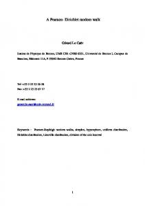
As expected, Fig. 3 shows that, < r ' (θ ci , Lsc ) > remains constant with the change in exit angle in the EBS regime (Lsc = 50 mm >> ls*). On the other hand, in low coherence regime (Lsc = 200 µm increases with the increase in exit angle.
(a)
It is also shown that the slope of the curve increases as the spatial coherence length Lsc decreases from 160 µm to 100 µm. Hence in low coherence regime, obtaining P(r) for different angles of collection significantly affects the width of LEBS peaks. Therefore, it is imperative to obtain a proper exit angle to accurately model the LEBS peak in the low order scattering regime. This aspect is further explored in the subsequent subsections. B. Importance of proper exit angle in accurate modeling of LEBS peaks The previous section clarifies that the exit angle of photons in the image plane is of critical importance for low coherence enhanced backscattering as LEBS probes low orders of scattering in a diffusive multiple scattering medium. This is due to the fact that the trajectories of the photons are clustered into a compact locus to exit at small angles for low orders of scattering, while the trajectories of the photons are less compact for higher orders of scattering. As can be seen from our simulations, the exit angle of the photons is sensitive to the depth from which the LEBS measurements are obtained. This fact is further illustrated in Figs. 4a and 4b, which show < r (θ ci , Lsc ) > calculated for different exit angles and different spatial coherence length Lsc. Lscj
< r (θ ci , Lsc ) >=
∫ rP
Lsc
(r , θ ci )dr ,
(16)
0
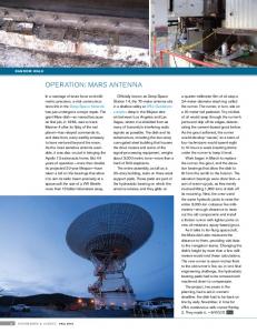
where i = 0 to 90 degrees and j = 20 to 140 µm. As shown in Fig. 4a, for a given Lsc,
< r (θ ci , Lsc ) > increases with the increase of the exit
angle from 0 to 90 degrees. Similarly, for a fixed exit angle, increase in Lsc from 20 to 140 µm leads to an increase in < r (θ ci , Lsc ) > as also shown in Fig. 4b. The relationship between < r (θ ci , Lsc ) > and the depth of penetration of the scattered photons are obtained using Monte Carlo simulation by tracking the propagation of photons along the z-direction. The simulation is performed in the low order scattering regime and the size of the grid tracking the photons in
(b)
< r (θ ci , Lsc ) > as a function of angle θci obtained for 4 different Lsc. (b) < r (θ ci , Lsc ) > as a function of Lsc obtained for 4 different θci. < r (θ ci , Lsc ) > FIG. 4.
(a)
is calculated using Monte Carlo simulation from a medium with ls* = 2 mm, and g = 0.9 (at λ = 520 nm) for θci varying from 1˚ to 90˚ and different Lsc varying between 35 and 140 µm. is proportional to both θci and Lsc.
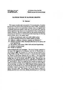
the z-direction is kept at 1 µm. Figure 5 shows this relationship which indicates that < r (θ ci , Lsc ) > is proportional to the depth from which the photons are scattered in the backward direction. In order to model LEBS, it is necessary to accurately determine the angle at which the photons are collected. The LEBS signals obtained from simulation are collected at θci ~ 1.5˚ which is close to
6
FIG. 5.
< r (θ ci , Lsc ) > as a function of r and depth of
penetration of scattered photons z for a fixed θci = 45˚ and a fixed Lsc = 600 µm. < r (θ ci , Lsc ) > is calculated from a medium with ls* = 2 mm, and g = 0.9 (at λ = 520 nm) using Monte Carlo simulation. < r (θ ci , Lsc ) > is proportional to
FIG. 6. Probability of exit angle Pex(θci) as a function of θci obtained for low order scattering regime. Pex(θci) is calculated using Monte Carlo simulation (ls* = 2 mm, g = 0.9 at λ = 520 nm) for a fixed Lsc = 200 µm by varying θci between 1˚ and 90˚. Pex(θci) converges at small angles of around 1˚-3˚ when Lsc