Nov 13, 1991 - Activity In Vivo. BRUCE J. MAYER,' PETER K. JACKSON,2t RICHARD A. VAN ETTEN,2t AND DAVID BALTIMORE'.2* ..... major tyrosine-phosphorylated protein that associates with ..... I. A. Wilson, R. A. Lerner, and M. Wigler.
MOLECULAR AND CELLULAR BIOLOGY, Feb. 1992, p. 609-618 0270-7306/92/020609-10$02.00/0 Copyright C 1992, American Society for Microbiology
Vol. 12, No. 2
Point Mutations in the abl SH2 Domain Coordinately Impair Phosphotyrosine Binding In Vitro and Transforming Activity In Vivo BRUCE J. MAYER,' PETER K. JACKSON,2t RICHARD A. VAN ETTEN,2t AND DAVID BALTIMORE'.2* The Rockefeller University, 1230 York Avenue, New York, New York 10021,' and The Whitehead Institute for Biomedical Research, 9 Cambridge Center, Cambridge, MA 021422 Received 29 July 1991/Accepted 13 November 1991
We have constructed a series of point mutations in the highly conserved FLVRES motif of the src homology 2 (SH2) domain of the abl tyrosine kinase. Mutant SH2 domains were expressed in bacteria, and their ability to bind to tyrosine-phosphorylated proteins was examined in vitro. Three mutants were greatly reduced in their ability to bind both phosphotyrosine itself and tyrosine-phosphorylated cellular proteins. All of the mutants that retained activity bound to the same set of tyrosine-phosphorylated proteins as did the wild type, suggesting that binding specificity was unaffected. These results implicate the FLVRES motif in direct binding to phosphotyrosine. When the mutant SH2 domains were inserted into an activated abl kinase and expressed in murine fibroblasts, decreased in vitro phosphotyrosine binding correlated with decreased transforming ability. This finding implies that SH2-phosphotyrosine interactions are involved in transmission of positive growth signals by the nonreceptor tyrosine kinases, most likely via the assembly of multiprotein complexes with other tyrosine-phosphorylated proteins.
The src homology 2 (SH2) domain is an approximately 100-amino-acid region found in a variety of proteins implicated in growth control, including nonreceptor proteintyrosine kinases, phospholipase C--y, the ras GTPase activator protein (GAP), the crk oncogene product, and the 85-kDa phosphatidylinositol 3-kinase subunit (p85) (reviewed in reference 29). Because SH2 domains are found in a wide variety of contexts and are not required for catalytic activity, it has been suggested that SH2 domains confer a modular function on the proteins in which they are found (29, 35, 44). Evidence from a number of groups has suggested that SH2 domains bind with high affinity to tyrosine-phosphorylated cellular proteins (1, 32-34, 36, 38, 39, 51). It is thought that the SH2 domains of phospholipase C-y, GAP, and p85 mediate the binding of these proteins to growth factor receptors of the tyrosine kinase class, which rapidly autophosphorylate on tyrosine upon ligand binding (1, 32, 39; reviewed in references 29 and 55). While the specific role of SH2-mediated association with growth factor receptors is still under investigation, altered access to substrates or increased enzymatic activity due to phosphorylation by the associated receptor are likely possibilities (16, 26, 40, 41). In nonreceptor tyrosine kinases, mutations in the SH2 domain modulate biological activity. In most cases, SH2 mutations decrease the transforming activity of activated kinases (references 8, 18, 24, 30, and 58 and references therein), although several point mutations in SH2 have been shown to activate the transforming potential of normally nontransforming proto-oncogene products (18, 42). Furthermore, SH2 mutations in nonreceptor tyrosine kinases have been reported to confer host-dependent transforming activ-
ity (8, 9, 19, 49, 56) and to generate dominant-negative inhibitors of transformation (20), suggesting a role for SH2 interactions with cellular factors. The mechanism whereby SH2 mutations modulate the activity of tyrosine kinase oncogene products is unclear. While the clearest activity of SH2 domains is phosphotyrosine binding, it is not obvious how this property is involved in the regulation of biological activity. The possibility remains that SH2 domains confer a crucial function that is independent of phosphotyrosine binding. In this work, we have generated a series of conservative point mutations in the SH2 domain of the abl oncogene product, a nonreceptor tyrosine kinase (17, 48). We show that several mutations in the highly conserved FLVRES motif drastically reduce in vitro binding to phosphotyrosine itself and to tyrosine-phosphorylated proteins, implicating this region in binding to phosphotyrosine. Reduced binding to tyrosine-phosphorylated proteins in vitro correlates with decreased transforming activity in vivo when the mutations are present in a retrovirally expressed activated abl protein. These data suggest that binding to phosphotyrosine is involved in transformation by nonreceptor tyrosine kinases. MATERIALS AND METHODS
Mutagenesis. The dut ung bacterial strain and protocols from the Muta-Gene kit (Bio-Rad) were used to generate oligonucleotide-directed point mutations in the abl SH2 domain. For mutants in the glutathione S-transferase (GST)SH2 expression vector, the BamHI-EcoRI fragment from wild-type pGEX-SH2 (38) was subcloned into M13mpl8, and mutagenesis was performed on uracil-substituted viral DNA. Mutant BamHI-EcoRI fragments were excised from M13 replicative-form DNA and reinserted into pGEX-2T (52). For SH2 mutations in full-length abl, the 2.5-kbp HincIl fragment containing the SH2 and catalytic domains of c-abl was subcloned into pTZ19U (Bio-Rad), and mutagenesis was performed on uracil-substituted phagemid DNA. Mutant
* Corresponding author. t Present address: Department of Biochemistry and Biophysics, University of California, San Francisco, San Francisco, CA 94143. t Present address: Center for Blood Research, Department of Genetics, Harvard Medical School, Boston, MA 02115.
609
610
MAYER ET AL.
HincIl fragments were subcloned into pPL-AXB-HA (see below). Mutagenic primers were 28 to 51 nucleotides in length with one to three mismatches. All resulting mutants were sequenced to verify their structures. Construction and expression of mutant abl genes. Mutant SH2 domains were inserted into pPL-AXB-HA, a derivative of the retroviral expression vector pPL-AXB, which encodes an SH3-deleted type IV c-abl (21). pPL-/XB-HA differs from its parental vector by the addition of a C-terminal epitope tag from the influenza virus HA protein (11; details to be published elsewhere). pPL-AXB-K290M-HA differs from pPL-AXB-HA by the presence of a point mutation in the ATP binding site, changing lysine 290 to methionine (23). pPL-AXB-HA-derived plasmids were transfected into NIH 3T3 fibroblasts by calcium phosphate coprecipitation along with pZAP, which encodes a Moloney murine leukemia virus helper, as described previously (21). Briefly, 10 ,ug of abl-encoding plasmid and 0.5 ptg of pZAP were mixed in calcium phosphate buffer, and dilutions of the mixture were transfected onto duplicate plates. Foci were scored from the 1:5 dilution plates 12 days posttransfection. Three independent plasmid isolates were analyzed for the V170L mutant, two were analyzed for R171K and S173C, and one was analyzed for the wild type (wt), E172Q, E174Q, and S175C. For transient transfections, 4 Rg of pPL-AXB-HA-derived plasmids was transfected by calcium phosphate into subconfluent human 293 cells, and lysates were made 44 h posttransfection. Bacterial expression. All SH2 domains were expressed in Escherichia coli NB42 by using the GEX expression system and purified by using glutathione-agarose as described previously (52). To generate cleaved SH2 peptides for nuclear magnetic resonance (NMR) spectroscopy, bacterial lysates containing GST-SH2 fusion proteins were incubated with glutathione-agarose beads (Molecular Probes), and then the beads were washed with TN buffer (50 mM Tris [pH 8.0], 150 mM NaCl) and incubated at room temperature for 2 h in TN buffer with 2.5 mM CaCl2 and 125 U of human thrombin (Calbiochem) per liter of original bacterial culture. The eluate and wash from the cleaved beads were concentrated with a Centriprep-10 (Amicon), and contaminating proteins were removed by gel filtration on Superose-12 (Pharmacia). Cloning and expression of GAP SH2. The N-terminal SH2 domain of murine GAP was cloned from NIH 3T3 total RNA by using reverse transcription and polymerase chain reaction. The amplified fragment contained sequences corresponding to amino acids 175 to 280 of human GAP (54) as well as N-terminal BamHI and C-terminal EcoRI sites in the proper reading frame and orientation for expression from pGEX-2T. The amplified fragment was digested with EcoRI and BamHI and cloned into pGEX-2T, and the resulting clones were sequenced to confirm their identities. The murine SH2 sequence encodes a protein that is identical in amino acid sequence to the published human clone. SH2 binding assays. Phosphotyrosine-agarose and phosphoserine-agarose were made by coupling phosphotyrosine or phosphoserine (Sigma) to Affi-Gel 15 beads (Bio-Rad) according to the manufacturer's recommendations. Briefly, 20 mM phosphoamino acids were prepared in 100 mM morpholine propanesulfonic acid (MOPS; pH 7.5), 50 ,mol of phosphoamino acid was added per ml of Affi-Gel, and the mixture was incubated at room temperature for 2 h. To assay binding of GST-SH2 fusion proteins to phosphotyrosine, 20 ,ug of fusion protein in 100 ,ul of phosphate-buffered saline (PBS) plus 1% Triton X-100 was added to 20 [lI of phos-
MOL. CELL. BIOL.
phoamino acid-agarose or 10 ,ul of glutathione-agarose bead slurry (50% beads by volume). The mixture was incubated at 4°C for 40 min, then the beads were quickly washed twice with 1 ml of cold PBS and resuspended in sample buffer, and half of the sample was subjected to sodium dodecyl sulfatepolyacrylamide gel electrophoresis (SDS-PAGE) (13% polyacrylamide gels). Filter-binding assays were performed with biotinylated GST-SH2 fusion proteins (1 ,ug/ml) as described previously (38) except that 1% ovalbumin was used for blocking and peptide binding. All incubations and washes were carried out at 4°C, and filters were incubated with biotinylated SH2 peptides overnight. For titration experiments, tyrosinephosphorylated proteins were purified from an abl-transformed pre-B cell line (AXB-BM [20a, 21]) by immunoaffinity chromatography on monoclonal antibody PY20agarose beads as described previously (14, 15). Binding assays were performed as described above at various GST fusion peptide concentrations, with the exceptions that the biotinylated probes were diluted fivefold with unbiotinylated peptide to decrease their specific activity and dithiothreitol was present at 10 mM in the blocking and binding reactions. The GST-SH2 fusion proteins were labeled with biotin as described previously (38) under identical conditions. To ensure that labeling efficiencies were similar, the relative specific activity of the biotinylated probes was assessed by side-by-side comparison of both the intensity of Coomassie blue staining and labeling with avidin-conjugated alkaline phosphatase. In all cases, the specific activities were indistinguishable. Immunoblotting. Immunoblotting was performed by standard protocols as described elsewhere (38), using monoclonal antibodies PY20 (15), 19-84 (50), and 12CA5 (11), which recognize phosphotyrosine, abl protein, and influenza virus HA protein epitope tag, respectively. Bound antibody was detected with alkaline phosphatase-conjugated anti-mouse immunoglobulins. For coprecipitation experiments, 293 cells were lysed in Triton X-100 lysis buffer containing 1 mM sodium orthovanadate, and equal amounts of protein were immunoprecipitated with polyclonal anti-abl serum pEX-4 (31). Immune complexes were collected on protein A-Sepharose beads and washed three times with lysis buffer, and aliquots were immunoblotted and probed with monoclonal antibody PY20 or 19-84 as described above. NMR spectroscopy. NMR spectra were acquired on a General Electric OMEGA 500-MHz NMR system operating at 500.115 MHz at 25°C. Native samples (see Fig. 6A) were approximately 1 mM in PBS plus 10% D20. A spectral width of 7,017.54 Hz was used with a water-presaturating singlepulse sequence of 1.3-s presaturation and an acquisition time of 1.17 s in 8,192 data locations for 512 cycles. The wt sample contained approximately 200 ,iM 3-(trimethylsilyl)1-propanesulfonate, whose principal resonance appears at 0.0 ppm in the upper spectrum of Fig. 6A. Spectra were apodized by applying an exponential function with a line broadening of 2 Hz. The denatured sample (see Fig. 6B) was 110 ,uM wt abl SH2-6 M guanidinium-HCl in 99.96% D20. A total of 2,048 scans were acquired with a sweep width of 6,006 Hz. No apodization function was applied. RESULTS We have previously shown that the abl SH2 domain, when expressed in E. coli as a fusion protein with GST, binds with high affinity to tyrosine-phosphorylated cell proteins (38). To
abl SH2 DOMAIN MUTANTS
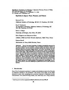
VOL. 12, 1992 SH3 SH2
Kinase
M 1142 J a.a.
1 2
3
4
5
6 7
611
8
A
U
q
._i
>l
mutants
GINGSFLVRESESSPGQ S
S
xxxGFLVRESlExxGx
type IV c-abl, a.a. 163-179
1 2 3 4 5 consensus
FIG. 1. SH2 mutations used in this study. The diagram at the top is a schematic representation of type IV c-abl protein, showing to scale the locations of the SH3 and SH2 domains, conserved kinase catalytic domain, and major nuclear localization signal (K5). Below is shown the sequence of the abl SH2 domain in the vicinity of the highly conserved FLVRES motif. Amino acids (a.a.) 170 through 175 were individually mutated to the those shown in the line above (arrowheads). The consensus sequence consists of amino acids present in at least 50% of all SH2 domains; X denotes lessconserved residues.
identify residues critical for phosphotyrosine binding and to probe the biological relevence of this property to abl gene function, we generated a series of conservative amino acid changes within the SH2 domain. We chose to mutate residues within the FLVRES motif of SH2, because this is the best-conserved region among all SH2 domains (29). Oligonucleotide-directed mutagenesis was used to create single amino acid changes designed to maintain the approximate size and hydrophilicity of the wt residues. The six mutants used in this study are shown in Fig. 1. These mutations were initially constructed into the GST expression vector to study the properties of the isolated, bacterially expressed SH2 domains in vitro. We first examined whether the mutant SH2 domains bound to phosphotyrosine itself. We have previously shown that a fraction of tyrosine-phosphorylated proteins bound to the wt abl SH2 domain could be eluted with high concentrations of phosphotyrosine (38), suggesting that phosphotyrosine is directly involved in binding; other studies, however, have found no effect of phosphotyrosine (33). When the bacterially expressed GST-SH2 fusion protein was incubated with agarose beads covalently linked to phosphotyrosine, a significant fraction bound to the beads, while none bound to similar beads linked to phosphoserine (Fig. 2B and C, lanes 2). GST protein lacking SH2 sequences did not bind (Fig. 2B, lane 1). These results demonstrate that the abl SH2 domain can bind directly and stably to phosphotyrosine and not to phosphoserine under these conditions. From experiments in which phenyl phosphate, a phosphotyrosine analog, was used to block association of tyrosine-phosphorylated cell proteins with the abi SH2 domain, we estimate the Ki for phenyl phosphate to be between 1 and 10 mM (not shown). This low-affinity binding to phosphotyrosine is probably a -common property of functional SH2 domains, because we have also observed binding to phosphotyrosine beads for bacterially expressed src and GAP SH2 domains (not shown). When the mutant abl SH2 domains were examined for phosphotyrosine binding, three of the mutants were indistinguishable from wt: V170L, E172Q, and E174Q (by convention, mutants are designated by wt amino acid, residue number, and mutant amino acid) (Fig. 2B). Three of the mutants, R171K, S173C, and S175C, showed no detectable
B
6 7 8
I._
_
Om
_
I... .
1 2 3 4 5 6 7 8 I
C
FIG. 2. Binding of bacterially expressed SH2 domains to phosphotyrosine-agarose. Purified GST-SH2 fusion proteins were incubated with glutathione-agarose (A), phosphotyrosine-agarose (B) or phosphoserine-agarose (C), and the bound fraction was separated by SDS-PAGE and visualized by staining with Coomassie blue. Lanes: M, molecular weight markers (45, 29, and 18 kDa apparent molecular mass); 1, GST alone; 2, wt abl SH2; 3, V17OL; 4, R171K; 5, E172Q; 6, S173C; 7, E174Q; 8, S175C.
binding to phosphotyrosine, suggesting that these mutations affect residues directly involved in interaction with phosphotyrosine. The fusion proteins were all present at the same concentration and could be bound by glutathione agarose with the same efficiency (Fig. 2A). The arginine at wt position 171 of abl is absolutely conserved in all known SH2 domains; substitution of lysine at this position (which maintains the positive charge) eliminated stable phosphotyrosine binding, suggesting that the specific structure of this arginine residue is critical to the binding interaction. To examine whether the SH2 mutants retained highaffinity binding to tyrosine-phosphorylated cell proteins, we used biotinylated GST-SH2 fusion proteins at a low concentration (approximately 25 nM) to bind to total protein from abl-transformed fibroblasts which had been separated by SDS-PAGE and transferred to nitrocellulose filters (38). Consistent with results from binding to phosphotyrosine, three mutants (V17OL, E172Q, and E174Q) bound to tyrosine-phosphorylated proteins as well as did the wt, while three mutants (R171K, S173C, and S175C) showed no specific binding (Fig. 3, lanes 1). No changes were seen in the profile of binding to various tyrosine-phosphorylated proteins for any of the mutants that retained activity; like wt SH2, these mutants bound well to a subset of tyrosinephosphorylated proteins and relatively poorly to others, such as abl protein itself and a prominent protein of approximately 34 kDa. As is the case with wt SH2, all of the mutants specifically bound few proteins in lysates from untransformed 3T3 cells, in which the levels of tyrosinephosphorylated proteins are low (Fig. 3, lanes 2).
612
MAYER ET AL. Probe:
=,:. ',:.
}*:-_W..s=g;g|.,x