Rijn for printing the micrographs, and Eli Heidt for typing the manuscript. .... Sando, G. N., P. Titus-Dillon, C. W. Hall, and E. F. Neufeld. 1979. Inhibition of.
Possible Pathways for Lysosomal Enzyme Delivery HANS J. GEUZE,* JAN W. SLOT,* GER J. A. M. STROUS,* ANDREJ HASILIK,* and KURT VON FIGURA* *Laboratory of Cell Biology, Medical School, University of Utrecht, The Netherlands; and *PhysiologischChemisches Institut der Westf/~lischen Wilhelms-Universitlat MEmster, Mt]nster, Federal Republic of Germany.
ABSTRACT Immunogold double-labeling and ultrathin cryosections were used to compare the subcellular distribution of albumin, mannose 6-phosphate receptor (MPR), galactosyltransferase, and the lysosomal enzymes cathepsin D, beta-hexosaminidase, and alpha-glucosidase in Hep Gz cells. MPR and lysosomal enzymes were found throughout the stack of Golgi cisternae and in a trans-Golgi reticulum (TGR) of smooth-surfaced tubules with coated buds and vesicles. The trans-Golgi orientation of TGR was ascertained by the co-localization with galactosyltransferase. MPR was particularly abundant in TGR and CURL, the compartment of uncoupling receptors and ligands. Both TGR and CURL also contained lysosomal enzymes, but endogenous albumin was detected in TGR only. The coated buds on TGR tubules contained MPR, lysosomal enzymes, as well as albumin. MPR and lysosomal enzymes were also found in coated pits of the plasma membrane. CURL tubules seemed to give rise to smooth vesicles, often of the multivesicular body type. In CURL, the enzymes were found in the lumina of the smooth vesicles while MPR prevailed in the tubules. These observations suggest a role of CURL in transport of lysosomal enzymes to lysosomes. When the cells were treated with the lysosomotropic amine primaquine, binding of antiMPR to the cells in culture was reduced by half. Immunocytochemistry showed that MPR accumulated in TGR, especially in coated buds. Since these buds contain endogenous albumin and lysosomal enzymes also, these data suggest that coated vesicles originating from TGR provide for a secretory route in Hep G2 cells and that this pathway is followed by the MPR system as well. Mannnose 6-phosphate receptors (MPR) 1 mediate the selective targeting of newly synthesized lysosomal enzymes to lysosomes. The receptors recognize mannose 6-phosphate residues which are added to the nascent enzyme molecules in, or in association with, the Golgi complex (1). After receptor-ligand binding, the complexes travel via an unknown pathway to the lysosomes. Before enzyme delivery, MPR and ligands uncouple in the acidic internal milieu of some prelysosomal compartment. Uncoupling permits free receptors to be re-used (2). The idea of recycling MPR stems mainly from studies on enzyme uptake by cells. These studies also indicate Abbreviations used in this paper. ASGP,asialoglycoprotein;CHO, Chinese hamster ovary; CURL, compartment of uncoupling receptors and ligands; GERL, region of smooth endoplasmic retieulum at the inner or trans-face of the Golgi stack; MPR, mannose 6-phosphate receptors; mvb's, multivesicular bodies; TGR, trans-Golgl reticulum. THE JOURNAL OF CELL BIOLOGY - VOLUME 101 DECEMBER 1985 2253-2262 © The Rockefeller University Press - 0 0 2 1 - 9 5 2 5 / 8 5 / 1 2 1 2 2 5 3 / 1 0 $1.00
that a relatively large pool of intracellular receptors is in equilibrium with the surface receptors (for reviews see references 1 and 3). The sites where lysosomal enzymes are sorted out from secretory proteins and are delivered to lysosomes have not yet been identified. The same holds for the place from where unoccupied MPR enter the recycling route. As a matter of fact, it is still poorly understood which pathway plasma membrane proteins in general, including the receptors, take when traveling (back) to the cell surface. This is partly due to the difficulty in localizing the receptors themselves. The elaborate enzyme cytochemical literature on MPR ligands (mainly acid phosphatase and aryl sulphatase) has not unequivocally defined the presumed acidic compartment wherein receptors and ligands uncouple and from where ligand is transferred to lysosomes. 2253
During the last few years, more information on receptor distribution and morphology of the prelysosomal compartment has become available. Developments in immunoelectron microscopical technology has allowed several authors to localize MPR in situ. Unfortunately, at the crucial level of the Golgi complex, reports are conflicting. Willingham et at. (4) were the first to show that MPR occurs in trans-Golgj tubules, in prelysosomal receptosomes, but not in lysosomes of Chinese hamster ovary (CHO) cells. In agreement with and extending these observations, we found MPR present in the cisternal membranes of the Golgi complex, in trans-GolD tubules, and in CURL of rat liver parenchymal cells (5, 6). CURL was defined as a prelysosomal compartment of uncoupling receptors and ligands, consisting of tubules and detaching vesicles (7). CURL tubules are rich in MPR, whereas CURL vesicles, which are probably equivalent to endosomes (8) and receptosomes (9), have only little MPR (5). An essentially different MPR distribution was reported by Brown and Farquhar (10). They found MPR predominantly located in the cis-Golg~ cisternae, prelysosomal endosomes, and in lysosomes of a variety of rat tissues including liver. Thus, one is left with data that on one hand are in agreement with an important post-Golgj functioning of MPR in ligand processing (4-6) and on the other hand with observations indicating the cis-Gol~ as the sorting element for lysosomal enzymes (10). Biochemical data are generally in support of the concept that MPR-ligand complexes travel through the Golgi complex, or at least pass through trans-Golgi elements containing transferases for terminal glycosylation. Typical lysosomal enzymes like cathepsin D and beta-hexosaminidase contain complex oligosaccharides (11, 12) and, in addition, MPR itself is terminally glycosylated (l 3). The enzyme equipment to manufacture the mannose 6-phosphate signal presumably resides in the Golgi complex (14, 15). Limitations to subfractionating the Golgi complex, however, have so far hampered a reliable dissection of cis- and trans-Golgfi membranes biochemically. We felt that high resolution immunocytochemistry could shed more light on the precise Golgi localization of MPR and, more importantly, on the organelles involved in ligand uncoupling and delivery and in receptor pathways. Using our immunogold double-labeling technique (16, 17), we found that lysosomal enzymes and MPR were located in CURL and in a tubulo-vesicular system at the trans-side of the Golgi complex as was concluded from co-localization with galactosyltransferase. Immunogold double-labeling indicated that the lysosomal enzymes and MPR present in CURL were uncoupled as was previously demonstrated for asialoglycoprotein receptors and ligands in liver (7).
MATERIALS AND METHODS
Cells: The human hepatoma cell line Hep G2 was cultured in Eagle's minimum essential medium (MEM) containing 10% fetal bovine serum. For immunocytochemistry, the calf serum was replaced by 10% rabbit serum 16 h before use (18). The lysosomotropic agents, primaquine (25, 100, 200, and 300 /~M) and NH4C1 (10 raM), were added to the culture medium 30 or 60 rain before use. Antibodies: All antibodies used were affinity-purified rabbit IgG's. The characterization of the antibodies against MPR, cathepsin D, and beta-hexosaminidase has been described before (19). Anti-alpha glucosidase was a kind gift of Dr. A. I. J. Reuser, Department of Cell Biology and Genetics, Erasmus University, Rotterdam, The Netherlands (20). Anti-human galactosyltransferase and albumin have been described before (21, 22). Immunocytochemistry: Cellswere fixed in a mixture of 1% acrolein and 2% glutaraldehyde in 0.1 M phosphate buffer at pH 7.4 for 1 h. After 2254
THE JOtJRNAI- OF CELL BIOLOGY • VOLUME 101, 1985
buffer washes, cells were embedded in 10% gelatin which was cross-linked with glutaraldehyde. Gelatin blocks containing cells were stored in 2.3 M sucrose. Ultrathin cryosections were indirectly double-labeled with colloidal gold particles complexed to protein A as described before (16, 17). In the present study, however, we used gold probes prepared by reduction of chloroauric acid with tannic acid and sodium citrate (23). This method enables the preparation of gold sols with any optional particle size between 3 and 17 rim, and with a size variability of < 10%. Thus, double-labeling with particles smaller than 10 nm could be achieved, which is important because the smallest probes are the most sensitive ones. We mostly used 6- and 9-nm particles. Cryosections were stained and embedded as described before (17).
Determination of Receptor Density: Immunogold labeling for MPR was quantitated in cryosections of normal cells and cells treated with 300 uM primaquine for 30 min. From sections of 12 normal cells and 14 primaquine-treated cells showing profiles of stacked Golgi cisternae and of transGolgi reticulum (TGR), electron micrographs were taken and printed at a magnification of 115,000. MPR gold particles were counted and attributed to smooth-surfaced and coated domains of TGR membranes. A total of 482 gold particles was counted in normal cells and 543 in primaquine-treated cells. A transparent sheet with a squared array of 1-cm-spaced parallel lines was superimposed on the prints and the number of intersections with smooth and coated TGR membranes was determined. The ratio of gold (%) to intersections (%) was then used as a measure of MPR density in the TGR membrane domains. Since TGR is an isotropic structure, primaquine-induced shifts in the number of intersections through smooth and coated TGR membranes were considered to indicate changes in relative membrane surface areas. Binding of Anti-Receptor [~2Sl]lg: Hep G2 cells were incubated for 30 rain at 37°C with primaquine (as indicated in Table I) and then placed for 15 rain on ice water. The cells were incubated for 1 h at 0°C with 0.5 ml of medium supplemented with amine and 500 ng of anti-receptor [~2Sl]Ig as described before (24). Cell-associated radioactivity was referred to cell protein (25).
RESULTS The main ultrastructural features of Hep G2 cells have been described before (26). Briefly, the flat to dome-shaped cells grow in monolayer and enclose at their lateral surfaces bile canalicular-like spaces bordered by extensive junctional complexes. The cells exhibit prominent Golgi complexes with, at their trans-side, an anastomosing system of narrow tubules from which numerous small coated vesicles seem to detach. The branches of the tubules are often so frequent that the impression of fenestrated lamellae is obtained. This system has been called the trans-Gol[~ reticulum (TGR) (27). The TGR coated membrane domains will be referred to as coated buds of TGR. For the morphology of TGR see Figs. l and 4-7. Hep G2 cells contain a well-differentiated endocytic apparatus including coated pits at the plasma membrane facing the culture medium, peripheral coated vesicles, and a CURL (compartment of uncoupling receptors and ligands) which consists of tubules and attached vesicles (7). The morphology of CURL tubules was very similar to that of TGR tubules. However, TGR had more coated buds and vesicles and showed more anastomoses. In uranyl-stained cryoscctions, both CURL and TGR had electron-dense contents, which made them easy to identify. As a rule, CURL had a more peripheral distribution than TGR. However, occasionally Golgi complexes were found very close to the plasma membrane, which made a sharp morphological distinction between TGR and CURL impossible. Endocytic structures are shown in Figs. 12-16. Numerous multivesicular bodies (mvb's) were present adjacent to the Golgi complex (Figs. 9-11) and in the peripheral cytoplasm (Fig. 16). Lysosomes were mostly located in the Golgi area. Most immunocytochemical data were collected from colloidal gold double-labeled ultrathin cryosections. The following combinations of antigens were studied: MPR + lysosomal
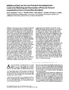
FIGURES 1-3 All electron micrographs were taken from ultrathin cryosections of acrolein-glutaraldehyde-fixed normal Hep G2 cells and cells treated with a lysosomotropic amine. The sections were labeled with 6- and 9-nm colloidal gold particles, further indicated as small and large gold, respectively. Unless mentioned otherwise, lysosomal enzymes were localized with a cocktail of antibodies against cathepsin D, beta-hexosaminidase, and alpha-glucosidase. (Fig. 1) Normal cell. Golgi complex (G) showing a clear cis to trans polarity. The section was double-labeled for galactosyltransferase (small gold) and MPR (large gold). Galactosyltransferase is present in one or two trans-Golgi cisternae and MPR is confined to a trans-Golgi reticulum (TGR) of tubules and vesicles, x 100,000. (Fig. 2) In this normal Hep Gz cell, galactosyltransferase (small gold) and MPR (large gold) are present in the same complex of TGR tubules albeit in different domains. G, Golgi stack, x 118,000. (Fig. 3) Hep G2 cell treated with 300 ~M primaquine for 30 min. Especially the galactosyltransferase (small gold) positive trans-Golgi cisternae are swollen. G, Golgi stack, x 118,000. All bars, 0.1 #m. 2255
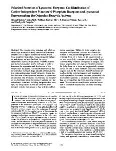
FIGURES 4-7 Electron micrographs prepared as described in legend to Fig. 1. (Fig. 4) Golgi stack (G) and TGR (at the right-hand side) of normal cell, showing MPR (small gold) in TGR and albumin (large gold) in both the Golgi cisternae and TGR. Arrowheads indicate coated buds of TGR. x 100,000. (Fig. 5) Normal cell. Labeling of cathepsin D, beta-hexosaminidase, and alpha-glucosidase (large gold) and of MPR (small gold) is almost entirely restricted to TGR (at the left). G, Golgi stack. N, nucleus, x 100,000. (Fig. 6) Normal cell. Section through Golgi stack (G) and TGR at the right, showing albumin (small gold) and some lysosomal enzyme labeling (large gold) in the Golgi cisternae and tubules and coated vesicles (arrowhead) of TGR. x 90,000. (Fig. 7) Main part of the figure shows a Golgi stack (G) and TGR (upper left-hand corner) of a cell treated with 100 #M primaquine for 30 min. The section was single-labeled for MPR. Note the coated buds of the TGR tubules. These buds are smaller than those in Figs. 4-6. x 90,000. (Inset) Cell treated for 30 rain with 10 mM NH4CI MPR (large gold) and lysosomal enzymes (small gold) are present in a cluster of TGR coated pits. × 140,000. All bars, 0.1 ~tm.
2256
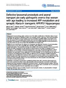
FIGURES 8-11 Electron micrographs prepared as described in legend to Fig. 1. (Fig. 8) MPR (large gold) in a TGR tubule attached to a vesicle which only contains cathepsin D (small gold). At the left is a tangential aspect of an mvb. x 125,000. (Fig. 9) Large mvb in the trans-Golgi area with associated tubular profiles. Lysosomal enzymes (small gold) can be seen in both the mvb and the tubules, whereas the MPR (large gold) prevails in the tubules, x 102,000. (Fig. 10) Large mvb in the Golgi region with abundant lysosomal enzyme labeling (small gold) and some MPR labeling (large gold), x 100,000. (Fig. 11) Section single-labeled for lysosomal enzymes. At the left a large mvb, at the right a lysosome. Note that part of the internal vesicles of the mvb seems to be in a process of desintegration, x 63,000. All bars, 0.1 #m.
enzymes, MPR + galactosyltransferase, MPR + albumin, and lysosomal enzymes + albumin. The sequence of antibodies and gold probes was varied, but usually anti-albumin was the first in its combinations and the smallest and most sensitive probe (6 nm gold) was used to tag lysosomal enzymes or galactosyltransferase which were present in relatively low concentrations. To enhance the immunocytochemical signal for lysosomal enzymes, we mostly used cocktails of antibodies against cathepsin D, beta-hexosaminidase, and alpha-glucosidase.
The Golgi Complex Hep G2 Golgi complexes consisted of stacks of five to six cisternae (Fig. 1) all containing albumin (Figs. 4 and 6). Both MPR and lysosomal enzymes occurred in low amounts throughout the Golgi stacks (Figs. 4-6). Galactosyltransferase was located in one or two cisternae of the Golgi stacks (Fig. 1).
Trans-Golgi Reticulum T G R was located adjacent to the galactosyltransferasepositive Golgi cisternae (Fig. 1). In several cases we found galactosyltransferase present in TGR instead of in the transmost Golgi cisternae (Fig. 2). The most prominent immunocytochemical feature of TGR was its enrichment in MPR
(Figs. 1, 2, and 5). MPR occurred in both smooth-surfaced and coated TGR membranes (Figs. 1, 2, 4, and 5). Coated and smooth TGR also contained albumin (Figs. 4 and 6) and lysosomal enzymes (Figs. 5 and 6). Thus, in the TGR tubules and coated buds no clear segregation of albumin and the MPR system was found. At the trans-lateral sides of the Golgi stacks, numerous smooth-surfaced vesicles were observed, which often contained vesicles inside (Figs. 8-11). These structures were clearly mvb's (Figs. 9-11). The vesicles were often accompanied by tubules which occasionally showed membrane continuities with them (Fig. 8). The origin of these tubulo-vesicles remained unclear. Lysosomal enzymes were found in both the tubules, the smooth vesicles, and the mvb's (Figs. 9-11). Often MPR and lysosomal enzymes seemed to be segregated at the junction of the tubules and smooth vesicles (Figs. 8 and 9).
Endocytotic Apparatus Coated pits, coated vesicles, and CURL showed small numbers of gold particles when labeled for MPR and lysosomal enzymes (Figs. 12-16). The number of coated pits and vesicles at the plasma membrane containing lysosomal enzyme labeling (Fig. 12) was low, but MPR-positive coated structures were regularly encountered. GEUZEETAL. Lysosomal Enzyme Delivery Pathways
2257
FIGURES 12-16 Electron micrographs prepared as described in legend to Fig. 1. Figs. 12-16 show structures of the endocytic apparatus in the peripheral cytoplasm of normal cells. (Fig. 12) Coated vesicle at the plasma membrane showing cathepsin D labeling, x 105,000. (Fig. 13) Section labeled for MPR (large gold) and lysosomal enzymes (small gold). The labels are present in a CURL tubule adjacent to the plasma membrane (arrowheads). x 130,000. (Fig. 14) CURL structures containing cathepsin D (small gold) and MPR (large gold), x 130,000. (Fig. 15) CURL vesicle with lysosomal enzymes (large gold), whereas MPR (small gold) is confined to adjacent CURL tubules, x 120,000. (Fig. 16) Section double-labeled for MPR (small gold) and albumin (the large gold particle at the lower edge of the figure). Albumin is absent from the large CURL vesicle that contains some internal vesicles. The membrane of the CURL vesicle is continuous with that of adjacent CURL tubules at several places. The majority of MPR is present in the tubules, but some is also found in association with internal vesicles. P, plasma membrane, x 120,000. At] bars, 0.1 pm.
The morphology of CURL tubules was almost similar to TGR tubules. Endogenous albumin, however, was abundant in TGR (Figs. 4, 6, and 8-10) but was below detectable levels in CURL (Fig. 16). MPR was predominantly found in CURL tubules, but occurred also in association with internal vesicles of mvb's (Figs. 15 and 16). CURL vesicles, however, showed an enrichment of lysosomal enzymes free in their lumen (Fig. 15).
Effects of Lysosomotropic Amines Treatment of Hep 62 cells with primaquine resulted in a dose-dependent reduction of MPR at the plasma membrane 2258
THE JOURNAL OF CELL BIOLOGY • VOLUME 101, 1985
as measured by binding at 0*C of anti-receptor [1251]Ig(Table I). After 30 min of treatment with 300 ~M primaquine, >40% of the surface receptors had disappeared. Primaquine and NH4C1 caused swelling of Golgi cisternae (Fig. 3). The morphology of the Golgi stacks was disturbed because of swelling of the trans-cisternae. These could then only be identified as such using galactosyltransferase as a marker (Fig. 3). After NH4CI and especially after primaquine treatment, TGR showed more coated buds. By morphometry, we determined the relative surface area of smooth-surfaced and coated TGR membranes in normal cells and cells treated with 300 ~M primaquine for 30 min. As can be seen in Table
II, primaquine caused a doubling of the relative surface area of coated TGR membrane. Most of this membrane occurred in coated buds of TGR, suggesting that primaquine prevented these buds from detaching (see Fig. 7 and inset). The percentage of MPR gold in coated TGR membrane after primaquine treatment was more than twice of that in normal cells (Table II). It follows that receptor density, as expressed as the ratio of MPR gold (%) to relative membrane surface area (% of intersections with the superimposed grid) in coated and smooth TGR was unaffected by primaquine. NI-LC1 and primaquine caused the formation of many vesicles with varying forms and sizes. Some of these vesicles were clearly associated wtih TGR tubules. Others had a more TABLE I.
Binding of [ T251]Anti-ReceptorIg to Cell Surface MPR
~tM
Primaquine
30 100 300
Bound [12Sl]lg % of control* 91 71 57
* Control Hep G2 cells bound 4.4 ng of anti-receptor [12Sl]IgJmgcell protein. Presenceof an excessof unlabeled anti-receptor Ig reduced the amount of cell-associated radioactivity by >90%. TABLE II. RelativeSurface Area of Smooth and Coated TGR Membranes and Distribution of MPR Gold Labeling of TGR Domains in Normal and Primaquine-treated Hep G2 Cells Normal
Primaquine
Relative Average Relative Average surface % of surface % of area gold area gold Smooth TGR Coated TGR SEM
89 11 1.9
89 11 2.9
73 27 3.3
74 26 4.9
peripheral distribution, were often clustered, and had no apparent morphological relationship to either TGR or CURL (Figs. 17 and 18). These vesicles were positive for albumin (Fig. 17), lysosomal enzymes (Figs. 17 and 18) and MPR (Fig. 18). Lysosomal enzyme labeling was mostly associated with the internal aspect of the limiting membranes, suggesting that the enzymes were bound to the receptors (Fig. 18). DISCUSSION
Comparative immunocytochemical observations per se, as described in this study, at best represent a series of static images depicting the instantaneous distribution of proteins. For many of the compartments described in this study it was therefore difficult to decide whether they were endocytotic or part of the biosynthetic pathway. We have overcome to some extent this problem of identification by making the following assumptions and by using certain markers. (a) All structures showing albumin immunoreactivity were considered to be part of the secretory pathway. Hep G2 cells actively synthesize and secrete albumin (28). Though the formal possibility exists that some of the secreted albumin was reinternalized by the cells, the presence of an excess of immunologically nonreactive rabbit albumin in the culture medium ruled out the possibility that such albumin was demonstrated. (b) Coated pits at the plasma membrane were considered to be endocytic. (c) Following coated pits and vesicles, CURL is the subsequent endocytic compartment in Hep G2 cells (26). (d) Galactosyltransferase was used as a trans-Golgi marker (21), defining the exit face of the Golgi stack. As judged from the distribution of albumin, the biosynthetic pathway in Hep G2 cells included the entire stack of Golgi cisternae and TGR with coated buds and vesicles. The presence of albumin in these coated buds and vesicles suggests that they mediate constitutive secretion in Hep G2 cells. The
FIGURES 17 and 18 Electron micrographs prepared as described in legend to Fig. 1. (Fig. 17) Hep G2 cell treated with 10 mM NH4Cl for 1 h. A cluster of smooth vesicles is present in the cell periphery. The limiting membrane of the vesicles shows label for cathepsin D and beta-hexosaminidase (small gold) and some for albumin (large gold). Both occur at the limiting membrane of the vesicles, x 90,000. (Fig. 18) Cell treated as in previous figure. The large vesicle in the periphery of the cell shows parietal label for cathepsin D (small gold) and MPR (large gold), indicating that ligand is still receptor bound, x 80,000. All bars, 0.1 #m. Geuze ET AL. LysosomalEnzyme Delivery Pathways
2259
cells did not exhibit marked storage of secretory proteins. Only occasionally were larger granules with dense albumin labeling found. It is possible that redistribution due to inadequate cross-linking had caused the presence of albumin in TGR coated buds, but this seems unlikely, since rigorous fixation in 4% glutaraldehyde for several hours gave the same results (not shown). The distribution of MPR, lysosomal enzymes, and albumin in the Golgi complex and TGR showed many similarities and a few important differences. All were found in the Golgi cisternae. Our data argue against an exclusive cis-Golgi localization of MPR, as has been reported in immunoperoxidase studies on a variety of tissues and culture cells (10, 29). Using anti-rat MPR antibodies, we have previously described that in rat liver cells, MPR and cathepsin D co-distributed throughout the Golgi stack (5, 6). These and our present observations suggest that newly synthesized lysosomal enzymes pass through the entire Golgi complex. This would be in good agreement with the fact that many lysosomal enzymes are terminally glycosylated. Since no segregation between albumin, MPR, and lysosomal enzymes was apparent at the level of the Golgi stack, sorting probably occurs after passage of the Golgi cisternae.
TGR and GERL The double-labeling patterns of MPR and galactosyltransferase demonstrates that, in Hep G2 ceils, the majority of MPR is located in a tubulo-vesicular system at the trans-side, which is properly indicated with the descriptive term TGR. This is in accordance with findings in CHO cells (4). Moreover, we found MPR and galactosyltransferase occasionally present in the same TGR tubules. MPR, lysosomal enzymes, and albumin co-distributed in TGR tubules and coated buds and vesicles. Whether or not the lysosomal enzymes in these structures were bound to MPR could not be elucidated because of the small diameters of the tubules relative to the label. Because lysosomal enzyme labeling was relatively low, a possible segregation of albumin and enzymes in TGR tubules and coated buds or in TGR tubule sub-domains may have been overlooked. Similarly, subpopulations of coated vesicles enriched in either lysosomal enzymes or albumin could easily have been missed. The observed co-localization of albumin, MPR, and enzymes suggests a simultaneous transport to the plasma membrane. However, proof that such TGR-derived vesicles are indeed exocytotic, is lacking. The tubulo-vesicular organelle TGR is morphologically very reminiscent of GERL. GERL was defined as a special region of smooth endoplasmic reticulum at the inner or trans-face of the Golgi stack. It was introduced as an organelle separate from the Golgi complex and was thought to receive lysosomal enzymes and secretory proteins directly from the rough endoplasmic reticulum. GERL was further suggested to give rise to several types of lysosomes such as coated vesicles (presumed primary lysosomes), residual bodies, and autophagic vacuoles including mvb-type vesicles (see, e.g., references 30 and 31). Although it is now generally accepted that the Golgi complex is an obligatory station for newly synthesized secretory and probably also lysosomal proteins (32, 33), several of the original observations of Novikoff et al. (30, 31) closely correspond to our present immunocytochemical data. The distribution of albumin, MPR, and lysosomal enzymes in TGR supports the notion that this structure is part of the intracellular pathway of both lysosomal enzymes and secretory pro2260
THE JOURNAL Of CELL BIOLOGY • VOLUME 101, 1985
teins. On the other hand, since TGR coated vesicles contain enzymes as well as MPR, they cannot be considered primary lysosomes. Furthermore, the presence of galactosyltransferase in TGR relates this organelle to Golgi stacks rather than to smooth endoplasmic reticulum.
Lysosomal Enzymes in CURL The presence of MPR and lysosomal enzymes in CURL suggests some role of CURL in lysosomal enzyme transport. The enzymes appeared to be localized in the contents of CURL vesicles where they are probably not receptor bound. On the other hand, MPR occurred predominantly in CURL tubules. We have previously described a similar situation for the asialoglycoprotein (ASGP) receptor system. After endocytosis, ASGP receptor and ligand were demonstrated to uncouple in the acidic (34) content of CURL (7, 26). ASGPcontaining CURL vesicles are supposed to fuse with lysosomes for ligand degradation. Whether the free lysosomal enzymes in CURL vesicles represent newly uncoupled MPR ligand or were liberated from MPR at some other intracellular site remains undecided. Similarly, no evidence exists as to the immediate pre-CURL route of the enzymes. CURL is known to be involved in targeting of endocytosed ligands to lysosomes. The presence of lysosomal enzymes in coated vesicles at the plasma membrane indeed suggests that at least part of the enzymes in CURL originates from the cell surface (35). However, transfer of (most of) the enzymes to CURL from other intracellular compartments remains of course well possible. Many of the CURL vesicles contained internal vesicles, even if they were still attached to tubules. These mvb's usually contained lysosomal enzymes in their matrices. Recently Harding et al. (36), studying the transport of transferrin and mannose-terminal proteins from coated pits to CURL in several cell types, also showed lysosomal enzyme activity in CURL vesicles and mvb's. Our observations provide no clue as to the origin of the numerous mvb's at the trans-lateral sides of the Golgi stacks. The observation that the majority of mvb's finally become loaded with exogenous ligands (26, 37) indicates that many of them derive from the endocytic apparatus, i.e., CURL.
Effects of NH4CI and Primaquine Lysosomotropic amines have been shown to inhibit receptor-mediated uptake of a variety of ligands (for a review see 38) including those recognized by MPR (2, 39, 40). For the receptors for ASGP (41), epidermal growth factor (42), mannose-glycoconjugates (43), and lysosomal enzymes (2), it has been reported that such an inhibition is reversible and results from blocking the re-utilization of receptors. The amines are known to become protonated within acidic compartments, thereby neutralizing the low pH. In the case of MPR it was thought that the neutral pH would prevent the release of ligands from the receptors. With time, all MPR would then become occupied (2). It is not clear whether occupied receptors are less capable of returning to the plasma membrane. In the case of the ASGP receptor, where the uncoupling is low pH dependent, receptors accumulate intracellularly even in the absence of ligand (41). Thus, other factors besides ligand occupation may control return of receptors to the plasma membrane. The present study shows that a 30-min treatment with 300 #M primaquine almost halves the number of surface
MPR. Thus accumulation of MPR by primaquine might be a result of other factors than neutralization of pH alone. It was not known yet where in the cell the receptors accumulate after lysosomotropic amine treatment. Because CURL and TGR are the main intracellular locations of MPR, these are important candidates. Preliminary observations on the ASGP receptor support this hypothesis. Our present morphometric and quantitative immunocytochemical data show that the primaquine treatment resulted in a doubling of MPR-containing TGR coated membrane area, This suggests that primaquine impairs formation of coated vesicles. Since many of the coated buds contained albumin, a blockade of coated vesicle formation is envisaged to result in inhibition of albumin secretion as well. This is exactly what we have found previously (18) and supports the idea that the coated TGR buds are precursor secretory vesicles. Another striking effect of the amines was the appearance of large smooth vesicles in the cell periphery. The origin of these vesicles is unclear. Some of them showed albumin reactivity, suggestive of their relation to TGR. The vesicles were reminiscent of MPR-positive vesicles found by Brown et al. (29) after chloroquine treatment of hepatocytes.
Are CURL and TGR Related? In Hep O2 cells, the Oolgi complex and TGR sometimes closely approached the cell surface. In such cases, CURL and TGR could not reliably be distinguished on morphological criteria alone. In liver, however, where the Golgi complex and TGR are situated in a different cell pole (at the bile canaliculus) than CURL (at the space of Disse), such a distinction is sharper (5, 7, 44). Although TGR in Hep G2 cells contained endogenous albumin and CURL did not, and TGR tubules were more branched and showed more coated buds than CURL, several observations suggest at least a functional relationship between TGR and CURL. (a) After receptor-mediated endocytosis, epidermal growth factor, alpha 2-macroglobulin, insulin, mannose-terminal proteins, and transferrin have been shown to be transferred from peripheral endocytotic structures to TGR (36, 37, 45, 46). (b) TGR and CURL are similarly enriched in MPR and ASGP receptors (preliminary observations) and constitute the main intracellular receptor pool in Hep G2 cells. The internal pools of both receptors exchange with the surface pools (19, 24, 47, 48). (c) Primaquine induces a shift of surface MPR to TGR from where recycling is blocked. These receptors most likely enter TGR via CURL. (d) Both CURL and TGR have been shown to be acidic (36, 49). A further morphological and biochemical characterization of TGR and CURL seems to be worthwhile since cytochemical data from us and others indicate an important role in transport, sorting, and targeting of both intracellular and exogenous proteins. We thank Janice Griffith for excellent technical assistance, Tom van Rijn for printing the micrographs, and Eli Heidt for typing the manuscript. This work was supported in part by grants from Foundation for Medical Research and Netherlands Cancer Foundation, the Netherlands. Received for publication 1985.
10 J u n e 1 9 8 5 , a n d in r e v i s e d f o r m
29 July
REFERENCES
1. Sly, W. S., and H. D. Fischer. 1982. The phosphomannosyl recognition system for
intracellular and intercellular transport of lysosomal enzymes. J. Cell. Biochem. 18:6785. 2. Gonzalez-Noriega, A., J. H. Grubb, V. Fallad, and W. S. Sly. 1980. Chloroquine inhibits lysosomal enzyme pinocytosis and enhances lysosomal enzyme secretion by impairing receptor recycling. J. Cell Biol. 85:839-852. 3. Creek, K. E., and W. S. Sly. 1984. In Lysosomes in Biology and Pathology. J. T. Dingle, R. T. Dean, and W. S. Sly, editors. 63-82. Elsevier/North Holland, Amsterdam. 4. Willingham, M. C., I. H. Paslan, and G. G. Sahaglan. 1983. Ultrastructural immunocytoehemical localization of the phosphomannosyl receptor in Chinese hamster ovary cells. J. Histochem. Cytochem. 31:1- 11. 5. Geuze, H. J., J. W. Slot, G. J. A. M. Strous, A. Hasilik, and K. von Figura. 1984. Ultrastruetural localization of the mannose 6-phosphate receptor in rat liver. J. Cell Biol. 98:2047-2054. 6. Geuze, H. J., J. W. Slot, G. J. A. M. Strous, J. P. Luzio, and A. L. Schwartz. 1984. A eycloheximide-resistant pool of receptors for asialoglycoproteins and mannose 6-phosphate residues in the Golgl complex of hepatocytes. EMBO (Eur Mol. Biol. Organ.) J. 3:2677-2685. 7. Geuze, H. J., J. W. Slot, G. J. A. M. Strous, H. F. Lodish, and A. L. Schwartz. 1983. lntracellular site of asialoglycopmtein receptor-ligand uncoupling: double-label immunoelectron microscopy during receptor-mediated endoeytosis. Cell. 32:277-287. 8. Helenius, A., 1. Mellman, D. Wall, and A. Hubbard. 1983. Endosomes. Trends Biochem. Sei. 7:245-249. 9. Pastan, I., and M. C. winingbam. 1983. Receptor-mediated endocytosis: coated pits, receptosomes, and the Golgl. Trends Biochem. Sci. 7:250-254. 10. Brown, W. J., and M. G. Farquhar. 1984. The mannose-6-phosphate receptor for lysosomal enzymes is concentrated in cis Golgl eisternae. Cell. 36:295-307. 11. Hasilik, A , and K. von Figura. 1981. Oligosaccharides in lysosomal enzymes. Distribution of high-mannose and complex oligosaccharides in cathepsin D and Beta-hexosaminidase. Eur J. Biochem. 121:125-129. 12. Varki, A., and S. Komfeld. 1983. The spectrum of annionic oligosaccharides released by ende-Beta-r~-acetylglucosaminidase H from glycoproteins../. BioL Chem. 258:28082818. 13. Sahagian, G. G., and E. F. Neufeld. 1983. Biosynthesis and turnover of the mannose 6phosphate receptor in cultured Chinese hamster ovary cells. Z BioL Chem. 258:71217128. 14. Pohlmann, R., A. Waheed, A. Hasilik, and K. von Figura. 1982. Synthesis of phosphorylated recognition marker in lysosomal enzymes is located in the eis part of Golgi apparatus. J. Biol. Chem. 257:5323-5325. 15. Goldberg, D. E., and S. Korofeld. 1983. Evidence for extensive subcenular organization of asparagme-linked oligosaccharide processing and lysosomal enzyme phosphorylation. J. Biol. Chem. 258:3159-3165. 16. Geaze, H. J., J. W. Slot, P. A. van der Ley, and R. C. T. Scheffer. 1981. Use of colloidal gold particles in double-labeling immuncelectron microscopy of ultrathin frozen sections. J. Cell Biol. 89:653-665. 17. Slot, 3. W., and H. J. Geuze. 1984. Gold markers for single and double immunolabeling of altrathin cryosections. In Immunolabeling for Electron Microscopy. J. M. Polak and I. M. Varndell~ editors. Elsevier Science Publ. B. V. Amsterdam. 129-142. 18. Strous, G. J., A. Du Maine, J. E. Zijderhand-Bleekemolen, J. W. Slot, and A. L. Schwartz. 1985. Effect of lysosomotrupic amines on the secretory pathway and on the recycling of the asialoglycoprotein receptor in human hepatoma ceils. J. Cell Biol. 101:531-539. 19. von Figura, K., V. Gieselmann, and A. Hasilik. 1984. Antibody to mannose 6-phosphate specific receptor induces receptor deficiency in human fibroblasts. EMBO (Eur. Mol. Biol. Organ.) J. 3:1281-1286. 20. Van Dongen, J. M., R. A. Barneveld, H. J. Geuze, and H. Galjaard. 1984. Immunocytochemistry of lysosomal hydrolases and their precursor forms in normal and mutant human cells. Histochem. J. 16:941-954. 21. Roth, J., and E. G. Berger. 1982. Immunocytochemical localization of galactosyltransferase in HeLa cells: codistribution with thiamine pyrophosphatase in trans43olgi cisternae. J. Cell Biol. 93:223-229. 22. Strous, G. L, P. van Kerkhof, R. Willemsen, H. J. Geuze, and E. G. Berger. 1983. Transport and topology of galactosyltransferase in endomembranes of HeLa cells. J. Cell Biol. 97:723-727. 23. Slot, J. W., and H. J. Geuze. 1985. A new method of preparing gold probes for multiple labeling cytochemistry. Eur. J. Cell Biol. 38:87-93. 24. Gartung, C., T. Braulke, A. Hasilik, and K. von Figura. 1985. Internalization of blocking antibodies against mannose 6-phospbate specific receptors. EMBO (Eur. MoL BioL Organ.) J 4:1725-1730. 25. Lowry, O. H., N. J. Rosebrough, A. L. Farr, and R. J. Randall. 1951. Protein measurement with the Folin phenol reagent. ,L Biol. Chem. 193:265-275. 26. Geaze, H. J., J. W. Slot, G. J. A. M. Strous, and A. L. Schwartz. 1983. The pathway of the asialoglyeoprotein-ligand during receptor-mediated endocytosis: a morphological study with colloidal gold/ligand in the human hepatoma cell line, Hep G2. Eur Z Cell Biol. 32:38-44. 27. Willingham, M. C., and 1. Pastan. 1984. Endoc~,¢osis and exocytosis: Current concepts of vesicle traffic in animal cells. Int. Rev~ Cytol. 92:51-92. 28. Strous, G. J. A. M., R. Willemsen, P. van Kerkhof, J. W. Slot, H. J. Geuze, and H. F. Lodish. 1983. Vesicular stomatitis virus glycoprotein, albumin, and transferrin are transported to the cell surface via the same Golgi vesicles. J. CellBioL 97:1815-1822. 29. Brown, W. J., E. Constantinescu, and M. G. Farquhar. 1984. Redistribution ofmannose6-phosphate receptors induced by tunicamycin and chloroquine..L Cell Biol. 99:320326. 30. Novikoff, P. M., A. B. Novikoff, N. Quintaua, and J.oJ. Hauw. 1971. Golgi apparatus, GERL, and lysosomes of neurons in rat dorsal root ganglia, studied by thick section and thin section cytocbemistry. J. Cell BioL 50:859-886. 31. Novikoff, A. B., and P. M. Novikoff. 1977. Cytochemical contributions to differentiating GERL from the Golgi apparatus. Histochem. J. 7:525-551. 32. Farquhar, M., and G. E. Palade. 1981. The Golgl apparatus (complex)--( 1954-198 l)--from artifact to center stage. J. Cell Biol. 91 (3, Pt. 2):77s- 103s. 33. Slot, J. W., and H. J. Geuze. 1983. Immunoelectron microscopic exploration of the Golgl complex. J. Histochem Cytochem 31:1049-1056. 34. Schwartz, A. L., G. J. A. M. Strous, J. W. Slot, and H. J. Geuze. 1985. Immunoelectron microscopic localization of acidic inlracellular compartments in hepatoma cells. EMBO (Eur MoL Biol. Organ.) J. 4:899-904. 35. von Figura, K., and E. Weber. 1978. An alternative hypothesis of ceUular transport of lysosomal enzymes in fibroblasts. Biochem. J. 176:943-950. 36. Harding, C., M. A. Levy, and P. Stahl. 1985. Morphological analysis of ligand uptake and processing: the role of multivesieular endosomes and CURL in receptor-ligand GEUZE Er AL.
LysosornalEnzyme Delivery Pathways
2261
processing. Eur. Z Cell Biol. 36:230--238. 37. Hopkins, C. R. 1983. Intracellular routing of transferrin and transferrin receptors in epidermoid carcinoma A 431 cells. Cell. 35:321-330. 38. Dean, R. T., W. Jessup, and C. R. Roberts. 1984. Effects of exogenous amines on mammalian cells, with particular reference to membrane flow. BiQchem. Z 217:27-40. 39. Wiesman, U. V., S. DiDonato, and N. N. Het~chkowitz. 1975. Effect of chloroquine on cultured fibroblasts: release of lysosomal hydrolases and inhibition of their uptake. Biochem. Biophys. Res. Comm. 66:1338-1343. 40. Sando, G. N., P. Titus-Dillon, C. W. Hall, and E. F. Neufeld. 1979. Inhibition of receptor-mediated uptake of a lysosomal enzyme into fibroblasts by chloroquine, procaine, and ammonia. Exp. Cell Res. 119:359-364. 41. Schwartz, A. L., A. Bolognesi, and S. E. Fridovich. 1984. Recycling of the asialoglycoprotein receptor and the effect of lysosomolropic amines in hepatoma cells. Z Cell Biol. 98:732-738. 42. King, A. C., L. Hernaez-Davis, and P. Cuatmcasas. 1980. Lysomotropic amines cause intracallular accumulation of receptors for epidermal growth factor. Proc. Natl. Acad. Sci. USA 77:3283-3287. 43. Tietze, C., P. Schlcsinger, and P. Stahl. 1982. Chloroquine and ammonium ion inhibit receptor-mediated endocytosis of mannose-glycoconjugates by macrophages: apparent
2262
THF JOURNAL OF CELL BIOLOGY ' VOLUME 101, 1985
inhibition of receptor recycling. J. Cell BioL 92:417-424. 44. Geuze, H. J., J. W. Slot, G. J. A. M. Strous, J. Peppard, K. yon Figura, A. Hasilik, and A. L. Schwartz. 1984. lntracenular receptor sorting during endocytosis: comparative immunoeleclron microscopy of multiple receptors in rat liver. Cell. 37:195-204. 45. WiUingham, M. C., I. H. Pastan, G. G. Sahagian, G. W. Jourdian and E. F. Neufeld. 1981. Morphologic study of the internalization of a lysosomal enzyme by the mannose 6-phosphate receptor in cultured Chinese hamster ovary cells. Proc. Natl. Acad. Sci. USA. 78:6967-6971. 46. Ackerman, G. A., and K. W. Wolken. 1981. Histochemical evidence for the differential surface labeling, uptake, and intracellular transport of a colloidal gold-labeled insulin complex by normal human blood cells. J. Histochem. Cytochem. 29:1137-1149. 47. Sahagian, (3. G. 1984. The mannose 6-phosphate receptor, function, biosynthesis and translocation. Biol. Cell. 51:207-214. 48. Schwartz, A. L., S. E. Fridovich, and H. F. Lodish. 1982. Kinetics of internalization and recycling of the asialoglycoprotein receptor in a bepatoma cell line. 3~ Biol. Chem. 257:4230--4237. 49. Anderson, R. G. W., J. R. Falck, J. L. Goldstein, and M. S. Brown. 1984. Visualization of acidic organelles in intact cells by electron microscopy. Proc. Natl. Acad. Sci. USA. 81:4838-4842.