general base-facilitated (z-proton abstraction (William- son and Kagan ...... Rosenfield, C.L., Shah, S., Kagan, H.M. and Taubman, M.B.: Identification of lysyl ...
Lysyt Oxidase: Properties, ReguLation and MuLtipLe Functions in BioLogy LYNDA I. SMITH-MUNGO and HERBERT M. KAGAN Department of Biochemistry,Boston University School of Medicine, Boston, Massachusetts, USA
Abstract Lysyl oxidase (LO) is a copper-dependent amine oxidase that plays a critical role in the biogenesis of connective tissue matrices by crosslinking the extracellular matrix proteins, collagen and elastin. Levels of LO increase in many fibrotic diseases, while expression of the enzyme is decreased in certain diseases involving impaired copper metabolism. While the threedimensional structure of the enzyme is not yet available, many of its physical-chemical properties, its novel carbonyl cofactor, and its catalytic mechanism have been described. Lysyl oxidase is synthesized as a preproprotein, secreted as a 50 kDa, N-glycosylated proenzyme and then proteolytically cleaved to the 32 kDa, catalytically active, mature enzyme. Within the past decade, the gene encoding LO has been cloned, facilitating investigations of the regulation of expression of the enzyme in response to diverse stimuli and in numerous disease states. Transforming growth factor-[3, platelet-derived growth factor, angiotensin II, retinoic acid, fibroblast growth factor, altered serum conditions, and shear stress are among the effectors or conditions that regulate LO expression. New, LO-like genes have also been identified and cloned, suggesting the existence of a multigene family. It has also become increasingly evident that LO may have other important biological functions in addition to its role in the crosslinking of elastin and collagen in the extracellular matrix.
Introduction Lysyl oxidase (LO) (E.C. 1.4.3.13) is unique among the mammalian copper amine oxidases by catalyzing a critical post-translational modification essential to the biogenesis of connective tissue matrices. This enzyme initiates covalent crosslinking between and within the molecular units of elastin and of collagen by oxidizing peptidyl lysine in these proteins to peptidyl ¢t-aminoadipic-8-semialdehyde. The peptidyl aldehyde can then condense with neighboring amino groups or peptidyl Abbreviations used: IRF, interferon regulatory factor; LO, lysyl oxidase; RA, retinoic acid Matrix Biology Vol. 16/1997/98, pp. 387-398 © 1998 by Gustav FischerVerlag
aldehydes to form the covalent crosslinkages found in fibrillar collagen and elastin (Kagan, 1986). From the time of its discovery (Pinnell and Martin, 1968) until the mid-1980s, investigations on this enzyme focussed on its specificity toward collagen and elastin substrates, approaches to its purification, its physicalchemical properties, catalytic mechanism and alteration of its expression in a spectrum of fibrotic and genetic diseases. Much of this information has been reviewed (Kagan, 1986; Kagan and Trackman, 1991; Kagan et al., 1995a). While these areas continue to develop, recent research has emphasized investigations of the pathway of its biosynthesis and post-translational processing, the struc-
388
L.I. Smith-Mungo and H. M. Kagan
ture and regulation of the connective tissue LO gene, the discovery of LO-like genes, and the developing evidence that the biological role of LO may extend beyond that of the oxidation of lysine in elastin and collagen. The present review will be largely concerned with these recent findings.
Pathway of Biosynthesis Recent years have witnessed the discovery of an increasing number of predicted protein species which contain varied extents (48 to 100%) of homology to the amino acid sequence of LO initially predicted from rat aorta LO cDNA. Comparison of this protein sequence with the known features of the mature bovine catalyst revealed a discrepancy between the Mr of 46 kDa of the predicted protein with the Mr of 32 kDa determined by SDS-PAGE of the purified calf enzyme. Within the 46 kDa sequence, the 21 N-terminal residues agreed with consensus sequences for signal peptides of secreted proteins, while the oligopeptide sequences which had been directly determined from peptides of the calf enzyme were recognized in the C-terminal two-thirds of that sequence. In addition, two, and possibly three, Ngtycosylation sites were identified upstream of the presumed start of the mature catalyst within what was surmised to be a propeptide portion of this predicted preproprotein (Trackman et al., 1990, 1991 ). Characterization of m vitro translation products and of newly synthesized proteins of fibrogenic cells pulse-labeled in culture confirmed that LO was synthesized as a 46 kDa proproenzyme which undergoes signal peptide cleavage and N-glycosylation to yield a 50 kDa proenzyme. The proenzyme is secreted into the extracellular space, where it is cleaved to a 32 kDa species by an EDTA-inhibitable metalloproteinase also secreted by these cells (Trackman et al., 1992). A likely site of cleavage was first indicated by Cronshaw et al. (1995), who determined the N-terminal sequence of the mature enzyme isolated from pig aorta as Asp 169 of the precursor protein, numbered according to the human LO sequence. Cleavage of the Gly168-Asp169 peptide bond would place the two N-linked glycosylation consensus sequences within the propeptide portion of the precursor, consistent with earlier evidence that the mature calf enzyme appeared to lack glycosyl units. Consistent with this result, a recombinant fusion protein model of the 46 kDa proenzyme proved to be selectively cleaved by an EDTA-sensitive enzyme activity within conditioned media of cultured rat vascular smooth muscle cells. Se-
quence analyses confirmed the site of cleavage of the fusion protein as the Gly-Asp bond in the Gly-Asp-Asp sequence at residues 162-164 of the rat aorta enzyme (Panchenko et al., 1996), corresponding to the site predicted by the N-terminal analyses of the pig aorta enzyme (Cronshaw et al., 1995). The report of Cronshaw et al. (1995) noted that this site would be consistent with the specificity of procollagen C-proteinase. Indeed, this enzyme cleaved the recombinant fusion protein substrate precisely and exclusively at this Gly-Asp site (Panchenko et al., 1996). Cleavage of the propeptides from procollagen by the procollagen N- and C-proteinases must precede assembly of collagen molecules into quarter-staggered fibrils, which is the form of collagen required for oxidation by LO (Siegel, 1974). Thus, the procollagen C-proteinase may play a dual role in collagen crosslinking by generating the LO-susceptible collagen substrate and by converting latent proLO to the fully functional catalyst. The function of the propeptide remains elusive, since transfection of Chinese hamster ovary cells with cDNA encoding either the full preproLO or a truncated version lacking more than 90% of the propeptide sequence (but retaining one N-glycosylation site) resulted in each case in the appearance in the medium of catalytically functional, proteolytically processed recombinant enzyme species (Kagan et al., 1995b). Apparently, the proper folding, secretion and activation of the enzyme can occur in the absence of the bulk of the propeptide moiety. It is not certain that the proenzyme is completely catalytically inactive, since it has not been isolated in sufficient bulk to detect traces of LO activity. Nevertheless, conditioned media of cells expressing only recombinant forms of the proenzyme exhibit negligible enzyme activity (Trackman et al., 1992; Panchenko et al., 1996). It remains possible that the propeptide prevents premature oxidation of peptidyl lysine within nascent matrix macromolecules within the cell.
Cofactors LO contains one tightly bound copper(II) cofactor per mole of purified, 32 kDa enzyme which correlates with the maximum expression of enzyme activity. Once freed of copper, the resulting inactive apoenzyme was fully reactivated by reconstitution with copper(II) but not by divalent Ni, Cd, Zn, Co, Fe, Hg, Mg or Cd. The copper cofactor is bound in a tetragonally distorted, octahe&ally coordinated ligand field (Gacheru et al., 1990). LO also contains a covalently bound carbonyl prosthetic
Lysyl Oxidase OII
II
389
(:B H
]
~'~-
N'~H-R
O=C Lys 314
0 ~
HC - (CH2)4-HN I
-/-7 R N "-.,~" H:B
CH 2 ,~TN-CH-R
-NH-CH-COTyr 349
RN~
......
]
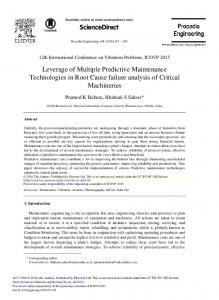
NH3~',,~ Figure 1. Structure of the carbonyl cofactor of lysyl oxidase, lysyl tyrosine quinone (Wang et al., 1996).
group recently identified as lysyltyrosine quinone (Wang et al., 1996). This contrasts with the finding that peptidyl trihydroxyphenylalanine quinone is the carbonyl cofactor in a variety of other copper amine oxidases which are mechanistically similar to LO (Janes et al., 1992). It appears likely that these carbonyl cofactors arise by autocatalytic hydroxylation and oxidation of specific peptidyl tyrosine residues requiring the copper cofactor present in newly synthesized forms of these enzymes (Matsuzaki et al., 1994). In the case of bovine aorta LO, Tyr 349 is the precursor of the quinone residue, while Lys 314 is linked to the quinone ring through its e-amino group, presumably as a result of a Michael addition (Wang et al., 1996; Fig. 1). An amine oxidase with LO activity has been identified in yeast and, like the plasma monoamine oxidase and diamine oxidase, was found to contain trihydroxyphenylalanine quinone as its carbonyl cofactor (Dove et al., 1996).
Mechanism of Action LO catalyzes primary amine oxidation through a ping pong bi ter kinetic mechanism (Williamson and Kagan, 1986). As shown in Figure 2, following initial Schiff base formation with the lysyltyrosine quinone cofactor (I --~ II), the bound substrate undergoes rate-limiting, general base-facilitated (z-proton abstraction (Williamson and Kagan, 1987). A histidine residue has been implicated as the general base in LO (Gacheru et al., 1988). Following stereospecific abstraction of the pro-S (z-proton (Shah et al., 1993), electrons migrating from the substrate carbanion reduce the carbonyl cofactor (II --~ III). Hydrolysis of the product imine intermediate releases the aldehyde product. The reduced enzyme, retaining the amino function of the substrate (IV), is r e o x i -
H20
o
o
C~
.NH RN" ~ H /
III
NH2 f v ~ H202
RN H
V
[
RCH=O
/ .......~......... IV
Figure 2. Mechanism of action of lysyl oxidase. The RNHsubstituent represents peptidyl lysine 314.
dized by molecular oxygen to produce hydrogen peroxide and ammonia, regenerating the oxidized enzyme and completing the catalytic cycle (IV -4 V --) I). While the chemical and kinetic approaches have permitted the development of the proposed mechanism of action shown in Figure 2, extension of the present level of understanding would benefit greatly from structure analysis by X-ray crystallography. This goal has yet to be achieved, and it is complicated by the limited quantities and unusual solubility properties of the purified enzyme. Efforts to express the recombinant enzyme in bulk would doubtlessly facilitate the achievement of this goal. Promising evidence of productive expression of LO in Escherichia coli has been reported (Ouzzine et al., 1996).
Substrate Specificity The sequences surrounding the substrate lysine residues within elastin and collagen differ dramatically, as do their primary and three-dimensional structures, suggesting that the specificity of LO may be flexible. Indeed, purified bovine enzyme oxidized peptidyl lysine in a variety of basic, globular proteins, including histone H1, although not in proteins with isoelectric points below pH 8 (Kagan et al., 1984). In view of its apparent preference for a net cationic charge in protein substrates in assays in vitro, it is of some interest that the oxidized
390
L.I. Smith-Mungo and H. M. Kagan
lysine residues in the telopeptidyl regions of types I, II and III collagens occur within -X-GIu-Lys-Y- sequences, while that in the N-telopeptide of the type I 0~1(I) collagen chain occurs in the -Tyr-Asp-Glu-Lys-Ser- sequence, thus vicinal to two sites of negative charge. Indeed, the efficiency of oxidation of synthetic, lysine-containing oligopeptides was markedly influenced by vicinal dicarboxylic amino acid residues (Nagan and Kagan, 1994). Thus, the -Glu-Lys- sequence within N-Acetyl-Gly4-GluI.ys-GlycAmide sequence was the most favorable substrate, while substituting -Lys- Glu- for -Glu-Lys- within these peptides decreased substrate potential considerably. Surprisingly, the -Asp-Glu-Lys- sequence within an l l-mer was not oxidized, although lysine is oxidized within this sequence in the c(1 (I) chain of type 1 collagen. This apparent anomaly may underlie the requirement that type I collagen must be pre-assembled into quarterstaggered, fibrillar arrays before it can be oxidized by 1~O (Siegel, 1974). Thus, intermolecular interactions between neighboring collagen molecules within this fibrillar structure are predicted to result in the approxmmtion of the cationic side chain of Arg 933 within the triple helix of one collagen molecule with the aspartate !3-carboxylate of the -Asp-Glu-Lys- sequence within the Ntelopeptide of the vicinal molecule. The resulting charge neutralization of this aspartic acid residue should then present effective ionic charges from the favorable -Glukys- sequence to the approaching enzyme molecule (Nagan and Kagan, 1994).
cDNA and Genomic CLoning A full length cDNA predicted to encode a protein of 409 amino acids (46 kDa) was first identified within a neonatal rat aorta cDNA ~,gt11 expression library using anti-bovine LO. The predicted molecular weight was consistent with that of the immunoprecipitated, cell-free translation product of rat aorta smooth muscle celt mRNA (Trackman et al., 1990, 1991), now known to represent the preproprotein form of the enzyme (Trackman et al., 1992). Human (Hamalainen et al., 1991; Mariani et al., 1992), chick (Wu et al., 1992) and mouse (Kenyon et al., 1991) LO cDNAs have now been cloned and sequenced, revealing the presence of both conserved and divergent sequence elements among the four predicted LO protein sequences. The greatest divergence exists within the N-terminal, 150 amino acid residue segment, containing the signal peptide and
tity between the chick and rat sequences. The propeptide domains (amino acids 22-162 in the rat sequence) are moderately conserved, exhibiting a 60-67'%, similarit), between species, while the C-terminal three-fifths of the preproprotein sequence (amino acids 163-411 in the rat) is highly conserved among the four species (90-95% identity). The presence of consensus copperbinding sequences and the recent observation that the carbonyl cofactor derives from tyrosine 349 within the rat sequence indicate that this conserved C-terminus contains the catalytic site of the enzyme (Wang et al., 1996). The LO gene has been mapped to human chromosome 5q23.3-31.2 (Hamalainen et al., 1991; Svinarich et al., 1992; Mariani et al., 1992). The mouse gene maps to chromosome 18 (Mock et al., 1992; Chang et al., 1993; Lossie et al., 1993). Genomic clones of the mouse (Contente et al., 1993) and human (Hamalainen et al., 1993; Boyd et al., 1995) LO genea have now been isolated. The genes of these two species are similar in size (14-15 kb) and exhibit similar exon/intron structure. The 15 kb human gene consists of seven exons and six introns, all of which have the consensus sequence (C/T)AG-exonGTA at their exon-intron boundaries. Beginning at the site proposed to be the major site of transcription initiation, the first exon encompasses 273 base pairs of untranslated and 631 bases of translated sequence. Thus, nearly half of the protein coding sequences are present in exon 1, while exon 7 codes for the last base of the codon for amino acid 416 and the full codon for the C-terminal amino acid, tyrosine 417. The rest of the translated sequence is encoded in exons 2-6, which range in size from 96 to 157 bases. Exon 7 also contains the termination codon and multiple potential poly A sites (Hamalainen et al., 1993). Messenger RNA species of 5.8 and 4.5 kb have been identified in rat aorta and lung (Trackman et al., 1990), while LO mRNA species of 4.8 and ,:;.8 kb occur in mouse NIH 3T3 cells (Contente et al., 1993), and at least four species were identified in Northern blots of human skin fibroblast mRNA (Hamalainen et al., 1991). This heterogeneity has been attributed to differential usage of the polyadenylation sites within the 3'-UTR of the mouse (Contente et al., 1993) and human (Boyd et al., 1995) transcripts. There also appears to be heterogeneity at the 5' end of LO transcripts, since multiple sites of transcription initiation were observed by S I nuclease mapping and primer extension analysis in human skin fibroblasts (Hamalainen et al., 1993), mouse NIH 3T3 cells (Contente et al., 100"}1
~.~A
. . . . .
£: I-_...
I_ 1 . . . .
;l-
. J
1
L~
I
Lysyl Oxidase
391
the human, mouse and rat genes, exists at -109, relative to the main transcription start site in the human gene (Hamalainen et al., 1993; see Fig. 3). In part, the preVarious lengths of the 5' flanking regions of the vailing uncertainty may be due to different numbering human (Hamalainen et al., 1993), mouse (Contente et systems of the various LO promoters, and to the fact al., 1993) and rat genes (Csiszar et al., 1996) have now that multiple transcription initiation sites may be differbeen cloned, sequenced, and subjected to computer analentially utilized. Additional consensus sequences for varyses for identification of consensus sequences for bindious transcription factors have been reported in the first ing of transcription and regulatory factors. Alignment of exon and intron of LO (Csiszar et al., 1996). the sequences upstream of the ATG codon (encoding the To date, a limited number of studies have been perAUG translation initiator) reveals a significant degree of formed to assess regulatory function associated with homology in these three species. Thus, putative binding specific regions of the LO promoter. Studies on LO prosites for Sp-l, AP-2, TFII/R, and the glucocorticoid remotet-CAT constructs transfected into human skin ceptor are completely conserved within the first kilobase fibroblasts, smooth muscle cells, and revertants of osupstream of the translation initiation site. MalT, bicoid teosarcoma cells revealed that the -30 to -895 fragment and silencer elements are also conserved (Csiszar et al., exhibited significantly greater promoter activity than did 1996). More recently, Tan et al. (1996) have described a two smaller fragments derived from this fragment. consensus sequence for interferon regulatory factor Moreover, the presence of intron 1 enhanced this probinding element (IRF-E) within the mouse promoter at moter activity two-fold, while the same domains in the position -886 to -898. Inspection of available sequences malignant c-H-ras promoter were not enhanced by this reveals that this element is completely conserved within intron in transformed osteosarcoma cells (Csiszar et al., the rat gene, while 10 of its 13 bases are conserved in the 1996). Significant luciferase reporter activity was inhuman gene. Putative binding sites for PEA3, AP-1, duced in transfected neonatal rat aorta smooth muscle MP1, as well as C C C C T C C C C boxes, and an MRE site cells by a lysyl oxidase promoter domain encompassing have been noted within the human gene (Hamalainen et 639 to +273 (the ATG codon), while a region between al., 1993). There is less agreement concerning the pres520 and -308 suppressed the stimulation of LO tranence of CAAT and TATA boxes. Five possible TATA scription induced by serum deprivation (Gacheru et al., boxes exist within the mouse gene, four of which can be 1997). Jourdan Le-Saux et al. (1997) cloned and secorrelated with CAP sites, although the upstream quenced approximately 2 kb of the Balb/C LO promoter CCAAT box lacks consensus in the flanking regions and studied its regulation in myofibroblast-like clones of (Contente et a]., 1993). A TATA box with one mismatch 3T6 fibroblasts. An area of the promoter spanning bases is present at -30, and a CCAAT box, conserved within -888 to -865 exhibited striking similarity (79%) to a 24 bp sequence conserved within the murine, rat and human COL1A1 promoters. This sequence of the LO ~:" : ~ ' :"1 ~ I I • I ATQ> 900 -800 700 600 500 -~0 300 -200 100 promoter was protected in DNAse 1 footprinting experiments and shown to bind nuclear factors from extracts of myofibroblast-like cell nuclei in gel mobility shift assays with specificity. Interestingly, this sequence is lo• m.,, A~E •~ OTF"/~ cated in a region of the promoter which was found to TATA I bicoid V Apl ~ Silencer down-regulate transcription in reporter assays, while se• CAAT B Ap2 I IRF-1 quences further upstream enhanced promoter activity. Further upstream, it was noted that a potential TGF-[3 response element [TC(X)4GCCAA] was present which Figure 3. Schematic representation of approximately 1 kb of the LO promoter upstream of the translation initiation (ATG) site. could correspond to a similar TGF-~ response element Relative locations of consensus binding sequences for transcripdescribed in the rat COL1AI (Ritzenthaler et al., 1991) tion and regulatory factors conserved in the human, rat and and mouse COL1A2 (Rossi et al., 1988) promoters. Furmouse LO genes are indicated as symbols. The human, rat and ther studies will undoubtedly be aimed at identifying mouse LO promoter sequences are numbered and aligned according to Csiszar et al. (1996). Assignment of nucleotide sespecific factors that alter LO gene expression in a multiquences as consensus binding sites is derived from the reports of tude of cell types in response to various stimuli and adCsiszar et al. (1996), Tan et al. (1996) and Hamalainen et al. dress the issue of coordinate regulation of LO and other (1993). The bold arrow indicates the major site for transcripextracellular matrix genes. tion initiation according to Hamalainen et al. (1993).
Regutatory Elements in the LO Promoter
4,
392
L.I. Smith-Mungo and H. M. Kagan
tysy[ Oxidase-Like Genes Evidence indicates that the DNA sequence encoding the connective tissue protein identified as lysyl oxidase stems from a single copy gene in each species examined thus far. A human cDNA species encoding a predicted lysyl oxidase-like (LOL) protein has been cloned and mapped to chromosome 15q24-q25. The homology of this LOL gene to LO begins at the exon 1-2 boundary in the mouse LO gene (Kenyon et al., 1993). Genomic cloning and more extensive analysis of the human LOL gene reveal that exons 2-6 are identical in size to the corresponding exons in LO (Kim et al., 1995). mRNA corresponding to the LOL gene has been detected by Northern blot analysis of adult human lung, kidney, placenta, liver, pancreas, heart and skeletal muscle (Kim et al., 1995; Kenyon et al., 1993). More recently, a novel cDNA with a predicted protein sequence which is 48% homologous to LO and LOL has been identified in senescent human fibroblasts (Saito et al., 1997). The mRNA level parallels procollagen type I,~ expression and is regulated by selected agents known to regulate LO expression (Saito et al., 1997), including TGF-]3 (Feres-Filho et al., 1995; Gacheru et al., 1997; Shanley et al., 1997) and retinoic acid (Dimaculangan et al., 1994). The existence of this series of highly related genes implies the existence of a lysyl oxidase gene family, additional members of which may yet be identified. However, the nature and catalytic function of the expressed protein product of these genes has been documented only for that coding for the known lysyl oxidase enzyme species of connective tissues.
Regulation of LO Expression LO expression is markedly responsive to a variety of physiological states, including growth, wound repair, ageing, genetic disease involving altered copper metabolism, and tumorigenesis. Not surprisingly, the expression of this enzyme is sensitively modulated by specific cytokines, growth factors and related intercellular molecular messengers.
Effects of transforming growth factor-~l (TGF-~I ) TGF-[31 is a fibrogenic cytokine known to activate the synthesis of collagen (Massague et al., 1994; Lawrence et al., 1994) and elastin (Davidson et al., 1993). The finding that TGF-]31 strongly promotes LO expression in fibroblasts of neonatal rat lung (Boak et al., 1994) and of
human embryos (Roy et al., 1996), as well as rat vascular smooth muscle cells (Gacheru et al., 1997; Shanley et al., 1997), suggests a coordinated fibrogenic response to this cytokine. It is of interest that the six- to seven-fold increase in LO mRNA induced by TGF-]31 in human lung fibroblasts is nearly completely prevented in the presence of PGE2. This prostanoid also decreases the basal level of enzyme message in these cells by 60 to 70% (Roy et al., 1996). Since PGE_, also induces the expression of cyclooxygenase 1 (Roy et al., 1996), the catalyst leading to PGE, production, these results suggest a coordinated, autocrine-like mechanism which could limit LO expression in inflammatory responses to connective tissue injury. The levels at which TGF-]31 exerts its regulatory effects on LO expression have been investigated in various cultured cell types. LO mRNA and protein levels increased in concert with the marked inhibition of proliferation of neonatal rat aorta smooth muscle cell cultures in response to this cytokine. The effect on mRNA levels was exerted predominantly at post-transcriptional levels. LO mRNA was unstable in these cultures, actively proliferating in the presence of 10% fetal bovine serum. Notably, the addition of TGF-]31 to this medium markedly stabilized the LO transcript and strongly reduced proliferative rates. Since the stimulation of mRNA expression by TGF-]31 was prevented by cycloheximide, a protein factor may be involved in the stabilization of the message (Gacheru et al., 1996). TGF-[31 also increased enzyme mRNA and protein expression in MC3T3-E1 osteoblastic cells. In this case, the proteolytic processing of the LO proenzyme appeared to be proportionately less stimulated by TGF-131, possibly accounting for the delay and slightly lower magnitude of the increase in LO enzyme activity in comparison to the peak of the increase in mRNA. This report thus raises the possibility that modulation of enzyme expression may involve altered rates of proteolytic processing of the proenzyme (Feres-Filho, 1995). This processing step did not appear to be affected on elevation of LO expression by TGF-[31 in cultures of neonatal rat lung fibroblasts, however (Boak et al., 1994).
Response to other effectors in various cell types (1) Vascular smooth muscle cells. Levels of LO mRNA and secreted enzyme activity were low in quiescent adult rat vascular smooth muscle cells and were markedly induced by PDGF, angiotensin II, or by increasing the serum concentration to 10% (Green et al., 1995). In contrast, expression of LO mRNA and secreted enzyme activity are elevated in quiescent neonatal rat vascular
Lysyl Oxidase smooth muscle cells but are markedly decreased on stimulation of proliferation by serum enrichment (Gacheru et al., 1997), suggesting age-specific mechanisms of regulation in these vascular cells. (2) Adipocyte differentiation. Levels of both LO mRNA and enzyme activity were markedly reduced within 24 h of exposure of 3T3-L1 preadipocytes to culture conditions inducing differentiation into adipocytes. Retinoic acid (RA), which suppresses the differentiation of preadipocytes to adipocytes, also blocked the differentiation-related reduction in LO gene expression, although RA had no independent stimulatory effect on LO expression in cells not exposed to differentiating conditions (Dimaculangan et al., 1994). Since RA acts by nuclear receptors which directly regulate gene expression, it is likely that primary targets of RA action in this system, seen here to prominently include LO, are genes which are regulated early in adipose conversion. (3) Osteoblastic cells. LO mRNA decreased by 70%, while that of COLIA1 was reduced more than 90% by treatment of MC3T3-E1 cells with basic fibroblast growth factor (bFGF). This growth factor stimulates osteogenic cell proliferation while inhibiting production and deposition of the collagen matrix of bone. At least 50% of the reduction in enzyme expression was accounted for at the post-trancriptional level. Thus, LO mRNA was quite stable in control MC3T3-E1 cells but was rapidly turned over in the presence of bFGF (Feres-Filho et al., 1996). It is of interest in this regard that TGF-[3 decreases proliferation and stimulates LO expression by stabilizing LO mRNA in neonatal vascular smooth muscle cells, while bFGF stimulates proliferation and reduces LO expression in part by destabilizing the transcript species in these osteogenic cells, raising the possibility that there may be common, coordinating mechanisms of control of the cell cycle and of LO expression in these cells. (4) Endothelial cells. Genes for human laminin B1 chain, H(÷)-ATP synthase coupling factor 6, LO, myosin light chain kinase, and interleukin-8 receptor were upregulated by shear stress (15 dynes/cm2) in cultured human umbilical vein. Approximately 4% of the total number of mRNAs expressed were responsive to this perturbation by shear stress (Ando et al., 1996), indicating that LO is among a select few of the genes responsive to this perturbation.
LO and Metal Ion Metabolism As previously reviewed, severe copper deficiency results in decreased crosslinking of connective tissue pro-
393
teins, consistent with decreased function of LO (Kagan, 1986). While enzyme activity levels were decreased in the skin of weanling rats fed a copper deficient diet, the basal, steady-state levels of LO specific mRNA or immunodetectable LO protein were not significantly reduced (Rucker et al., 1996). These results suggest both that the biosynthesis of the enzyme is not markedly affected by copper deficient diets and that the increasing percentage of copper-deficient, catalytically compromised enzyme molecules presumed to accumulate during this dietary treatment remain relatively stable. Notably, copper-deficient diets significantly reduced cardiac LO activity and induced cardiac pathology in male but not in female rats (Werman et al., 1995). LO expression, assessed at the mRNA, protein and activity levels, was markedly repressed in a population of cadmium resistant fibroblasts, compared to the cadmium-sensitive, parental population. The resistant phenotype exhibited markedly elevated levels of metallothionein and glutathione, each of which has the potential to scavenge copper. The partial restoration of enzyme expression by the addition of CuC12 suggested that the reduction of enzyme expression partially reflected a disturbance in intracellular copper metabolism in these cells (Li et al., 1995).
LO and Human Disease Cutis laxa, reported to have both autosomal dominant and recessive patterns of inheritance, is characterized by hyperextensible skin and a marked deficiency of skin elastic fibers. As previously reviewed, deficient LO expression has been noted in affected human skin fibroblasts derived from X-linked cases of cutis laxa which also exhibit abnormalities of copper metabolism (Kagan, 1986). A more recent report notes two phenotypically similar patients with primary cutis laxa associated with a deficiency of LO but in whom copper metabolism appears normal and who exhibit an autosoreal recessive inheritance of the LO gene (Khakoo et al., 1997). One report indicates that steady-state levels of LO mRNAs did not appear to differ from controls in fibroblasts of cutis laxa patients (Yeowell et al., 1994). Clearly, further clarification of the relationship of LO to the pathology of this disease is required. Menkes syndrome is an X-linked, recessively inherited disorder characterized in part by abnormal copper transport, cellular copper sequestration, and defective crosslinking of collagen and elastin. This disease appears attributable to a mutation in the gene coding for a cop-
394
L.I. Smith-Mungo and H. M. Kagan
per- transporting ATPase (Vulpe et al., 1993). In part, the phenotypic symptoms are likely to be due to indirect effects of altered copper metabolism. Reports previously reviewed note that LO activity is markedly reduced in Menke's fibroblasts (Kagan, 1986). More recently, decreased steady-state levels of LO mRNA (Gacheru et al. 1993; Kemppainen et al., 1996) and elastin mRNA (Gacheru et al., 1993) have been described, while enhanced levels of type I (Gacheru et al., 1993) and type lII (Kemppainen et al., 1996) procollagen mRNA have also been observed in Menke's fibroblasts. Thus, the Menkes syndrome may involve abnormalities in the expression of several genes coding for connective tissue proteins. There are several examples indicating that LO expression is significantly elevated in fibrosis induced by exposure to certain toxic environmental agents or as sequelae of a variety of disease states (Kagan et al., 1986). Fibrosis is a prominent symptom of scleroderma, in which immunohistochemical techniques have indicated that LO is markedly elevated in affected tissues of this disease (Chanoki et al., 1995). Elevations of LO mRNA and protein species were identified in myofibroblast-like cells derived from the liver granules in a murine model of schistosomiasis (Sommer et al., 1993). The possibility that LO levels may prove to be subject to chemotherapeutic control provides at least one potential pathway to the interruption of the fibrotic process in such disease states. In contrast to its elevation in non-malignant fibrotic conditions, LO expression at the protein (Kuivaniemi et al., 1986) and transcriptional (Hamalainen et al., 1995) levels is markedly down-regulated in malignant cell lines in culture. Asssessment of LO expression at the protein and mRNA levels by immtinohistochemistry and in situ hybridization, respectively, in breast carcinoma tissue revealed that maximal expression of LO and its type I collagen substrate was observed in myofibroblasts and myoepithelial cells around in situ tumors and in the fibrotic deposits facing the invasion front of infiltrating tumors. In contrast, there was a lack of LO, although collagen continued to be synthesized, in the loose stroma accompanying invading tumors, consistent with the apparent deficiency of stable, crosslinked fibers in the loose stromal tissue (Peyrol et al., 1997). The results of this study suggested synchrony between the loss of myofibroblast and epithelial cell differentiation and the loss of LO expression. It was further noted that the early development of the crosslinked matrix surrounding ductal breast carcinoma may represent a defense mechanism, whereas the subsequent lack of LO in the late stromal reaction would favor tumor dispersion (Peyrol et al., 1997).
LO in Development and in Wound Repair Within the developing chick embryo aorta, the mRNA species encoding LO increased between days 8 and 16 in reasonable concordance with those of its tropoelastin and type I collagen substrates, while that of LO then decreased by day 16 of embryonic age (Wu et al., 1992). An apparently similar pattern was observed in the developing human amnion, in which the high levels of LO mRNA, protein, and activity seen at 12-14 weeks gestation in human amnion declined ubruptly during the second trimester to minimum values at 20-24 weeks gestation, which then persisted to term (Casey and MacDonald, 1997). The seemingly coordinated increase in the expression of LO and its substrates in embryogenesis contrasts with events during wound healing in rat skin, in which the peak of the increase in LO mRNA following skin injury preceded that of type III collagen mRNA by several days. A similar discontinuity was observed in the early and marked increase in LO which preceded the increase in collagen protein synthesis in cadmium-exposed rat lungs (Sampson et al., 1984). Quagliano et al. (1993) determined the levels of saline-soluble and of matrix-associated, urea-soluble LO activity in aorta, lung and skin of developing and aging rats. The profiles of activity varied between the differently soluble enzyme fractions, with the tissue examined, and with the baseline used - that is, activity per gram of tissue or per cell actively producing collagen and/or elastin - confounding the generalities that might be drawn from the study. Nevertheless, the data suggested that crosslinking might be markedly increased in aging skin, slightly increased in aging lungs and reduced in aging aortas. Zhu et al. (1993) reported the reduced insolubilization of newly synthesized etastin in the ductus arteriosus in comparison to the aorta and pulmonary artery during fetal lamb development, although, here, again, corresponding differences in LO activity were not seen. The mechanisms regulating the differing temporal realtionships between the expression of LO and its substrates remain to be understood.
Novet Biological Rotes of LO It has traditionally been assumed that the role of LO is restricted to the oxidation of peptidyl lysine in extracellular collagen and elastin substrates, resulting in the deposition of insoluble fibrous deposits of these proteins. The report noting the potent chemotactic response of peripheral human blood mononuclear cells to purified
Lysyl Oxidase calf aorta LO (Lazarus et al., 1995) and the recent evidence that LO may act as a repressor of the oncogenic activity of mutant r a s species (Kenyon et al., 1991) indicate that this assumption should be reassessed. The chemotactic effect of LO, measured in Boyden chambers in v i t r o , was dependent on the catalytic activity of the enzyme. The molecular basis of this unusual effect of the enzyme has yet to be detailed. More than a decade ago, it was reported that a variety of malignantly transformed human cells exhibited very low levels of LO activity in culture when compared to their non-transformed counterparts. The enzyme deficiency was due to reduced levels of synthesis rather than to impaired secretion of the enzyme (Kuivaniemi et al., 1986), and it was subsequently shown to reflect low quantities of mRNA and low levels of transcription (Hamalainen et al., 1995). A molecular link between LO and transformation stemmed from the discovery of the r a s recision gene (rrg), identified as a cDNA species that was dramatically reduced upon transformation of mouse NIH-3T3 cells with LTR c - H a - r a s and re-expressed upon interferon-mediated reversion of the transformants. The apparent r a s recision function of r r g was demonstrated by the observation that stable transfection of a non-tumorigenic, revertant cell line (PR4) derived from the r a s transformants with a partial r r g cDNA antisense expression vector resulted in the reappearance of tumorigenic, transformed cells exhibiting decreased levels of the rrg. PR4 cells transformed with the sense counterpart did not appear significantly altered (Contente at al., 1990). A direct correlation was subsequently demonstrated between r r g mRNA expression and LO activity in the culture media of the mouse NIH 3T3 cells and the derived transformed and revertant cell lines. Moreover, the cDNA sequences of mouse r r g and rat LO proved to be identical, indicating that r r g , a modulator of r a s expression, encodes LO (Kenyon et al., 1991). I n t o t o , these studies suggest that LO has anti-oncogenic activity (Contente et al., 1990). Csiszar et al. (1996) have observed reduced transcription of LO in r a s transformed osteoblast cells, and implicated a regulatory element located between -796 and 274 of the LO promoter in this down-regulation. Further support for this possible role of LO has been provided by Tan et al. (1996), who demonstrated by differential display that LO is down-regulated in embryonic fibroblasts deficient for the anti-oncogenic transcriptional activator, interferon regulatory factor 1 (IRF-1). An element capable of binding recombinant IRF-1 was identified in the mouse LO promoter, and promoter fragments of LO linked to a reporter gene were activated
395
and repressed by cotransfected cDNA expression vectors containing IRF-1 and IRF-2, respectively. Additionally, cotransfection of LO cDNA into ras transformed IRF-1deficient embryonic fibroblasts inhibited colony formation three-fold. I n t o t o , these results implicate LO as a target gene for IRF-1 in tumor suppression (Tan et al., 1996). The mechanism by which LO modulates cellular transformation by r a s has yet to be determined, although recent studies suggest a role for LO in the condensation of nuclear chromatin. Thus, r a s transformed NIH3T3 cells exhibit increasingly condensed regions of nuclear chromatin (Mello and Russo, 1990), while the chromatin of interferon-induced, non-tumorigenic revertants of these transformants, as well as that of revertants transfected with an LO sense cDNA construct, was packed significantly less tightly. Notably, the extent of chromatin packing in revertants transfected with LO antisense DNA resembled that of the r a s transformed cells. It has been hypothesized that changes in the supraorganization of chromatin may be associated with limited transcription of the genome necessary to maintain the transformed phenotype. (Mello and Russo, 1990; Mello et al., 1995). Among the considerations raised by the r a s suppressor effect of LO is the intriguing possibility that LO may function within the cell. Consistent with that possibility, Wakasaki and Ooshima (1990)localized LO within fibroblasts, chondrocytes, smooth muscle cells and in a variety of non-fibroblastic cells, including endothelial, basal, biliary epithelial and glomerular epithelial cells. Interestingly, immunodeposits were found in association with fine, filamentous structures in the cytoplasm of cultured fibroblasts consistent with cytoskeletal protein. Since the molecular weights of these intracellular proteins were not assessed, it is not known whether they represented proenzyme and/or mature forms of the enzyme. The apparent LO-dependent alteration in chromatin structure noted by Mello et al. (1995) raises the possibility that LO may directly or indirectly exert effects on nuclear components. Indeed, Li et al. (1997) have recently described the localization of LO by immunocytochemistry within nuclei of rat vascular smooth muscle cells and 3T3 fibroblasts. Western blot analyses of extracts of nuclei isolated from the rat vascular smooth muscle cells revealed the presence of immunoreactive 32 kDa protein, consistent with the presence of the mature catalyst. Moreover, lysinonorleucine, the adduct formed between the peptidyl aldehyde product of LO action and the e-amino group of peptidyl lysine, was detected in a hydrolysate of protein extracted from nuclei purified from
396
L.I. Smith-Mungo and H. M. Kagan
these vascular smooth muscle cells. The formation of this crosslink was largely prevented by culturing these cells in the presence of [3-aminopropionitrile, an irreversible inhibitor of LO (Li et al., 1997). The identity of the nuclear substrate of LO has yet to be determined, as have the functional consequences of its intranuclear activity. It should be noted in this regard that the oxidative deamination of peptidyl lysine results in the loss of the positive charge of the side chain. This chemical change is analogous to that resulting from the enzyme-catalyzed N-acetylation of lysine side chains of bistones. The subsequent deacetytation of these residues by histone deacetylase has been correlated with the changes in transcription of nucleosomal D N A (Wollfe, 1996). In toto, these reports suggest that LO has unusual and important roles to play in cellular homeostasis.
Acknowtedgements The original research of the authors' laboratory cited in this review was supported by National Institutes of Health grants HL 13262 and R37 AR 18880.
References Ando, J., Tsuboi, H., Korenaga, R., Takahashi, K., Kosaki, K., Isshiki, M., Tojo, T., Takada, Y. and Kamiya, A.: Differential display and cloning of shear stress-responsive messenger RNAs in human endothelial cells. Biochem. Biophys. Res. Commun. 225: 347-351, 1996. Boak, A.M., Roy, R., Berk, J., Taylor, L., Polgar, E, Goldstein, R H. and Kagan, H.M.: Regulation of lysyl oxidase expression in lung fibroblasts by transforming growth factor-beta 1 and prostaglandin E2. Am. J. Resp. Cell Mol. Biol. 11: 751-755, 1994. Boy& C.D., Mariani, T. J., Kim, Y. and Csiszar, K.: The size heterogeneity of human lysyl oxidase mRNA is due to alternate polyadenylation site and not alternate exon usage. Mol. Biol. Reports 21: 95-103, 1995. Casey, M i . and MacDonald, P.C.: Lysyl oxidase (ras recision gene) expression in human amnion: ontogeny and cellular localization. J. Clin. Endocrin. Metab. 82: 167-172, 1997. Chang, Y.S., Svinarich, D.M., Yang, T.P. and Krawetz, S.A.: The mouse lysyl oxidase gene (Lox) resides on chromosome 18. Cytogenet. Cell Genet. 63: 47-49, 1993. Chanoki, M., lshii, M., Kobayashi, H., Fushida, H., Yashiro, N., Hamada, T. and Ooshima, A.: Increased expression of lysyl oxidase in skin with scleroderma. Br. J. Dermatol. 133: 710-715, 1995. Contente, S., Csiszar, K., Kenyon, K. and Friedman, R.M.: Structure of the mouse lysyl oxidase gene. Genomics 16: 395-400, 1993. Contente, S., Kenyon, K., Rimoldi, D. and Friedman, R.M.: Expression of gene rrg is associated with reversion of NIH 3T3 transformed by LTR-c-H-ras. Science 249: 796-798, 1990. Croushaw, A.D., Fothergill-Gilmore, L.A. and Hulmes, D.J.S.: The proteolytic prosessing of the precursor of LO. Biochem. J. ~06: 279-284, 1995.
Csiszar, K., Entersz, I., Trackman, EC., Samid, D. and Boyd, C.D.: Functional analysis of the promoter and first intron of the human lysyl oxidase gene. Mol. Biol. Reports 23: 97-108, 1996. Davidson, J.M., Zoia, O. and Liu, J.M.: Modulation of transforming growth factor-J31 stimulated elastin and collagen production and proliferation in porcine vascular smooth muscle cells and skin fibroblasts by basic fibroblast growth factor-c~ and insulin-like growth factor-I. J. Cell. Phys. 155: 149-156, 1993. Dimaculangan, D.D., Chawla, A., Boak, A., Kagan, H.M. and Lazar, M.A.: Retinoic acid prevents downregulation of ras recision gene/lysyl oxidase early in adipocyte differentiation. Differentiation 58: 47-52, 1994. Dove, J.E., Smith, A.J.,. Kuchar, J., Brown, D.E., Dooley, D.M. and Klinman, J.P.: Identification of the quinone cofactor in a lysyl oxidase from Pichia pastoris. FEBS Lett. 398:231-234, 1996. Feres-Filho, E.J., Choi, Y.J., Han, X., Takala, T.E. and Trackman, EC.: Pre- and post-translational regulation of lysyl oxidase by transforming growth factor-beta 1 in osteoblastic MC3T3-E1 cells. J. Biol. Chem. 270: 30797-30803, 1995. Feres-Filho, E.., Menassa, G.B. and Trackman, P.C: Regulation of lysyl oxidase by basic fibroblast growth factor in osteoblastic MC3T3-E1 cells..I. Biol. Chem. 271: 6411-6416, 1996. Gacheru, S., McGee, C., Uriu-Hare, J.Y., Kosonen, T., Packman, S., Tinker, D., Krawetz, S.A., Reiser, K., Keen, CL. and Rucker, R.B.: Expression and accumulation of LO, elastin, and type I procollagen in human Menkes and mottled mouse fibroblasts. Arch. Biochem. Biophys. 301: 325-329, 1993. Gacheru, S.N., Thomas, K.M., Murray, S.A., Csiszar, K., Smith-Mungo, L.I. and Kagan, H. M.: Transcriptional and post-transcriptional control of lysyl oxidase expression in vascular smooth muscle cells: effects of TGF-beta 1 and serum deprivation. J. Cell. Biochem. 65: 395-407, 1997. Gacheru, S.N., Trackman, P.C. and Kagan, H.M.: Evidence for a functional role for histidine in lysyl oxidase catalysis. J. Biol. Chem. 263: 16704-16708, 1988. Gacheru, S.N., Trackman, EC., Shah, M.A., O'Gara, C.Y., Spacciapoli, E, Greenaway, ET. and Kagan, H.M.: Structural and catalytic properties of copper in lysyl oxidase. J. Biol. Chem 265:19022-19027, 1990. Green, R.S., I,ieb, M.E., Weintraub, A.S., Gacheru, S.N., Rosenfield, C.L., Shah, S., Kagan, H.M. and Taubman, M.B.: Identification of lysyl oxidase and other platelet-derived growth factor-inducible genes in vascular smooth muscle cells by differential screening. Lab. Invest. 73: 476-482, 1995. Hamalainen, E.-R., Jones, T.A., Sheer, D., Taskinen, K., Pihlajaniemi, T. and Kivirikko, K.: Molecular cloning of human lysyl oxidase and assignment of the gene to chromosome 5q23.3-31.2. Genomics 1 I: 508-516, 1991. Hamalainen, E.R., Kemppainen, R., Kuivaniemi, H., Tromp, G., Vaheri, A., Pihlajaniemi, T. and Kivirikko, K.I.: Quantitative polymerase chain reaction of lysyl oxidase mRNA in malignantly transformed human cell lines demonstrates that their low lysyl oxidase activity is due to low quantities of its mRNA and low levels of transcription of the respective gene../. Biol. Chem. 270: 21590-21593, 1995. Hamalainen, E.-R., Kemppainen, R., Pihlajaniemi, "E and Kivirikko, K.: Structure of the human lysyl oxidase gene. Genomics 17: 544-548, 1993. Janes, S.M., Palcic, M.M., Scaman, C.H., Smith, A.,I., Brown, D.E., DooleB D.M., Mure, M. and Klinman, J.P.: Identifica-
Lysyl Oxidase tion of topaquinone and its consensus sequence in copper amine oxidases. Biochemistry 31: 12147-12147, 1992. Jourdan-Le Saux, C., Gleyzal, C., Garnier, J.M., Peraldi, M., Sommer, P. and Grimaud, J.A.: Lysyl oxidase cDNA of myofibroblast from mouse fibrotic liver. Biochem. Biophys. Res. Commun. 199: 587-592, 1994. Jourdan-LeSaux, C., Gleyzal, C., Raccurt, M. and Sommer, P.: Functional analysis of the lysyl oxidase promoter in myofibroblast-like clones of 3T6 fibroblast. ,1. Cell. Biochem. 64328-341, 1997. Kagan, H.M.: Characterization and regulation of LO. In: Biology of the Extracellular Matrix, ed. by Mecham, R.P., Vol h Regulation of Matrix Accumulation, Academic Press, Orlando, FL, 1986, pp: 321-398. Kagan, H.M., Redd~, V.B., Narasimhan, N. and Csizsar, K.: Catalytic properties and structural components of LO. In: The Molecular Biology and Pathology of Elastic Tissues,
Wiley, Chichester (Ciba Foundation Symposium 192), 1995a, pp. 100-121. Kagan, H.M., Reddy, V.B., Panchenko, M., Thomas, K., Narasimhan, N., Boak, A.M. and Gacheru, S.N.: Expression of lysyl oxidase from cDNA constructs in mammalian cells: The propeptide region is not essential to the folding and secretion of the functional enzyme. J. Cell. Biochem. 58: 1-10, 1995b. Kagan, H.M. and Trackman, EC.: Properties and function of LO. Am. J. Resp. Cell Mol. Biol. 5: 206-210, 1991. Kagan, H.M., Williams, M.A., Williamson, P.R. and Anderson, J.M.- Influence of sequence and charge on the specificity of lysyl oxidase toward protein and synthetic peptide substrates. J. Biol. Chem. 259:11203-11207, 1984. Kemppainen, R., Hamalainen, E.R., Kuivaniemi, H., Tromp, G., Pihlajaniemi, T. and Kivirikko, K.I.: Expression of mRNAs for lysyl oxidase and type III procollagen in cultured fibroblasts from patients with the Menkes and occipital horn syndromes as determined by quantitative polymerase chain reaction. Arch. Biochem. Biophys. 328:101-106, 1996. Kenyon, K., Contente, S., Trackman, P.C., Tang, J., Kagan, H. and Friedman, R.M.: A suppressor of the phenotypic expression of ras encodes lysyl oxidase. Science 253: 802, 1991. Kenyon, K., Modi, W.S., Contente, S. and Friedman, R.M.: A novel human cDNA with a predicted protein similar to lysyl oxidase maps to chromosome 15q24-q25.J. Biol. Chem. 268: 18435-18437, 1993. Khakoo, A., Thomas, R., Trompeter, R., Duffy, P., Price, R. and Pope, EM.: Congenital cutis laxa and lysyl oxidase deficiency. Clin. Genet. 51: 109-114, 1997. Kim, Y., Boyd, C.D. and Csiszar, K.: A new gene with sequence and structural similarity to the gene encoding human lysyl oxidase. J. Biol. Chem. 270: 7176-7182, 1995. Kuivaniemi, H., Korhonen, R.M., Vaheri, A. and Kivirikko, K.I.: Deficient production of lysyl oxidase in cultures of malignantly transformed human cells. FEBS Lett. 195: 261-264, 1986. Lawrence, R., Hartmann, D.J. and Sonenshein, G.E.: Transforming growth factor-[B1 stimulates type V collagen expression in bovine vascular smooth muscle cells. J. Biol. Chem. 269: 9603-9609, 1994. Lazarus, H.M., Cruikshank, W.W., Narasimhan, N., Kagan, H.M. and Center, D M.: Induction of human monocyte motility by LO. Matrix Biol. 14:727-731, 1995. Li, W., Chou, I.N., Boak, A. and Kagan, H.M.: Downregulation of lysyl oxidase in cadmium-resistant fibroblasts. Am. J. Respir. Cell Molec. Biol. 1.3: 418-425, 1995.
397
Li, W., Nellaiappan, K., Strassmaier, T., Graham, L., Thomas, K.M. and Kagan, H.M.: Localization and activity of lysyl oxidase within nuclei of fibrogenic cells. Proc. Natl. Acad. Sci. USA, 94: 12817-12822, 1997. Lossie, A.C., Buckwalter, M.S. and Camper, S.A.: Lysyl oxidase (Lox) maps between Grl-1 and Adrb-2 on mouse chromosome 18. Mamm. Genome 4: 177-178, 1993. Mariani, T.J., Trackman, EC., Kagan, H.M., EddB R.L., Shows, T.B., Boyd, C.D. and Deak, S.B.: The complete derived amino acid sequence of human lysyl oxidase and assignment of the gene to chromosome 5 (extensive sequence homology with the murine ras recision gene). Matrix 12: 242-248, 1992. Massagu~, J., Attisano, L. and Wrana, J i . : The TGF-]3 family and its composite receptors. Trends Cell Biol. 4: 172-177, 1994. Matsuzaki, R., Fukui, T., Sato, H., Ozaki, Y. and Tanizawa, K.: Generation of the topa quinone cofactor in bacterial monoamine oxidase by cupric ion-dependent autooxidation of a specific tyrosyl residue. FEBS l,ett. ,351: 360-364, 1994. Mello, M.I,., Contente, S., Vidal, B.C., Planding, W. and Schenck, U.: Modulation of ras transformation affecting chromatin supraorganization as assessed by image analysis. Exp. Cell Res. 220: 374-382, 1995. Mello, M.L.S. and Russo, J.: Image analysis of Feulgen-stained c-H-ras-transformed NIH/3T3 cells. Biochem. Cell Biol. 68: 1026-1031, 1990. Mock, B.A., Contente, S., Kenyon, K., Friedman, R.M. and Kozak C.A.: The gene for lysyl oxidase maps to mouse chromosome 18. Genomics 14: 822-823, 1992. Nagan, N. and Kagan, H.M.: Modulation of lysyl oxidase activity toward peptidyl lysine by vicinal dicarboxylic amino acid residues. Implications for collagen crosslinking..1. Biol. Chem. 269: 22366-22371, 1994. Ouzzine, M., Boyd, A. and Hulmes, D.J.: Expression of active, human lysyl oxidase in Escherichia coli. FEBS Lett. 399: 215-219, 1996 Panchenko M.V., Stetler-Stevenson, W.G., Trubetskoy, O.V., Gacheru, S.N. and Kagan, H.M.: Metalloprnteinase activity secreted by fibrogenic cells in the processing of prolysyl nxidase. Potential role of procollagen C-proteinase. J. Biol. Chem. 271: 7113-7119, 1996. Peyrol, S., Raccurt, M., Gerard, E, Gleyzal, C. Grimaud, J.A. and Sommer, E: Lysyl oxidase gene expression in the stromal reaction to in situ and invasive ductal breast carcinoma. Am. J. Path. 150: 497-507, 1997. Pinnell, S.R. and Martin, G.R.: The cross-linking of collagen and elastin: enzymatic conversion of lysine in peptide linkage to a-aminnadipic-d-semialdehyde (allysine) by an extract from bone. Proc. Natl. Acad. Sci. USA 61: 708-714, 1968. Quaglino, D., Fornieri, C., Nanney, L.B. and l)avidson, J.M.: Extracellular matrix modifications in rat tissues of different ages. Correlations between elastin and collagen type I mRNA expression and lysyl-oxidase activity. Matrix 1.3: 481-490, 1993. Ritzenthaler, J.D., Goldstein, R.H., Fine, A., I,ichtler, A., Rowe, D.W. and Smith, B.D.: Transforming growth factor B activation elements in the distal promoter regions of the rat a I type I collagen gene. Biochem. J. 280: 157-162, 1991. Rossi, P., Karsenty, G., Roberts, A.B., Roche, N.S., Sporn, M.B. and de Crombrugghe, B.: A nuclear factor 1 binding site mediates the transcriptional activation of a type I collagen promoter by transforming growth factor B. Cell 52: 405-414, 1988.
398
L.I. Smith-Mungo and H. M. Kagan
Roy, R., Polgar, P., Wang, Y., Goldstein, R.H., Taylor, L. and Kagan, H.M.: Regulation of lysyl oxidase and cyclooxygenase expression in human lung fibroblasts: interactions among TGF-[3, IL-liB, and prostaglandin E. J. Cell. Biochem. 62: 411-417, 1996. Rucker, R.B., Romero-Chapman, N., Wong, T., Lee, J., Steinberg, EM., McGee, C., Clegg, M.S., Reiser, K., Kosonen, T., Uriu-Hare, J.Y., Murphy, J. and Keen, C.L.: Modulation of lysyl oxidase by dietary copper in rats. J. Nutr. 126: 51-60, 1996. Saito, H., Papaconstantinou, J., Sat(), H. and Goldstein, S.: Regulation of a novel gene encoding a lysyl oxidase-related protein in cellular adhesion and senescence. J. Biol. Chem. 272: 8157- 8160, 1997. Sampson, C.E., Chichester, C.O., Hayes, J.A. and Kagan, H.M.: Alterations in collagen biosynthesis and in metallothionein in lungs of rats acutely or repeatedly exposed to CdCl2 aerosol. Am. Ret: Resp. Dis. 129: 619-624, 1984. Shah, M.A., Scaman, C.H., Palcic, M.M. and Kagan, H.M.: Kinetics and stereospecificity of the lysyl oxidase reaction. J. Biol. Chem. 268:11573-11579, 1993. Shanley, C.J., Gharaee-Kermani, M., Sarkar, R., Welling, T.H., Kriegel, A., Ford, J.W., Stanley, J.C. and Phan, S.H.: Transforming growth factor-beta 1 increases lysyl oxidase enzyme activity and mRNA in rat aortic smooth muscle cells..1. Vasc. Surg. 25: 446-452, 1997. Siegel, R.C.: Biosynthesis of collagen crosslinks: increased activity of purified lysyl oxidase with reconstituted collagen fibrils. Proc. Natl. Acad. Sci. USA 71: 4826-4830, 1974. Sommer, E, Gleyzal, C., Raccurt, M., Delbourg, M., Serrar, M., Joazeiro, E, Peyrol, S., Kagan, H., Trackman, P.C. and Grimaud, J.A.: Transient expression of lysyl oxidase by liver myofibroblasts in murine schistosomiasis. Lab. Invest. 69: 460-470, 1993. Svinarich, D.M., Twomey, T.A., Macauley, S.P., Krebs, C.J., Yang, T.P. and Krawetz, S.A.: Characterization of the human lysyl oxidase gene locus. J. Biol. Chem. 267: 14382-14387, 1992. Tan, R.S.-P., Taniguchi, T. and Harada, H.: Identification of the lysyl oxidase gene as target of the antioncogenic transcription factor, IRF-1, and its possible role in tumor suppression. Canc. Res. 56: 2417-2421, 1996. Trackman, EC., Bedell-Hogan, D., Tang, J. and Kagan, H.M.: Post-translational glycosylation and proteolytic processing of a lysyl oxidase precursor..1- Biol. Chem. 267: 8666-8671, 1992. Trackman, P.C, Pratt, A.M., Wolanski, A., Tang, S.-S., Offner, G.D., Troxler, R. and Kagan, H.: Cloning of rat aorta lysyl
oxidase cDNA: complete codons and predicted amino acid sequence. Biochemistry 29: 4863-4870, 1990. Trackman, EC., Pratt, A.M., Wolanski, A., Tang, S.-S., Offner, G.D., Troxler, R. and Kagan, H.: Chining of rat aorta lysyl oxidase cDNA: complete codons and predicted amino acid sequence. Biochemistry 30: 8282, 1991. Vulpe, C., Levinson, B., Whitney, S., Packman, S. and Gitschier, J.: Isolation of a candidate gene for Menkes disease and evidence that it encodes a copper-transporting ATPase. Nat. Genet..3: 7-13, 1993. Wakasaki, H. and Ooshima, A.: lmmunohistochemical local ization of lysyl oxidase with monoclonal antibodies. Lab. Invest. 63: 377-384, 1990. Wang, S.X., Mute, M., Medzihradszky, K.E, Burlingame, A i . , Brown, D., Dooley, D.M., Smith, A.J., Kagan, H.M. and Klinman, J.P.: A crosslinked cofactor in LO: redox function for amino acid side chains. Science 273: 1078-1064, 1996. Werman, M.J., Barat, E. and Bhathena, S.J.: Gender, dietary copper and carbohydrate source influence cardiac collagen and lysyl oxidase in weanling rats..I. Nutr. 125: 857-863, 1995. Williamson, P.R. and Kagan H.M.: Reaction pathway of bovine aortic LO.J. Biol. Chem. 261: 9477-9482, 1986. Williamson, ER. and Kagan, H.M.: a-Proton abstraction and carbanion formation in the mechanism of action of LO../. Biol. Chem. 262: 8196-8201, 1987. Wolffe, A.P.: Histone deacetylase- a regulator of transcription. Science 272: 371-372, 1996. Wu, Y., Rich, C.B., Lincecum, J., Trackman, EC., Kagan, H.M. and Foster, J.A.: Characterization and developmental expres sion of chick aortic LO..1. Biol. Chem. 267: 24199-24206, 1992. Yeowell, H.N., Marshall, M.K., Walker, L.C., Ha, V. and Pinnell, S.R.: Regulation of lysyl oxidase mRNA in dermal fibroblasts from normal donors and patients with inherited connective tissue disorders. Arch. Biochem. Biophys. 308: 299-305, 1994. Zhu, L., Dagher, E., Johnson, D.J., Bedell-Hogan, D., Keeley, EW., Kagan, H.M. and Rabinovitch, M.: A developmentally regulated program restricting insolubilization of elastin and formation of laminae in the fetal lamb ductus arteriosus. Lab. Invest. 68:321-331, 1993. Dr. Herbert M. Kagan, Department of Biochemistry, Boston University School of Medicine, Boston, MA 02118 USA. Received November 4, 1997