Jun 8, 2017 - Typically, for a specified biomarker, a threshold is chosen and HIV positive individuals with a measured optical density (OD) be-.
Quantifying the recency of HIV infection using multiple longitudinal biomarkers Loumpiana Koulai, Anne Presanis, Gary Murphy, Barbara Suligoi and Daniela De Angelis
arXiv:1706.02508v1 [stat.AP] 8 Jun 2017
June 9, 2017 Knowledge of the time at which an HIV-infected individual seroconverts, when the immune system starts responding to HIV infection, plays a vital role in the design and implementation of interventions to reduce the impact of the HIV epidemic. A number of biomarkers have been developed to distinguish between recent and long-term HIV infection, based on the antibody response to HIV. To quantify the recency of infection at an individual level, we propose characterising the growth of such biomarkers from observations from a panel of individuals with known seroconversion time, using Bayesian mixed effect models. We combine this knowledge of the growth patterns with observations from a newly diagnosed individual, to estimate the probability seroconversion occurred in the X months prior to diagnosis. We explore, through a simulation study, the characteristics of different biomarkers that affect our ability to estimate recency, such as the growth rate. In particular, we find that predictive ability is improved by using joint models of two biomarkers, accounting for their correlation, rather than univariate models of single biomarkers.
1
Introduction
Following infection with the Human Immunodeficiency Virus (HIV), the immune system responds by producing anti-HIV antibodies of different types at different stages from infection [5], culminating in what is known as seroconversion, i.e the time at which antibodies are detectable in blood serum. CD4 counts and viral load traditionally have been used as prognostic biomarkers of HIV progression [18, 22] but have been less successful for estimating time since infection, due to their non-monotonic behaviour and the difficulty of observing individuals at the early stages of infection . In recent years, focussing instead on the antibody response, a number of serological assays, able to detect different aspects of this diverse response, have been developed with the goal of distinguishing recent from long-standing infections. Typically, for a specified biomarker, a threshold is chosen and HIV positive individuals with a measured optical density (OD) below the threshold are classified as recently infected (see [11, 12, 13, 17, 26, 31] and references therein). This classification has been used to estimate HIV incidence at population level [10, 19]. At an individual level, however, this dichotomization does not allow a clear quantification of the recency of infection. The type of statement that can be made on recency can only be on average, based on knowledge of the mean time taken to cross the chosen threshold from seroconversion. The key question of interest is whether it is possible to make probabilistic statements at individual-level about the recency of HIV infection. Can biomarker measurements for a newly diagnosed individual, combined with knowledge of the natural evolution of the biomarker, be 1
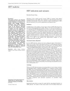
used to infer the probability, PX , that an individual has seroconverted in the X months prior to diagnosis? Antibody-response biomarkers increase monotonically and approach a plateau over time. An example is the Architect Avidity [31], whose growth pattern is shown in Figure 1. These data are routinely collected from HIV-positive patients attending clinics in Italy. These patients have known (or well-estimated) seroconversion times, and at each clinic visit following diagnosis, one or more biomarkers are measured. For such a panel of individuals, the growth pattern of each biomarker observed can be estimated. Figure 1 shows each individual’s OD values of Architect Avidity against time since seroconversion, with the estimated mean growth curve in blue. Given such growth curves and observations on a newly diagnosed individual, how well can we estimate the seroconversion probability PX ?
1
log(Architect Avidity)
0
−1
● ● ● ● ● ● ● ● ● ● ● ● ●● ● ● ● ● ●●● ● ● ● ● ● ● ●● ●●●● ● ● ● ● ● ● ●● ● ● ● ● ● ●● ● ● ● ●● ● ● ● ● ● ● ● ● ● ● ● ● ● ● ●● ●● ●● ● ● ●● ● ● ● ● ● ● ● ● ● ● ● ● ● ●● ● ● ● ● ● ●● ●●●● ● ● ●● ● ● ● ● ● ● ● ● ● ●● ● ●●●● ● ●● ● ●● ● ● ●●● ● ● ●● ● ● ● ● ● ●● ● ●●● ●●● ●●● ● ● ●●● ● ● ●● ● ● ● ● ● ● ● ● ●● ● ● ●● ● ● ● ● ● ● ●● ● ● ● ●● ● ●● ● ●●● ●● ● ●●● ● ● ● ●● ●● ●● ● ●● ● ● ●● ● ● ●
●
● ● ● ●● ● ● ●● ● ●● ●● ●
●
●
● ●●
−2
●
−3 0.0
0.5
1.0
1.5
2.0
Time since seroconversion (years)
Figure 1: Architect Avidity (on logarithmic scale) with the blue line representing the mean growth pattern. PX can be derived from the distribution of an individual’s seroconversion time. Up to now, little attention has been paid to developing methods for estimating individual seroconversion times. Traditionally, the midpoint between the last negative and the first positive HIV test date has been adopted as an estimate [1]. A number of authors have considered the use of markers of immune response to improve the estimation of seroconversion time. [23, 24] fit a Weibull model to the known seroconversion times of seroconverters, using CD4 counts as a covariate. The fitted model is then used to impute the unknown seroconversion times for seroprevalent individuals for whom CD4 counts are available. [7] develop a Bayesian model for estimating the conditional distribution of time since seroconversion given CD4 counts at the time of the first positive HIV test. More recently, [29] model the evolution of two non-linearly evolving biomarkers by using the same functional form for each biomarker, deriving parameter estimates through a maximum likelihood approach. Resulting estimates are then used via Bayes’ rule to estimate the distribution of infection time for each individual in their sample. Similarly, [15] uses a Bayesian bivariate non-linear mixed-effects model, with the same functional form for each antibody-response biomarker, to estimate the average time spent in the recent infection state. More recently, [2] models separately the level of a measured biomarker and presence/absence of 2
Tij∗ tij
τi t−ve i
sci
t+ve i
j th measurement
ti
Figure 2: Time-span of the sequence of measurements for each individual i. recency, assuming that they are independent. These two sources of information are combined into one conditional probability that the time of infection is recent. However, in reality, levels or presence of different biomarkers may be correlated and this correlation should be taken into account in the estimation process. Our aim is to explore the feasibility of using a limited number of serial measurements of one or more biomarkers to quantify the recency of HIV infection for any newly diagnosed patient. Univariate linear and non-linear mixed-effect models to describe the growth patterns of antibody response and viral load biomarkers are given in Section 2.1. Joint non-linear mixed-effects models of bivariate biomarkers are given in Section 2.2. We evaluate the performance of single and multiple intrinsically correlated biomarkers in estimating the probability PX of having seroconverted in the X months prior to HIV diagnosis through a simulation study (Section 3). Biomarkers with different growth patterns are investigated to evaluate the impact of particular characteristics, such as the growth rate, on the accuracy of the estimation. Results are reported in Section 4 and we end with a discussion in Section 5.
2
Biomarker models
Let yijk denote the observed measurement of the random variable Y k representing the k th biomarker, for the ith individual at the j th observation time, tij , where i = 1, . . . , n, j = 1, . . . , ni , k = 1, . . . , K. Assume that the available data for n individuals (see Figure 2) also include the dates of the last negative and the first positive HIV test, t−ve and t+ve respectively. The i i −ve +ve interval [ti , ti ] is the interval within which individual i has seroconverted, with length sci = t+ve − t−ve . Note that τi is the time from seroconversion to t+ve and Tij∗ = τi + tij is i i i the time from seroconversion to the j th measurement.
2.1
Single outcome models
Suppose that a single outcome k is measured on each individual i at each time point tij from the first positive HIV test date. The observed longitudinal trajectories of biomarker k can be modelled as yijk = g(tij + τi , β ki ) + �kij (1) where �kij ∼ N (0, σ�2k ) represent normally distributed measurement errors. Function g(·) represents the true underlying values of biomarker k and depends on the time since seroconversion (tij + τi ) and random effects β ki , that are normally distributed with mean 0 and variancecovariance matrix Σβ k . Different functional forms of g(·) can be used to capture the underlying evolution of a biomarker of interest. In what follows markers of antibody-response and viral presence will be considered.
3
2.1.1
Antibody response
Antibody response may evolve linearly over time since seroconversion [14]. Such evolution can k k be represented by a linear mixed-effects model with random intercept β1i and random slope β2i [6, 8, 27]: k k (tij + τi ) (2) + β2i g(tij + τi , β ki ) = β1i The intercept represents the value of the biomarker at seroconversion and the slope the growth rate. Alternatively, and more commonly, antibody response follows a non-linear trajectory [13, 17, 31? ]. The three-parameter non-linear function used by Sweeting [30] could be adopted to describe the growth of monotonically increasing biomarkers: � k k k k + (β2i − β1i ) ∗ exp − exp(β3i )(tij + τi ) (3) g(tij + τi , β ki ) = β1i k k over a period of time. The paand approaches an asymptote β1i This function has intercept β2i k rameter β3i is the logarithm of the rate constant, representing the growth rate for each individual i.
2.1.2
Viral presence
Viral presence is thought to be exponentially decreasing and approaching a plateau within a short period after seroconversion [16, 21, 22, 28], as the immune response starts controlling the infection. A two-parameter exponential decay function could be used to model such trajectories: � �� k k k (4) g(tij + τi , β i ) = β1i 1 + exp − β2i (tij + τi ) k k This non-linear function has decay rate β2i and plateau β1i .
2.2
Bivariate outcome models
Suppose now that two outcomes are measured on each individual i over time from the first positive HIV test date. The response vector for an individual i at time tij is (yij1 , yij2 )T with k T k k ) being the sequence of measurements for each biomarker k and t i = , yi2 , . . . , yin y ki = (yi1 i T (ti1 , ti2 , . . . , tini ) being the sequence of measurement times. A bivariate joint model for the response outcomes is: � 1� � � � 1� yi g1 (τi , t i , β 1i ) �i = + (5) 2 2 yi � 2i g2 (τi , t i , β i ) where the � 1i , � 2i are the within-subject measurement errors of the first and second biomarker respectively and gk (ti1 + τi , β ki ) g (t + τ , β k ) i i k i2 k gk (τi , t i , β i ) = k = 1, 2. , .. . gk (tini + τi , β ki ) The measurement errors are assumed �to be independent � and normally distributed with mean 0 σ�21 I n1i 0 and variance-covariance matrix Σ� = where I ni denotes the ni × ni identity 0 σ�22 I n2i matrix. The random effects β 1i , β 2i follow the joint multivariate normal distribution with mean 4
� Σ1β Σ12 β vector µ β and variance covariance matrix Σ β = , which is partitioned into four Σ21 Σ2β β sub-matrices: (a) Σ1β includes variances and covariances of the random effects for biomarker 1, 21 (b) Σ2β includes variances and covariances of the random effects for biomarker 2, (c) Σ12 β = Σβ includes covariances between random effects of each biomarker, allowing for correlation between the two biomarkers. We first consider a bivariate outcome consisting of a linearly and a non-linearly evolving antibody-response biomarker, for example two different avidity assays [4, 31]. Additionally, a bivariate outcome of a non-linearly evolving antibody-response biomarker and viral load is examined. �
3
Simulation study
Assume data from a panel of 100 HIV-infected “in-sample” individuals with known seroconversion times (τi ) are available. For each individual, measurements of biomarkers of antibody response and viral load are taken at HIV diagnosis and regularly thereafter. The information provided by the “in-sample” individuals is used to model the dynamics of biomarkers of interest. A new “out-of-sample” individual, with unknown seroconversion time in the seroconversion interval [t−ve , t+ve ], is diagnosed in a healthcare facility. For this new individual, a number of i i biomarkers are measured at HIV diagnosis and every few weeks thereafter.
3.1
Generating simulated datasets
We simulate 100 datasets, each consisting of 100 “in-sample” and 5 “out-of-sample” individuals. All “in-sample” individuals are assumed to be observed every three months from the first positive HIV test date to two years thereafter, resulting in nine observed values of univariate and bivariate outcomes. For each new individual we generate single and bivariate measures at HIV diagnosis, two weeks and one month afterwards. We use smaller time intervals between consecutive measurements for new individuals to investigate the feasibility of recency quantification within a reasonably short period after HIV diagnosis. The length of the seroconversion interval is assumed to be one year for both the “in-sample” and “out-of-sample” individuals. The seroconversion time for the “in-sample” individuals is generated uniformly from the seroconversion interval, τi ∼ U (0, 1), i ∈ 1, . . . , 100. The seroconversion time for the “out-of-sample” individuals is set to be five days (0.014 years), three months (0.250 years), six months (0.500 years), nine months (0.750 years) and 360 days (0.986 years) respectively before the first positive HIV test date. Univariate and bivariate realizations of antibody response and viral load are generated by using equations (1) and (5) with values of growth parameters as in Table 1. In this “realistic scenario”, the mean and variance of the random effects for univariate outcomes are chosen so as to resemble the log-transformed trajectories of existing biomarkers of antibody response, such as the Avidity Index, LAg Avidity and viral load [1, 13, 14, 17, 31? ]. We generate one linearly (AR1) and three non-linearly (AR2, AR3, AR4) evolving biomarkers of antibody response with their mean trajectories shown in Figure 3. The asymptote of each non-linearly evolving antibodyresponse biomarker is a fixed effect for all individuals. Biomarkers AR2, AR3, and AR4 differ in terms of their growth rates, asymptotes and intercepts. In particular, biomarker AR4 is steeper compared to biomarkers AR2 and AR3 and has a higher asymptote. Bivariate outcomes of two antibody-response biomarkers are assumed to have positively correlated intercepts (ρ = 0.1) and growth rates (ρ = 0.5, see Table 1). High initial viral load may 5
Table 1: Parameter values for generating univariate and bivariate outcomes under the realistic scenario. Model
Mean
Variance-Covariance matrix of random effects
Measurement error
Antibody Response AR1
µβ AR1 = (5, 2)
AR2
µβ AR2 = (0, −1, 1)
AR3
µβ AR3 = (0, −1.5, 0.5)
AR4
µβ AR4 = (1.5, −1.5, 0.8)
Joint model AR1 & AR4
µ β = (1.5, −1.5, 0.8, 5, 2)
0.0000 0.0000 Σβ = 0.0000 0.0000 0.0000
� � 0.5000 −0.1900 Σβ AR1 = σ�AR1 −0.1900 0.2000 0.0000 0.0000 0.0000 Σβ AR2 = 0.0000 0.2000 −0.0850 σ�AR2 0.0000 −0.0850 0.4000 Σβ AR3 = Σβ AR2 σ�AR3 0.0000 0.0000 0.0000 Σβ AR4 = 0.0000 0.4000 −0.1470 σ�AR4 0.0000 −0.1470 0.6000 0.0000 0.0000 0.0000 0.0000 � 0.4000 −0.1470 0.0450 −0.0280 0.0025 −0.1470 0.6000 −0.0550 0.1730 Σ� = 0 0.0450 −0.0550 0.5000 −0.1900 −0.0280 0.1730 −0.1900 0.2000
= 0.1000 = 0.0500 = 0.0500 = 0.0500
� 0 0.0100
Viral load VL
Joint model AR4 & VL
µβ V L = (3, 2)
µ β = (1.5, −1.5, 0.8, 3, 2)
� 1.0000 0.3536 0.0000 0.0000 0.0000 0.0000 0.0000 0.4000 −0.1470 0.0630 Σβ = 0.0000 −0.1470 0.6000 0.2320 0.0000 0.0630 0.2320 1.0000 0.0000 0.1340 0.0550 0.3536 Σβ V L =
� 0.3536 0.5000 0.0000 0.1340 0.0550 0.3536 0.5000
σ�V L = 0.2000
Σ� =
� � 0.0025 0 0 0.0400
trigger rapid growth of antibodies; conversely, high initial antibody response may induce a rapid decline in viral load. We therefore assume that viral load intercepts and antibody growth rates are positively correlated (ρ = 0.3); as are antibody intercepts and viral load declines (ρ = 0.3). Subject-specific trajectories of bivariate outcomes, generated using equation (5) and parameter values presented in Table 1, are shown in Figure 4. The growth model parameters may affect the ability of antibody-response biomarkers and viral load to quantify the recency of HIV infection. For linearly evolving biomarkers, where the slope defines the change in biomarker values over time, a steep slope results in mean values differing at consecutive time points. Thus, the mean at each time point may be strongly related to a particular seroconversion time. For non-linearly evolving biomarkers, given a fixed asymptote, the intercept along with the growth rate define the time that the asymptote will be approached. In particular, a rapidly evolving biomarker will approach the asymptote within a short period after seroconversion. After the asymptote is approached, such a biomarker will no longer be discriminative of recency. In contrast, a slowly evolving biomarker will approach the asymptote a long time after seroconversion. However, the mean of such a biomarker may be very similar at different time points, with values that are challenging to relate to particular seroconversion times.
3.2
Analysing simulated datasets
The analysis is conducted in a Bayesian framework, using a Markov chain Monte Carlo (MCMC) algorithm as implemented in OpenBUGS 3.2.3 [20] to obtain the joint posterior distribution of the parameters of interest. Let π(·) and p(·) denote the prior and posterior distribution respectively. k k k T For each individual i we have a vector y ki = (yi1 , yi2 , . . . , yin ) of ni responses of biomarker k i generated from equation (1). The joint probability density function for Yik = (Yi1k , Yi2k , . . . , Yink i )T , 6
10 4 −2
0
2
Biomarker
6
8
AR1 AR2 AR3 AR4 VL
0.0
0.5
1.0
1.5
2.0
Time from seroconversion (in years)
Figure 3: Mean dynamics of univariate outcomes. AR1 is linearly-evolving, whereas AR2, AR3 and AR4 represent non-linearly evolving antibody-response biomarkers. conditional on parameters, is given by: k k k f ( y ki | β ki , t i , τi , σ�k ) = f (yi1 , yi2 , . . . , yin | β ki , t i , τi , σ�k ) i
�T � k k ni 1 1 k k −1 = (2π)− 2 |Σi | 2 e− 2 y i − gk (τi , t i , β i ) (Σi ) y i − gk (τi , t i , β i )
(6)
where Σi = σ�2k I nki . For univariate mixed-effects models, the generic form of the joint posterior distribution is: p( Θ1 | y k ) ∝
n n o Y f ( y ki | β ki , t i , τi , σ�k )π( β ki |µβ k , Σβ k )π(τi ) × π(µβ k )π(Σβ k )π(σ�k )
(7)
i=1
� where Θ1 = β ki , µβ k , Σβ k , σ�k , τi . When two outcomes are measured at the same time, for each individual i we have a vector Yi = (Yi11 , Yi21 , . . . , Yin1 i , Yi12 , Yi22 , . . . , Yin2 i )T . The joint probability density function for Yi conditional on parameters, can be expressed as: � ni 1 1 T −1 (8) f ( y 1i , y 2i | βi 1 , βi 2 , t i , τi , Σ� ) = (2π)− 2 |Σ� | 2 e− 2 Qi Σ� Qi � 2 � σ�1 I n1i 0 where Qi = − − and Σ� = . 0 σ�22 I n2i The generic form of the joint posterior distribution for a bivariate outcome is given by: y 1i
p( Θ2 | y 1 , y 2 ) ∝
g1 (τi , t i , β 1i ),
y 2i
�T g2 (τi , t i , β 2i )
n n o Y f ( y 1i , y 2i | βi 1 , βi 2 , t i , τi , Σ� )π(τi )π( β 1i , β 2i |µ β , Σ β ) π(µ β )π(Σ β )π(Σ� ) i=1
(9) where Θ2 = { βi 1 , βi 2 , µ β , Σ β , Σ� , τi }. For each simulated dataset, the first 100 individuals are considered as the “in-sample” individuals for whom the seroconversion time (τ1:(n−1) ) is known. The analysis is conducted by assuming that we have only one new individual (n = 101) in each dataset, with unknown seroconversion time, as shown in Figure 5. 7
12 6 AR1 0
−3
2
−2
4
−1
AR4 0
8
1
2
10
3
AR4 AR1
0.2
0.4
0.6 0.8 1.0 1.2 1.4 Years since seroconversion
1.6
1.8
2.0
0
−3
2
−2
4
−1
6
VL
AR4 0
8
1
10
2
12
3
AR4 VL
14
0.0
0.0
0.2
0.4
0.6 0.8 1.0 1.2 1.4 Years since seroconversion
1.6
1.8
2.0
Figure 4: Subject-specific trajectories of bivariate outcomes as generated for one simulated dataset.
8
µβk
Σβ k
βk
βn k
βi k
µkij
tij
tnj
τi
yijk
µknj
τn
k ynj
j = 1, . . . , ni
j = 1, . . . , nn
i = 1, . . . , n − 1
σ�2k
Figure 5: Directed acyclic graph depicting the univariate model for the k th biomarker as shown in equation (1) by assuming that the first n − 1 are the in-sample individuals and the nth is the new individual. Data are represented by shaded circles and the rest of the nodes are unknown random variables. The joint posterior distribution of the univariate model displayed in Figure 5 is therefore: k
p(τn , Θ|y , t, τ1:(n−1) ) ∝
n−1 Yn
o
f (y ki |βi k , ti , τi , σ�2k )π(β ki |µβk , Σβk )
i=1
× f (y kn |βn k , tn , τn , σ�2k )π(β kn |µβk , Σβk )π(τn )π(µβ k )π(Σβ k )π(σ�k )
(10)
where Θ = {β ki , µβ k , Σβ k , σ�2k }. Similarly, the joint posterior distribution for a bivariate outcome is obtained by replacing y k with y 1 , y 2 and Θ = {β 1i , β 2i , µβ , Σβ , Σ� }.
Priors Each new individual is assumed to have unknown seroconversion time occurring in their sero+ve conversion interval, τn ∈ [t−ve n , tn ]. Our a priori belief is therefore that τn ∼ U nif orm(0, 1), since each seroconversion interval is of length 1 year. Vague Gaussian priors, N (0, 106 ), are placed on the means of the random effects (µβ k , µβ ), while σ�2k is given an inverse-Gamma prior, π(σ�2k ) ∼ IG(2, 0.01). Each variance-covariance matrix of the random effects (Σβ k , Σβ ) and of the measurement error (Σ� ), is given an inverse-Wishart prior with degrees of freedom equal to one plus the matrix dimension. These priors effectively place a uniform distribution on each of the correlation parameters [9]. For each simulated dataset, MCMC samples from the marginal posterior distribution of the seroconversion time p(τn |y, t, τ1:(n−1) ) are obtained. These samples are used to derive posterior probabilities, PX = P r(τn ≤ X|y, t, τ1:(n−1) ), of a new individual having seroconverted in the last X years before HIV diagnosis. We evaluate the predictive ability of the proposed models in quantifying the recency of HIV infection by calculating these probabilities for X = 0.167, 0.333 and 0.5 years before HIV diagnosis (P2 , P4 and P6 respectively, corresponding to 2, 4 and 6 months).
9
4 4.1
Results Probability of recent seroconversion
The simulated data are initially analysed assuming that the new individual contributes only a single measurement taken at HIV diagnosis. The analysis is repeated including consecutive measurements of the new individual taken either two weeks or one month after HIV diagnosis. No significant differences are observed when we add consecutive measurements to the estimation process. We therefore present results based only on the measurements taken at HIV diagnosis. The distributions of the probabilities P2 , P4 , P6 are summarized over the 100 simulated datasets in Figure 6 for each of the 5 “out-of-sample” individuals. For a perfectly discriminatory biomarker, we would expect these probabilities to lie near 0 or 1 depending on the truth. For instance, P2 should lie around 0 for all patients with true seroconversion occurring more than two months before HIV diagnosis. 4.1.1
Single outcome
The linearly evolving biomarker AR1 leads to very similar estimates of P2 , P4 and P6 for each new patient. In particular, P2 is estimated to lie below 0.05 even for a new patient with true seroconversion occurring five days before HIV diagnosis (see Figure 6). A possible explanation might be that biomarker AR1 is only gradually evolving, so that its observed values are too similar across time. The non-linear biomarkers of antibody response with a low asymptote, such as AR2 and AR3, perform worse compared to AR4 (see Web Appendix C, Figure 8). They lead to flat posterior distributions of seroconversion time (see Web Appendix B Figures 3-7). On the other hand, AR4 and viral load seem to be quite discriminative, providing strong information on seroconversion time, especially for seroconversions occurring a few days before HIV diagnosis. However, as both biomarkers approach their asymptotes, their ability to discriminate the seroconversion time vanishes. For instance, P6 is greater than 0.6 for patients with long-standing infections, using the univariate model of viral load. 4.1.2
Bivariate outcomes
The quantification of recency is improved by using joint models of two biomarkers of interest. In particular, the joint model of AR4 and VL is able to accurately estimate P2 for all new patients. It leads to estimates above 0.9 and below 0.1 for very recent and long-standing infections respectively. Similar results are obtained when we use a bivariate outcome of two antibody-response biomarkers. Notably, the combination of AR4 and AR1 leads to the most accurate estimates of P2 , P4 and P6 for those individuals with long-standing infections (see Figure 6). More specifically, it gives estimates of P6 below 0.05 for a new patient with τn = 0.986 years before HIV diagnosis, compared to 0.2 when a univariate model of AR4 is used. This improvement might be due to the fact that AR1 is linear and so does not plateau, providing some information on recency even if HIV diagnosis takes place a long time after infection. The accuracy of the estimation clearly depends on particular characteristics of biomarkers of interest, as well as on the timing of the first measurement. If we had to choose only a single biomarker to quantify recency, we would prefer a rapidly evolving biomarker such as AR4. If viral load is available, a joint model of antibody response and viral load will lead to more accurate estimates of the probability of having seroconverted recently. However, even a joint model of two biomarkers lacks the ability to provide reliable estimates of the recency for all new patients. 10
1.00
P2
0.75
0.50
0.25
0.00 0.168
3.000
6.000
9.000
11.832
Time of seroconversion (in months) Scenario
AR1
AR4
VL
AR1+AR4
AR4+VL
1.00
P4
0.75
0.50
0.25
0.00 0.168
3.000
6.000
9.000
11.832
Time of seroconversion (in months) Scenario
AR1
AR4
VL
AR1+AR4
AR4+VL
1.00
P6
0.75
0.50
0.25
0.00 0.168
3.000
6.000
9.000
11.832
Time of seroconversion (in months) Scenario
AR1
AR4
VL
AR1+AR4
AR4+VL
Figure 6: Probability of having seroconverted in the last two, four, and six months before HIV diagnosis under the realistic scenario. The black dot represents the median and the upper and lower limits of the error bars show the 25th and 75th percentile of the distribution over the simulated datasets respectively.
11
4.2
Ideal Scenario
A further simulation exercise (see Web Appendix A) reveals that the magnitude of betweenindividual heterogeneity has a significant effect on the discriminatory ability of each biomarker or combination of biomarkers. To investigate this effect, we generated and analysed data as shown previously, but with the variance of all the random effects being set to 0.01, in an “ideal scenario”. We obtain more accurate estimates of the probabilities P2 , P4 and P6 for any new individual (see Figure 7), when univariate and bivariate outcomes are generated under the ideal compared to the realistic scenario. 4.2.1
Single outcome
Univariate models of AR4 and viral load lead to high values of P2 for recently infected individuals, and very low values for long-standing infections. The same pattern is observed for P4 when univariate outcomes are used. However, for a patient with true seroconversion occurring exactly six months before HIV diagnosis, all univariate outcomes lead to P6 below 0.6 (See Web Appendix Figure 9). 4.2.2
Bivariate outcomes
Bivariate joint models improve the estimates of P2 , P4 and P6 compared to their univariate counterparts. In particular, for a new patient who has seroconverted a few days (τn = 0.014 years) before HIV diagnosis, the bivariate joint models lead to P2 ≥ 0.95. Furthermore, for patients who have seroconverted more than two months before HIV diagnosis, both bivariate models lead to estimates of P2 ≤ 0.05. For τn = 0.5, all models give a small probability of having seroconverted in the last six months, with the joint models leading to the smallest estimates. A possible explanation might be that the non-linear biomarkers of antibody response usually approach their asymptote around the first six months from seroconversion [4, 30, 31? ]. Therefore, all measurements taken six months after seroconversion are very similar and are indicative of long-standing infections, but cannot discriminate the actual time of seroconversion.
5
Discussion
We have investigated a fully Bayesian approach to quantify the recency of HIV infection for a newly diagnosed individual, using values of one or more biomarkers and information on biomarker evolution obtained from a panel of HIV-infected individuals. This is the first study to investigate the ability of biomarkers of both antibody response and viral presence in quantifying recency at an individual level. We have also explored the characteristics that affect the discriminatory ability of such biomarkers to provide reliable estimates of the probability of having recently seroconverted. Linear and non-linear mixed-effects models are used to describe the growth/decline trajectories of biomarkers. We have introduced a bivariate non-linear mixed-effects model which allows for different non-linear trends to be modelled simultaneously. To our knowledge, few studies with their main interest being the estimation of the seroconversion time have been published, and usually the number of CD4 T-cell counts is used as a biomarker of interest [7, 23, 24]. Bivariate linear mixed-effects models have been proposed for markers of immunological and virological status [3, 25, 32] but examples of multivariate nonlinear mixed-models are less common in the context of HIV [15, 29]. The bivariate non-linear 12
1.00
P2
0.75
0.50
0.25
0.00 0.168
3.000
6.000
9.000
11.832
Time of seroconversion (in months) Scenario
AR1
AR4
VL
AR1+AR4
AR4+VL
1.00
P4
0.75
0.50
0.25
0.00 0.168
3.000
6.000
9.000
11.832
Time of seroconversion (in months) Scenario
AR1
AR4
VL
AR1+AR4
AR4+VL
1.00
P6
0.75
0.50
0.25
0.00 0.168
3.000
6.000
9.000
11.832
Time of seroconversion (in months) Scenario
AR1
AR4
VL
AR1+AR4
AR4+VL
Figure 7: Probability of having seroconverted in the last two, four, and six months before HIV diagnosis under the ideal scenario. The black dot represents the median and the upper and lower limits of the error bars show the 25th and 75th percentile of the distribution over the simulated datasets respectively.
13
Density plot of posterior distribution for seroconversion time truth prior
10 5
Density
10
Density
5
0.2
●
0
0
●
0.0
0.4
0.6
0.8
1.0
0.0
0.2
0.4
N = 4000 Bandwidth = 0.02
0.6
0.8
1.0
N = 4000 Bandwidth = 0.02
(a) VL ideal scenario
(b) VL realistic scenario
Density plot of posterior distribution for seroconversion time
Density plot of posterior distribution for seroconversion time
truth prior
truth prior
10 5
5
Density
10
15
●
15
●
Density
truth prior
15
●
15
●
Density plot of posterior distribution for seroconversion time
0.2
●
0
0
●
0.0
0.4
0.6
0.8
1.0
0.0
0.2
0.4
N = 4000 Bandwidth = 0.02
0.6
0.8
1.0
N = 4000 Bandwidth = 0.02
(c) AR4 ideal scenario
(d) AR4 realistic scenario Density plot of posterior distribution for seroconversion time
Density plot of posterior distribution for seroconversion time
15
truth prior
truth prior
10
Density
5
5
10
Density
15
●
●
0
●
0.0
0
●
0.0
0.2
0.4
0.6
0.8
0.2
0.4
0.6
0.8
1.0
N = 4000 Bandwidth = 0.02
1.0
N = 4000 Bandwidth = 0.02
(f) VL & AR4 realistic scenario
(e) VL & AR4 ideal scenario
Figure 8: Density plots of the posterior distribution of seroconversion time over the simulated datasets, when we use the antibody response biomarker AR4, the viral load, and their joint distribution.
14
mixed-effects models proposed in Section 2 can be used when the aim of the study is to explore the association between the evolutions of two non-linear outcomes. The proposed method can be easily extended to a multivariate non-linear model if more than two outcomes are available. The results of the simulation study suggest that we are able to learn about the probability of having recently seroconverted from longitudinal data on biomarkers of recent infection. The accuracy of the estimation is highly influenced by particular characteristics of markers, as well as the time of HIV diagnosis. The magnitude of the growth or decline rate plays a vital role in the estimation, with rapidly-evolving biomarkers (e.g. 3-6 months) providing more precise estimates of recency. The results indicate also that the level of the asymptote of the non-linear biomarkers affects their ability to discriminate the recency of infection. In practice, physicians are interested in using a single biomarker to quantify the recency of HIV infection, especially if multiple biomarker measures are challenging to obtain due to time and cost restrictions. Biomarker AR4 seems to provide reliable estimates of the probability of having recently seroconverted when used in a univariate model. As shown in Web Appendix A: Figure 1, the distribution of AR4 at different time points overlaps less compared to all the other single biomarkers. Therefore, we suggest using an antibody-response biomarker similar to AR4, such as LAg Avidity, if restricted to a single biomarker. However, we have demonstrated that the use of bivariate joint models improves the quantification of recency. The resulting posterior distributions of seroconversion time for each new individual that have narrower 95% highest posterior density (HPD) intervals compared to the univariate models, having accounted for the correlation between biomarkers. A combination of antibody response and viral load seems to perform slightly better for those seroconverting up to six months before HIV diagnosis. By contrast, for seroconversions occurring nine months or almost one year before HIV diagnosis, we obtain marginally better estimates when two antibody-response biomarkers are used in the estimation process. This result may be due to both non-linear antibody-response biomarkers and viral load approaching their asymptotes six months after seroconversion, when they are no longer discriminative. On the other hand, the antibody-response biomarker AR1, which is linearly evolving, allows the bivariate joint model to discriminate the seroconversion time for long-standing infections. Overall, we recommend a combination of two antibody-response biomarkers with different growth patterns, such as AR1 and AR4, to quantify recency of HIV infection. Surprisingly, no significant differences were found in the quantification of recency when we use additional bivariate outcomes taken two weeks or one month after HIV diagnosis for each new individual. It seems that only the first measurement of the bivariate outcome is adequate to distinguish recency, especially if it is taken soon after seroconversion. On the other hand, the univariate models become slightly more discriminative with additional information taken every two weeks after diagnosis (results not shown). The crucial finding to emerge from the analysis is that the heterogeneity between individuals plays a vital role in the discriminatory ability of biomarkers of interest. When the betweensubjects variability is reduced to a minimum, the values of the bivariate outcome are indicative of the seroconversion time. As we increase the between-subjects variability, the biomarkers of recency become less discriminative, leading to very flat posterior distributions of seroconversion time. However, it is challenging to find currently existing biomarkers that are as homogeneous as those generated under the ideal scenario. Researchers developing new and/or alternative biomarkers for recent infection should aim to find markers that have minimum heterogeneity, if they are to be valuable for estimation at individual level. The proposed method is based on specific assumptions about the number and time of measurements, the length of seroconversion intervals, the distribution of random effects and our 15
prior beliefs about the parameters of interest. Further work might consider different design of observation times, leading to unbalanced longitudinal data, or wider seroconversion intervals. The seroconversion time for the new individual is given a uniform prior which reflects our belief that seroconversion is equally likely to occur at any time between the last negative and the first positive HIV test date. If information on testing behaviour is available, it may be incorporated in the choice of a different prior distribution. Throughout this paper, we assume that all the information for the in-sample individuals is available. In practice, we may not have access to all data for the in-sample individuals but we may only know the posterior distribution of the growth model parameters. In this case, a two-stage analysis would be more applicable, where this posterior distribution is used as a prior distribution in the second stage. Despite the further research required, we have provided a valuable proof-of-concept that fully Bayesian linear and non-linear mixed effects models for multiple biomarkers may be combined in joint models to improve estimation of the recency of HIV infection.
Acknowledgements The authors would like to thank Dr Shaun Seaman, Dr Brian Tom, Dr Alex Welte, Dr Eduard Grebe, and the CEPHIA group for helpful discussions. This work was supported by the Medical Research Council [Unit Programme Number U105260566]; Public Health England; and the NIHR HPRU in Evaluation of Interventions.
References [1] Alcabes, P., Munoz, A., Vlahov, D., & Friedland, G. H. (1993). Incubation period of human immunodeficiency virus. Epidemiologic Reviews, 15(2), 303-318. [2] Borremans, B., Hens, N., Beutels, P., Leirs, H. & Reijniers, J. (2016). Estimating Time of Infection Using Prior Serological and Individual Information Can Greatly Improve Incidence Estimation of Human and Wildlife Infections. PLOS Computational Biology, 12(5). [3] Chakraborty, H., Helms, R. W., Sen, P. K., & Cohen, M. S. (2003). Estimating correlation by using a general linear mixed model: evaluation of the relationship between the concentration of HIV-1 RNA in blood and semen. Statistics in medicine, 22(9):1457-1464. [4] Chawla, A., Murphy, G., Donnelly, C., Booth, C. L., Johnson, M., Parry, J. V., ... & Geretti, A. M. (2007). Human immunodeficiency virus (HIV) antibody avidity testing to identify recent infection in newly diagnosed HIV type 1 (HIV-1)-seropositive persons infected with diverse HIV-1 subtypes. Journal of Clinical Microbiology, 45(2):415-420. [5] Chen, J., Wang, L., Chen, J. J. Y., Sahu, G. K., Tyring, S., Ramsey, K., ... & Cloyd, M. W. (2002). Detection of antibodies to human immunodeficiency virus (HIV) that recognize conformational epitopes of glycoproteins 160 and 41 often allows for early diagnosis of HIV infection. Journal of Infectious Diseases, 186(3), 321-331. [6] Diggle, P., Heagerty, P., Liang, K.Y. & Zeger, S. (2013). Analysis of longitudinal data. Oxford University Press.
16
[7] Dubin, N., Berman, S., Marmor, M., Tindall, B., Jarlais, D. D., & Kim, M. (1994). Estimation of time since infection using longitudinal disease marker data. Statistics in medicine, 13(3):231-244. [8] Fitzmaurice, G., Davidian, M., Verbeke, G. & Molenberghs, G. (2008). Longitudinal data analysis. CRC Press. [9] Gelman, A., & Hill, J. (2007). Data analysis using regression and multilevel/hierarchical models. Cambridge University Press: Cambridge. [10] Karon, J. M., Song, R., Brookmeyer, R., Kaplan, E. H., & Hall, H. I. (2008). Estimating HIV incidence in the United States from HIV/AIDS surveillance data and biomarker HIV test results. Statistics in medicine, 27(23), 4617-4633. [11] Kassanjee, R., Pilcher, C. D., Keating, S. M., Facente, S. N., McKinney, E., Price, M. A., ... & Welte, A. (2014). Independent assessment of candidate HIV incidence assays on specimens in the CEPHIA repository. AIDS, 28(16): 2439-2449. [12] Kassanjee, R., De Angelis, D., Farah, M., Hanson, D., Labuschagne, P., Laeyendecker, O., Le Vu, S., Tom, B., Wang, R., & Welte, A. (2016). Cross-sectional HIV incidence surveillance: A benchmarking of approaches for estimating the mean duration of recent infection. Statistical Communications in Infectious Diseases. Under revision. [13] Keating, S. M., Hanson, D., Lebedeva, M., Laeyendecker, O., Owen, S. M., Stramer, S. L., ... & Busch, M. P. (2012). Lower-sensitivity and avidity modifications of the vitros antiHIV 1+ 2 assay for detection of recent HIV infections and incidence estimation. Journal of clinical microbiology, 50(12):3968-3976. [14] Keating, S. M., Kassanjee, R., Lebedeva, M., Facente, S. N., MacArthur, J. C., Grebe, E., ... & Price, M. A. (2016). Performance of the Bio-Rad Geenius (TM) HIV1/2 supplemental assay in detecting ’recent’ HIV infection and calculating population incidence. Journal of Acquired Immune Deficiency Syndromes. [15] Konikoff, J. (2015). Cross-Sectional HIV Incidence Estimation, (Doctoral Dissertation). Retrieved from Electronic Thesis and Dissertations UCLA. [16] Ledergerber, B., Egger, M., Opravil, M., Telenti, A., Hirschel, B., Battegay, M., & Swiss HIV Cohort Study. (1999). Clinical progression and virological failure on highly active antiretroviral therapy in HIV-1 patients: a prospective cohort study. The Lancet, 353(9156): 863-868. [17] Laeyendecker, O., Brookmeyer, R., Oliver, A. E., Mullis, C. E., Eaton, K. P., Mueller, A. C., ... & Eshleman, for the Multicenter Aids Cohort Study, S. H. (2012). Factors associated with incorrect identification of recent HIV infection using the BED capture immunoassay. AIDS research and human retroviruses, 28(8):816-822. [18] Lange, N., Carlin, B. P., & Gelfand, A. E. (1992). Hierarchical Bayes models for the progression of HIV infection using longitudinal CD4 T-cell numbers. Journal of the American Statistical Association, 87(419):615-626. [19] Le Vu, S., Velter, A., Meyer, L., Peytavin, G., Guinard, J., Pillonel, J., ... & Semaille, C. (2012). Biomarker-based HIV incidence in a community sample of men who have sex with men in Paris, France. PloS one, 7(6), e39872. 17
[20] Lunn, D. J., Thomas, A., Best, N., & Spiegelhalter, D. (2000). WinBUGS-a Bayesian modelling framework: concepts, structure, and extensibility. Statistics and computing, 10(4), 325-337. [21] Mellors, J. W., Rinaldo, C. R., Gupta, P., White, R. M., Todd, J. A., & Kingsley, L. A. (1996). Prognosis in HIV-1 infection predicted by the quantity of virus in plasma. Science, 272(5265):1167-1170. [22] Mellors, J. W., Munoz, A., Giorgi, J. V., Margolick, J. B., Tassoni, C. J., Gupta, P., & Rinaldo, C. R. (1997). Plasma viral load and CD4+ lymphocytes as prognostic markers of HIV-1 infection. Annals of internal medicine, 126(12):946-954. [23] Munoz, A., Wang, M. C., Bass, S., Taylor, J. M., Kingsley, L. A., Chmiel, J. S., & Polk, B. F. (1989). Acquired immunodeficiency syndrome (AIDS)-free time after human immunodeficiency virus type 1 (HIV-1) seroconversion in homosexual men. American Journal of Epidemiology, 130(3), 530-539. [24] Munoz, A., Carey, V., Taylor, J. M., Chmiel, J. S., Kingsley, L., van Raden, M., & Hoover, D. R. (1992). Estimation of time since exposure for a prevalent cohort. Statistics in Medicine, 11(7), 939-952. [25] Pantazis, N., Touloumi, G., Walker, A. S., & Babiker, A. G. (2005). Bivariate modelling of longitudinal measurements of two human immunodeficiency type 1 disease progression markers in the presence of informative drop-outs. Journal of the Royal Statistical Society: Series C (Applied Statistics), 54(2):405-423. [26] Parekh, B. S., Hanson, D. L., Hargrove, J., Branson, B., Green, T., Dobbs, T., ... & McDougal, J. S. (2011). Determination of mean recency period for estimation of HIV type 1 Incidence with the BED-capture EIA in persons infected with diverse subtypes. AIDS research and human retroviruses, 27(3): 265-273. [27] Verbeke, G., Molenberghs, G. (2000). Linear mixed models for longitudinal data. Springer Series in Statistics. [28] Saag, M. S., Holodniy, M., Kuritzkes, D. R., O’Brien, W. A., Coombs, R., Poscher, M. E., & Volberding, P. A. (1996). HIV viral load markers in clinical practice. Nature medicine 1996, 2(6):625-629. [29] Sommen, C., Commenges, D., Vu, S. L., Meyer, L., & Alioum, A. (2011). Estimation of the distribution of infection times using longitudinal serological markers of HIV: implications for the estimation of HIV incidence. Biometrics 2011, 67(2):467-475. [30] Sweeting, M. J., De Angelis, D., Parry, J., & Suligoi, B. (2010). Estimating the distribution of the window period for recent HIV infections: a comparison of statistical methods. Statistics in Medicine 2010, 29(30), 3194-3202. [31] Suligoi, B., Rodella, A., Raimondo, M., Regine, V., Terlenghi, L., Manca, N., ... & Galli, C. (2011). Avidity Index for anti-HIV antibodies: comparison between third-and fourthgeneration automated immunoassays. Journal of clinical microbiology 2011, 49(7):26102613.
18
[32] Thibaut, R., JacqminGadda, H., Babiker, A., & Commenges, D. (2005). Joint modelling of bivariate longitudinal data with informative dropout and left-censoring, with application to the evolution of CD4+ cell count and HIV RNA viral load in response to treatment of HIV infection. Statistics in medicine, 24(1):65-82.
19