cephalograms (Pirttiniemi et al., 1989; Huggare et al., 1991) is therefore of interest. The reference points and lines for lateral cephalometry proposed by Solow ...
1998 European Orthodontic Society
European Journal of Orthodontics 20 (1998) 694–699
Reference structures for assessment of frontal head posture I. Zepa and J. Huggare Department of Orthodontics, Karolinska Institutet, Huddinge, Sweden
The aim of the study was to test (a) the validity of the supra-orbital line as a substitute for the interpupillary line, and (b) the reproducibility of two different approaches of assessing cranio- and cervico-vertical relationships on frontal cephalograms. The material consisted of natural head position frontal cephalograms of 21 healthy Finnish students and 11 Danish young adults prepared according to a method described previously, modified in the Danish sample by addition of spectacles with a wire to indicate the interpupillary line. The cephalograms were analysed manually twice by two investigators with an interval of one week. The following reference lines were selected: (1) orbital line (ORB): a tangent to the extreme cranial point on the supra-orbital margins; (2) cranial line (CR): the line drawn through crista galli and anterior nasal spine; (3) cervical line (CER): a line drawn through the midpoints of atlas (half the distance between the most median points on the tubercle of the transverse ligament), and C4 (half the distance between the most concave points of the lateral masses); (4) the main course of the upper cervical spine (SPINE): a subjective impression of the spinal inclination; (5) the interpupillary line (IP); and (6) the true vertical and horizontal lines (VER, HOR). The angles were measured to the nearest 0.5 degrees. Intra- and inter-examiner reproducibility was calculated. The supra-orbital line almost coincided with the interpupillary line as the mean inclination between the lines was only 0.5 degrees. Both intra- and inter-examiner errors were less for ORB and CER than for CR and the line indicating the main course of the upper cervical spine (SPINE). The intra-examiner reproducibility s (i ) for ORB/HOR and CR/VER was 0.4 and 0.8 degrees respectively, and for CER/VER and SPINE/VER 0.8 degrees and 1.0 degrees. Based on these results, the supra-orbital line and the cervical line (defined as the mid-transversal line between the atlas and the fourth cervical vertebra) are recommended as reference lines for assessing frontal head posture. SUMMARY
Introduction Many studies of head posture, based on lateral cephalometric data, describe only the sagittal head orientation (Solow and Tallgren, 1971; Solow et al., 1982; Kylämarkula and Huggare, 1985; Fjellvang and Solow, 1986; Tallgren and Solow, 1987; Huggare and Raustia, 1992; Sandikçioˇglu et al., 1994). Although some postural disorders presumably affect frontal head posture (Sage, 1980; Morrison and MacEwan, 1982; Keller et al., 1986), this aspect has not been extensively studied. Assessment of individual
head posture recorded on postero-anterior (PA) cephalograms (Pirttiniemi et al., 1989; Huggare et al., 1991) is therefore of interest. The reference points and lines for lateral cephalometry proposed by Solow and Tallgren (1971) also allowed head posture analysis. These have been extensively adopted in cephalometric analyses for diagnostic procedures, and in research dealing with head posture and its relationship to craniofacial morphology. In contemporary literature there is no such consensus about which reference lines should be used for analyses of PA cephalograms. Ferguson
R E F E R E N C E S T RU C T U R E S F O R F RO N TA L H E A D P O S T U R E
(1993) described a system of cephalometric assessment of facial asymmetry in congenital torticollis patients. Without presenting any data he concluded that the supra-orbital line was an acceptable reference line to depict the cranial base. He also established that the interpupillary line could be used as a reference line as it is parallel to the supra-orbital line. By superimposing 1:1 black and white transparencies of the face over the PA cephalograms he found the variance in the angle between these lines to be less than 0.5 degrees. In another investigation natural head posture was studied on PA cephalograms of 22 dental students in terms of the craniovertical, craniocervical, and cervicohorizontal angles (Huggare, 1989). The craniovertical angle was determined as the angle between the true vertical and a line drawn through crista galli and the anterior nasal spine. This line was called the cranial central line. The craniocervical angle was determined as the angle between the cranial central line and a line indicating the main course of the uppermost part of the cervical spine, and the cervicohorizontal angle as the angle between the main course of the cervical spine and the true horizontal. The line describing the cervical spine inclination was derived from a subjective assessment of the main course of the upper cervical spine and not from any clearly definable anatomical landmark. The aims of this study were to find relevant reference lines for assessing head posture and cervical spine relationship using PA cephalograms, and to assess the adequacy of the supraorbital line and interpupillary line from PA cephalograms. Materials and methods The material consisted of 21 PA cephalograms of healthy Finnish dental students aged 21–27 years (mean age 22 years) and 11 PA cephalograms of Danish subjects 12–35 years (mean age 23 years) with clinically detectable mandibular asymmetry, which did not involve the orbital area. The cephalograms of the Finnish group were obtained in natural head position using the method described by Huggare (1989). The radiographs were exposed at 75 kV/130 mAs with a
695
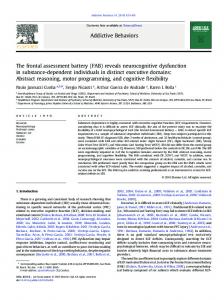
Figure 1 A Danish subject wearing spectacles with a wire indicating the interpupillary line.
190 cm film-focus distance and a 15-cm distance between the film and mid-frontal plane, the enlargement being 8 per cent. A pivot-mounted spirit-level was attached to the subject’s forehead with double-sided tape, and the subject stood in a relaxed position looking into the distance. The level was then adjusted horizontally and the subject positioned in the cephalostat to face the film, with the ear rods only slightly touching the external passage of the ear. The head position in the sagittal plane was corrected in accordance with the Frankfort plane, and that in the frontal plane in accordance with the position recorded previously with the level. To facilitate visibility of the level when the subject was standing in the cephalostat, a hand mirror was attached in front of the subject. A metal chain suspended from the film holder indicated the true vertical line on the roentgen films. The same basic method was used for the PA cephalograms of the Danish subjects, obtained with the addition of spectacles with a metal wire attached to the frames and adjusted to correspond with the interpupillary line (Figure 1). The cephalograms were analysed manually twice, with an interval of 1 week, by the two authors.
696
I . Z E PA A N D J. H U G G A R E
The reference points and lines used for the assessment of head posture and cervical spine relationship are shown in Figure 2. The angles between these lines were measured to the nearest 0.5 degrees. Intra- and inter-examiner reproducibility s(i) was calculated by the formula: s(i) =
∑d2
! 2n
where d is the difference between the two tracings and n is the sample size. The paired t-test was used to determine any systematic error. Results
Figure 2 Landmarks and points used in the study: (1) supra-orbital line (ORB)–tangent to the extreme cranial point on the supra-orbital margins; (2) cranial line (CR)–a line through crista galli and anterior nasal spine; (3) cervical line (CER)–a line through the mid-point of atlas (half the distance between the most median points on the tubercle of the transverse ligament) and C4 (half the distance between the most concave points of the lateral masses); (4) cervical spine line (SPINE): subjectively defined main course of the upper cervical spine; (5) interpupillary line (IP): a line indicated by the image of the metal wire on the radiograph; (6) true vertical and horizontal lines (VER, HOR).
The supra-orbital and interpupillary lines displayed a mean deviation of 0.5 degrees. There was no systematic error in the evaluated angles, except for one author (IZ) with regard to the spinal inclination (SPINE/VER; Table 1). Both intra- and inter-examiner errors were smaller for ORB and CER than for CR and the SPINE. The intra-examiner reproducibility s(i) for ORB/HOR and CR/VER was 0.4 degrees and 0.8 degrees, respectively, and for CER/VER and SPINE/VER 0.8 degrees and 1.0 degrees, respectively (Table 2). Discussion In assessing the suitability of radiographic landmarks of the head and upper part of the cervical spine for frontal cephalometric analyses, the main emphasis should be on the median structures of the skull (Svanholt and Solow, 1977; Pirttiniemi
Table 1 Means and standard deviations for differences between two repeated tracings of frontal head posture and spine inclination (– indicates a deviation to the left). CR/CER
CR/VER
ORB/HOR
CER/VER
SPINE/VER
Author
Mean
SD
Mean
SD
Mean
SD
Mean
SD
Mean
SD
IZ JH
–0.05 –0.24
1.46, NS 1.18, NS
0.19 0.57
1.07, NS 1.14, NS
–0.14 0.12
0.55, NS 0.63, NS
–0.14 –0.05
1.13, NS 1.12, NS
–0.6 –0.33
1.32* 1.46, NS
*P < 0.05; NS = not significant; paired t-test.
697
R E F E R E N C E S T RU C T U R E S F O R F RO N TA L H E A D P O S T U R E
Table 2 Intra- and inter-examiner reproducibility in two repeated calculations of frontal head posture and spine inclination.
Intra-examiner Inter-examiner
IZ JH
CR/VER s(i)
ORB/HOR s(i)
SPINE/VER s(i)
CER/VER s(i)
0.75 0.89 0.59
0.39 0.44 0.34
1 1.04 0.7
0.79 0.77 0.6
et al., 1996): if the patient’s skull rotates vertically the landmarks derived from the median craniocervical structures are less subject to deviation on the PA cephalograms (Svanholt and Solow, 1977). This is important in analysing radiographs of scoliotic patients, who tend to compensate the inclination of their deviated spine with a changed head posture, in order to maintain the visual axis in the horizontal plane (Huggare et al., 1991) and any lateral bend of the spine tends to cause concomitant head rotation. However, the drawback of such a cranial central line (for instance, a line from crista galli to anterior nasal spine) as an indicator of head posture, is that it could be influenced by an existing craniofacial asymmetry and thus give a false expression of a skewed head posture. As asymmetry of the face is usually due to suborbital torsional deformity (Ferguson, 1993), the suitability of the interpupillary line and the supraorbital line as alternative reference structures was tested. The perpendicular to the supraorbital line through crista galli could then be used as a cranial central line. However, this reference line should not be used to analyse radiographs of patients with asymmetry affecting the orbital area (orbital dystopia, etc.). The cervical spine has been assessed extensively on lateral cephalograms but there are few reports dealing with the frontal view of the spine as seen on PA cephalograms. This study therefore attempted to create a reference system analogous with that of Solow and Tallgren (1971) for lateral head posture. The principal movement in the upper part of the cervical spine takes place between the occiput and C2, but is regulated by the atlas (Penning, 1978). In lateral bending the atlas has a rigidly
prescribed position in any position of the C0–C2 junction. This is due to the shape of the lateral masses of the atlas as seen in anteroposterior projection (i.e. the wedge shaped lateral mass interposed between occipital condyles and lateral masses of C2). During movement the odontoid process of axis (C2) must remain midway between the occipital condyles due to its fixation by the alar ligaments. Thus, lateral bending in the atlanto-occipital segment is always combined with lateral bending in the atlanto-axial segment and vice versa. Lateral bending is also facilitated by simultaneous atlanto-axial rotation (Penning, 1978). Rotational movement between occiput and atlas, if at all possible, is so small that it is virtually impossible to measure radiologically (Penning, 1978). Movements between occiput and atlas occur around transverse and anteroposterior axes, but not around the vertical axis (Gray’s Anatomy, 1973). Therefore, radiographically, atlas serves as an appropriate reference structure unaffected by image distortion due to rotation. In practice, rotation takes place only in the C1– C2 segment with C2 as a rotating element. Lateral sliding of the atlas on the axis is a complex motion which cannot be achieved by lateral bending alone but requires a combination of lateral bending and rotation. Under these conditions a physiological lateral shift of the atlas on the axis of as much as 4 mm can easily occur (Clark, 1973). In the context of the present study, this shift is potentially important because it might undermine several conditions related to changed head posture due to changes in the cervical spine. For instance, extension of scoliosis into the cervical area results in compensatory tilting of the atlanto-occipital complex on the
698 axis to permit the head to assume an upright position. Congenital torticollis, which persists during growth, may produce an apparent lateral shift of the atlanto-occipital complex on the axis in adult life. Post-traumatic distorted neck balance, accompanied by unilateral muscle spasm and flattening of the cervical lordosis, may cause considerable lateral shift of the atlanto-occipital complex on the axis, which returns to normal as the spasm subsides. These conditions further support use of the mid-point of the atlas as an upper cervical reference structure. The distance between the most median points on the tubercle of the transverse ligament of the atlas describes more accurately the midline of the upper cervical spine than the midpoint of the odontoid process of the second vertebraaxis. The odontoid process of axis is fixed to the occiput by ligamentum apicis dentis and ligamenti alaria, and is usually located between the condyles of the occiput. It can be located asymmetrically or tilted from the midpoint between the lateral masses of atlas, as a result of asymmetry of the odontoid process itself or as a compensatory movement of the atlas on the axis (Hohl and Baker, 1964; Shapiro et al., 1973; Figure 3). As the lower cervical reference point the midpoint between the most concave surfaces of the lateral masses of the fourth cervical vertebra
I . Z E PA A N D J. H U G G A R E
(C4) was selected. This vertebra is readily distinguishable on a PA cephalogram and the resultant cervical line (CER) then constitutes the analogous line to CVT (cervical tangent line) in the lateral analyses by Solow and Tallgren (1971). Our results showed that this line was more accurately reproduced than the subjectively assessed main course of the cervical spine. It was observed that the supra-orbital line (ORB) was almost parallel to the interpupillary line (IP), thus confirming the results of Ferguson (1993). An interesting observation was that for all angles measured inter-examiner error was less than intra-examiner error (Table 2), indicating that the structures defined could easily be adopted by others, not only for research purposes, but also as clinical norms (Major et al., 1994). Conclusions For assessing frontal head posture the supraorbital line (ORB) and cervical line (CER), defined as the midtransversal line between the atlas and the fourth cervical vertebra, are recommended as appropriate radiographic references. Address for correspondence Professor Jan Huggare Department of Orthodontics Faculty of Odontology Karolinska Institutet Box 4064, S-141 04 Huddinge Sweden Acknowledgements We wish to thank Professor Birte Melsen, Institute of Dentistry, University of Århus, Denmark, for providing some of the radiographs. References
Figure 3 (A) Widening of the right odontoid lateral mass interspace (OLMI) with right rotation of the atlas. (B) Symmetric OLMI with the atlas in the neutral position. (C) Widening of the left OLMI with left rotation of the atlas (from Shapiro et al., 1973). (Reprinted with kind permission of W B Saunders & Co., Philadelphia.)
Clark K C 1973 Positioning in radiography. William Heinemann Medical Books Ltd, London, pp. 166–179 Ferguson J W 1993 Cephalometric interpretation and assessment of facial asymmetry secondary to congenital torticollis. The significance of cranial base reference lines.
R E F E R E N C E S T RU C T U R E S F O R F RO N TA L H E A D P O S T U R E
699
International Journal of Oral and Maxillofacial Surgery 22: 7–10
associated conditions, and results of treatment. Journal of Paediatric Orthopaedics 2: 500–505
Fjellvang H, Solow B 1986 Craniocervical postural relations and craniofacial morphology in 30 blind subjects. American Journal of Orthodontics and Dentofacial Orthopedics 90: 327–334
Penning L 1978 Normal movements of the cervical spine. American Journal of Roentgenology 2: 317–326
Gray’s anatomy 1973 Longman Group Ltd, Harlow, pp. 416–417 Hohl M, Baker H R 1964 The atlanto-axial joint. Roentgenographic and anatomical study of normal and abnormal motion. Journal of Bone and Joint Surgery 46-A: 1739–1752 Huggare J 1989 Natural head position recording on frontal skull radiographs. Acta Odontologica Scandinavica 47: 105–109 Huggare J, Raustia A 1992 Head posture and cervicovertebral and craniofacial morphology in patients with craniomandibular dysfunction. Journal of Craniomandibular Practice 10: 173–177 Huggare J, Pirttiniemi P, Serlo W 1991 Head posture and dentofacial morphology in subjects treated for scoliosis. Proceedings of the Finnish Dental Society 87: 151–158 Keller E E, Jackson I T, Marsh W R, Triplett W W 1986 Mandibular asymmetry associated with congenital muscular torticollis. Oral Surgery, Oral Medicine, Oral Pathology 61: 216–220 Kylämarkula S, Huggare J 1985 Head posture and the morphology of the first cervical vertebra. European Journal of Orthodontics 7: 151–156 Major P W, Johnson D E, Hesse K L, Glover K E 1994 Landmark identification error in posterior anterior cephalometrics. Angle Orthodontist 64: 447–454 Morrison D L, MacEwan G D 1982 Congenital muscular torticollis: observations regarding clinical findings,
Pirttiniemi P, Lahtela P, Huggare J, Serlo W 1989 Head posture and dentofacial asymmetries in surgically treated muscular torticollis patients. Acta Odontologica Scandinavica 47: 193–197 Pirttiniemi P, Miettinen J, Kantomaa T 1996 Combined effects of errors in frontal-view asymmetry diagnosis. European Journal of Orthodontics 18: 629–636 Sage F P 1980 Congenital anomalies. In: Edmonson A S, Greenshaw A H (eds) Campbell’s operative orthopedics, Vol. II. C V Mosby Co., St Louis Sandikçioglu ˇ M, Skov S, Solow B 1994 Atlas morphology in relation to craniofacial morphology and head posture. European Journal of Orthodontics 16: 96–103 Shapiro R, Youngberg A S, Rothman S L G 1973 The differential diagnosis of traumatic lesions of the occipitoaxial segment. Radiology Clinics of Northern America 11: 505–526 Solow B, Tallgren A 1971 Natural head position in standing subjects. Acta Odontologica Scandinavica 29: 591–607 Solow B, Barret M J, Brown T 1982 Craniocervical morphology and posture in Australian aboriginals. American Journal of Physical Anthropology 59: 33–45 Svanholt P, Solow B 1977 Assessment of midline discrepancies on the postero-anterior cephalometric radiograph. Transactions of the European Orthodontic Society, pp. 261–268 Tallgren A, Solow B 1987 Hyoid bone position, facial morphology and head posture in adults. European Journal of Orthodontics 9: 1–8