Molecular Cell 22, 851–868, June 23, 2006 ª2006 Elsevier Inc.
DOI 10.1016/j.molcel.2006.06.001
The Human and Mouse Complement Resource of SH2 Domain Proteins—Establishing the Boundaries of Phosphotyrosine Signaling Bernard A. Liu,1,3 Karl Jablonowski,1 Monica Raina,2 Michael Arce´,1 Tony Pawson,2,* and Piers D. Nash1,3,* 1 Ben May Institute for Cancer Research and the Committee on Cancer Biology The University of Chicago Chicago, Illinois 60637 2 Samuel Lunenfeld Research Institute Mount Sinai Hospital Toronto M5G 1X5 Canada
Summary SH2 domains are interaction modules uniquely dedicated to the recognition of phosphotyrosine sites and are embedded in proteins that couple protein-tyrosine kinases to intracellular signaling pathways. Here, we report a comprehensive bioinformatics, structural, and functional view of the human and mouse complement of SH2 domain proteins. This information delimits the set of SH2-containing effectors available for PTK signaling and will facilitate the systems-level analysis of pTyr-dependent protein-protein interactions and PTK-mediated signal transduction. The domainbased architecture of SH2-containing proteins is of more general relevance for understanding the large family of protein interaction domains and the modular organization of the majority of human proteins. Introduction Selective protein-protein interactions are important in organizing the regulatory processes of eukaryotic cells and are commonly mediated by modular protein domains, for which the prototype is the Src homology 2 (SH2) domain, a conserved sequence that controls signaling by protein tyrosine kinases (PTK). SH2 domains typically bind specific phosphotyrosine (pTyr)-containing motifs (Figure 1) and thereby couple activated PTKs to intracellular pathways that regulate many aspects of cellular communication in metazoans (Chervitz et al., 1998; Hunter, 2000; Pawson and Nash, 2000). The intimate relationship between tyrosine kinases and SH2 domains is supported by their coordinate emergence during eukaryotic evolution. Unicellular fungi such as yeast lack conventional PTKs and SH2 domains. However, pTyr binding SH2 domains are features of some amoebozoa. Notably, the social amoeba Distyostelium discoidum contains 12 proteins with SH2 domains, including polypeptides similar to STAT transcription factors and Cbl E3 protein-ubiquitin ligases, as well as dual specificity protein kinases with an N-terminal SH2 domain (SHKs) and proteins with unusual architectures in which the SH2 domain is linked to leucine rich repeats or to an F box and ankyrin repeats (Eichinger *Correspondence:
[email protected] (T.P.); pdnash@uchicago. edu (P.D.N.) 3 These authors contributed equally to this work.
et al., 2005). Such amoebae lack conventional PTKs but have a large set of tyrosine kinase-like (TKL-group) kinases, with the potential for tyrosine phosphorylation (Goldberg et al., 2006; Moniakis et al., 2001). Choanoflagellates are protazoa that may represent immediate predecessors of multicellular animals; one such organism, Monosiga breicollis, encodes not only SH2 domains but also bona fide PTKs and has the earliest identified example of an SH2 domain covalently linked to a tyrosine kinase domain (King et al., 2003). These observations suggest that the acquisition of pTyr-SH2 domain signaling facilitated metazoan evolution (Katzmann et al., 2004; Kawata et al., 1997). SH2 domains represent the largest class of known pTyr-recognition domains (Pawson et al., 2001). In addition to SH2 domains, the PTB domains of docking proteins within the IRS, SHC, and DOK subclasses also bind specific pTyr-containing motifs on activated receptor tyrosine kinases (RTKs) and are consequently phosphorylated at sites that recruit SH2 domains (Yaffe, 2002). However, only about one quarter of the 79 human PTB domains have apparently acquired the capacity for pTyr-dependent recognition, while the majority of PTB domains recognize nonphosphorylated peptide ligands, or phosphoinositides (Schlessinger and Lemmon, 2003; Uhlik et al., 2005). In addition, the C2 domain of PKCd binds a transmembrane Src-associated glycoprotein, CDCP1/SIMA135, in a pTyr-dependent manner, though this is likely an idiosyncratic property of this particular C2 family member (Benes et al., 2005). SH2 domains therefore appear to be uniquely dedicated to pTyr recognition and thus represent primary targeting and specificity elements in tyrosine kinase signaling. To fully appreciate the signaling events initiated by tyrosine kinases, it is important to clarify the set of genes that encode SH2 domains in different species. Here, we describe the SH2 domains specified in the genomes of both human and mouse, combined with an analysis of the SH2 domain families and a catalog of current SH2 domain structures, human diseases, and mouse mutants. This reveals a number of hitherto unstudied SH2 domain proteins, as well as gaps in our overall understanding of SH2 domain structure and biological function. It sets a benchmark for a more global approach to studying pTyr-nucleated signaling events. Interactive figures and additional information may be found at http://sh2.uchicago.edu/.
Results and Discussion The Human Complement of SH2 Domains We combined several approaches to identify the complete nonredundant set of recognizable human SH2 domains and their corresponding proteins (see the Supplemental Data available with this article online). After removal of duplicates, splice variants, and pseudogenes, this analysis afforded a total of 120 SH2 domains contained in 110 distinct proteins, of which 10 have dual SH2 domains. These 110 human proteins are outlined in
Molecular Cell 852
Figure 1. Modes of SH2 Binding (A) Structure of the Src SH2 domain bound to a pTyr-Glu-Glu-Ile peptide (PDB, 1SPS) (Waksman et al., 1993). The surface of the SH2 domain is shown as a translucent gray skin and the secondary structural elements of the SH2 domain in blue. The aA helix is to the right and aB helix to the left. The Arg bB5 residue critical for pTyr binding is in yellow. The N-terminal pTyr of the peptide (red) occupies the pTyr binding pocket. The peptide runs over the central b sheet of the SH2 domain, the +1 and +2 glutamates contact the surface of the domain, and the side chain of the +3 Ile (to the left) fits in a hydrophobic pocket. (B) Grb2 SH2 domain in complex with pYVNV (red) (PBD, 1BMB). Trp (green) stabilizes the b turn conformation essential for high affinity binding. (C) Two phosphotyrosine binding pockets are present on a single SH2 domain of APS. A single APS SH2 molecule is bound to pYETDpY (red) peptide of the activation loop of INSR. The pTyr-1158 interacts with the Arg-438 (yellow), while Lys-455 and Lys-457 (orange) create a second binding pocket for pTyr-1162 (PDB, 1RQQ). (D) SH2D1A/SAP in complex with a nonphosphorylated SLAM peptide KSLTIYAQVQK (red) (PBD, 1D4T). Ribbon and surface diagrams were generated using Pymol.
Table S1 along with the chromosomal location of each corresponding gene, a list of aliases or alternate symbols, and information on their mouse homologs. This is somewhat more than previous numbers; an early survey of the human genome identified 87 SH2 domain-containing proteins (Venter et al., 2001), and a more recent study identified 108 SH2 domains in 98 proteins (Jones et al., 2006). The present work clarifies previous curation efforts that generated similar total numbers but suffered from issues of redundancy and incomplete annotation (Huang et al., 2004; Lander et al., 2001). Mouse homologs were independently determined and the closest relatives established by BLAST, supported by information from UniGene (Wheeler et al., 2005) and manual examination of genetic loci, revealing a list of
120 SH2 domains in 110 proteins. The gene encoding SH2D1C (or ERT) is a functional gene in mouse, though it appears to be a pseudogene in humans. Murine SH2D1C is a signal regulator related to SH2D1B/EAT2 and SH2D1A/SAP, though its expression appears highly restricted to natural killer (NK) cells, in which it acts as a negative regulator of antigen signaling (Roncagalli et al., 2005). The human gene encoding SH2D3A appears to be absent in the rodent lineage, though it is present in primates. Human diseases associated with genetic lesions identified as corresponding to SH2 domain-containing proteins were assembled from OMIM (Hamosh et al., 2005) and from the literature (Table 1). A survey of targeted gene disruptions in mouse models was obtained from the literature (Table 2). These data
The Human and Mouse Complement of SH2 Domains 853
Table 1. Human Diseases Associated with Mutations in SH2-Encoding Genes Name
Summary of Mutations
Phenotype
ABL1
t(9;22) translocation results in fusion with the BCR gene
ABL2 BLNK
t(1;12)(q25;p13) translocation with ETV6/TEL Base pair substitution of a splice donor site results in reduction or loss of BLNK transcripts Complete loss or drastic reduction of expression due to incorporation of alternative exons Missense mutations, deletions, or splice site mutations Translocation with MLL t(4;11)(q21;q23) t(11;14)(q23;q32) t(11;22)(q23;q12) Deletions of 17p13.3
Chronic myelogeneous leukemia (CML), acute lymphoblastic leukemia (ALL), myelogenous leukemia (AML) (de Klein et al., 1982; Heisterkamp et al., 1983) Acute myeloid leukemia (Cazzaniga et al., 1999) Human immundeficiency (Minegishi et al., 1999)
BTK CBL
CRK ITK JAK2
JAK3 LCK
PTPN11
RASA1
SH2D1A SH3BP2 SHIP2 SRC STAT1 STAT5B
SYK TYK2 ZAP70
t(5;9)(q33;q22) translocation to SYK t(9;12)(p24;p13) fusion with TEL, results in a constitutive kinase t(9;15;12)(p24;q15;p13) fusion with TEL and ETV6 Dominant gain-of-function mutation V617F Nucleotide insertion, substitution, or deletion resulting in a frame shift or premature termination Translocation of the t(1;7)(p34;q34) that fuses LCK and TCRB Decrease expression of p56(lck), likely due to alternative splicing of exon 7 Missense mutations altering amino acids D61 at the N-SH2 domain, resulting in gain of function (Tartaglia et al., 2001) Missense mutation in exons 7, 12, and 13 95% of mutations are in exon 3 or a defect in exon 13 affecting the protein tyrosine phosphatase domain resulting in a gain of function Nonsense mutations within the SH2 domain A 2 bp deletion results in a frameshift and a premature stop codon; a missense mutation in the PH domain Mutations or deletions cause the protein to not fold and function correctly Point mutations in exon 9 affect three amino acids within the six amino acid sequence (RSPPDG) Mutation or deletions, SNPs Truncating mutation in the SRC codon 531 Nucleotide substitutions, homozygous deletions generating a premature stop codon Homozygous A630P mutation t(15;17)(q11.2; q21.1) translocation with RARA t(9;12)(q22;p12) translocation with TEL t(5;9)(q33;q22) translocation with ITK SNPs resulting in amino acid substitutions in the JAK homology (JH) regions A single base substitution mutation in a splice acceptor site
provide a census of SH2 domain-containing proteins encoded in the human genome. The SH2-ome SH2 domains are incorporated into proteins with a range of biochemical properties. We have classified SH2-containing proteins into 11 functional categories, based on their modular domain composition (Figure 2). Taken together, this information delimits the physiological SH2containing targets available for pTyr signaling in human cells. Since the functional output from any given PTK
Pre-B-cell acute lymphoblastic leukemia (ALL) (Jumaa et al., 2003) X-linked agammaglobulinemia (XLA) (Rawlings et al., 1993) Acute myeloid leukemia (Fu et al., 2003) Acute leukemia B cell lymphoma Ewing sarcoma (Savage et al., 1991) Isolated lissencephaly sequence (ILS) to Miller-Dieker syndrome (MDS) (Cardoso et al., 2003) Peripheral T cell lymphoma (Streubel et al., 2005) Acute lymphoblastic leukemia (Lacronique et al., 1997) Chronic myelogenous leukemia (Peeters et al., 1997) Polycythemia vera and myeloproliferative disorders (James et al., 2005; Kralovics et al., 2005) SCID, lymphopenia (Russell et al., 1995) T cell acute lymphoblastic leukemia (T cell ALL) (Burnett et al., 1991) SCID (Goldman et al., 1998) Noonan syndrome (Tartaglia et al., 2001)
Multiple lentigines (ML)/Leopard syndrome (LS) (Digilio et al., 2002) Juvenile myelomonocytic leukemia (JMML) (Tartaglia et al., 2003) Basal cell carcinoma (Friedman et al., 1993) Capillary malformation-arteriovenous malformation (Eerola et al., 2003) X-linked lymphoproliferative syndrome (XLP) (Sayos et al., 1998) Cherubism (Ueki et al., 2001) Type 2 diabetes, hypertension (Marion et al., 2002) Advanced colon cancer (Irby et al., 1999) Susceptibility to mycobacterial and viral disease (Dupuis et al., 2001) Growth hormone insensitivity with immunodeficiency (Kofoed et al., 2003) Acute promyeloyctic leukemia (APL) (Arnould et al., 1999) Myelodysplastic syndrome (MDS) (Kuno et al., 2001) Peripheral T cell lymphoma (Streubel et al., 2005) Systemic lupus erythematosus (SLE) (Sigurdsson et al., 2005) SCID (T cell defect) (Chan et al., 1994; Elder et al., 1994)
likely depends on the subset of SH2 signaling proteins it recruits, either directly or through phosphorylated scaffold proteins, these data define the predominant pathways through which PTKs modify cellular behavior. The groups of SH2 proteins indicate that pTyr-dependent interactions channel PTK signals into defined, if diverse, areas of cellular regulation. These include tyrosine phosphorylation itself (through cytoplasmic PTKs and tyrosine phosphatases), the control of phospholipid metabolism (by phosphatidylinositol 30 -kinase, PLCg and inositol phosphatases), the regulation of small
Molecular Cell 854
Table 2. Mouse Knockouts of SH2 Domain Proteins Knockout
Phenotype
Organs and Cell Types Affected
Source
Abl1
Death 1–2 weeks postbirth
Thymus, spleen
Abl2 Aps Bks Blk Blnk Bmx
Multiple behavioral abnormalities Viable and fertile; hypoinsulinemia Normal Normal High incidence of pre-B cell lymphoma Normal
Btk Cbl Cblb Cblc Crk Crkl Csk
XID phenotype Thymic phenotype Autoimmune encephalomyelitis Normal Normal Partial embryonic lethality Embryonic lethality at E10–E12
Brain B1 lymphocytes, adipocytes Hepatocytes None B lymphocytes Endocardium and arteries, endothelial cells B lymphocytes Thymus, T lymphocytes T lymphocytes None None Thymus Neural tube
(Schwartzberg et al., 1991; Tybulewicz et al., 1991) (Koleske et al., 1998) (Iseki et al., 2004; Minami et al., 2003) (Minoguchi et al., 2003) (Texido et al., 2000) (Flemming et al., 2003; Jumaa et al., 2003) (Rajantie et al., 2001)
Dapp1 Fer Fes Fgr Frk Fyn Gads Grap Grb2 Grb10 Grb14 Hck Itk Jak1
Normal Healthy and fertile (kinase-inactivating mutation) Viable; abnormal innate immunity Defective lung inflammation Normal Suckling abnormality Healthy Viable, fertile, and healthy Embryonic lethality 30% larger in size than wild-type Glucose tolerance improvement Normal Normal Perinatally lethal, nursing defect
Jak2
Embryonic lethality
Jak3 Lck Lnk
Thymus, spleen, lymph nodes Fetal thymus Hematopoietic progenitor cells, spleen B lymphocytes, mast cells None None None None B lymphocytes Muscle N/A
(Ueki et al., 2002) (Ji et al., 1997)
Plcg2 Ptpn6
Immunodeficient; lymphopenia Thymic atrophy Splenomegaly, defects in hematopoietic lineages Hyperglobulinemic, glomerulonephritis Normal Normal Normal Normal X-linked immunodeficiency; hypoglycemia Hypoglycemia Embryonic lethality between E8.5 and E9.0 Viable, B cell deficiency Immunity and hematopoiesis defects
(Neubauer et al., 1998; Parganas et al., 1998) (Nosaka et al., 1995; Thomis et al., 1995) (Molina et al., 1992) (Takaki et al., 2000; Velazquez et al., 2002) (Hibbs et al., 1995; Nishizumi et al., 1995) (Hamaguchi et al., 1996) (Utting et al., 2004) (Bladt et al., 2003) (Bladt et al., 2003) (Fruman et al., 1999; Suzuki et al., 1999)
B lymphocytes T lymphocytes
Ptpn11
Die peri-implantation
Rasa1
Embryonic lethality by E10.5
Rin1 Sh2b
Normal Growth retardation, fertility defect, insulinemia Recapitulates X-linked lymphoproliferative disease Susceptible to Lupus-like autoimmune disease Embryonic lethality by E11.5 Normal Normal Fail to thrive with splenomegaly
Trophoblast stem cell survival, node, notochord Vascular system, endothelial cells Amygdala Testis, ovaries, skeletal muscle, liver and fat T and (B) lymphocytes
(Wang et al., 2000) (Shultz et al., 1993; Van Zant and Shultz, 1989) (Arrandale et al., 1996; Saxton et al., 1997; Yang et al., 2006) (Henkemeyer et al., 1995)
Lyn Matk Mist Nck1 Nck2 Pik3r1 Pik3r2 Plcg1
Sh2d1a Sh2d2a Shc1 Shc2 Shc3 Ship1 Ship2
Resistant to dietary obesity and growth affects
B lymphocytes None Bone marrow, myeloid cells Tissue and airway eosinophilia None Olfactory bulb, hippocampus T lymphocytes Lymphocytes Endoderm Brain, liver Liver, skeletal muscle Macrophages T lymphocytes Neurons, fibroblasts, T and B lymphocytes Hematopoeitic cells
T lymphocytes Heart Sensory neurons None Spleen, lymphoid, erthryoid, myeloid cells Skeletal muscle, liver
(Kerner et al., 1995; Khan et al., 1995) (Naramura et al., 1998) (Chiang et al., 2000) (Griffiths et al., 2003) (Imaizumi et al., 1999) (Guris et al., 2001; Peterson et al., 2003) (Imamoto and Soriano, 1993; Nada et al., 1993) (Fournier et al., 2003; Han et al., 2003) (Craig et al., 2001) (Hackenmiller et al., 2000) (Lowell et al., 1994) (Chandrasekharan et al., 2002) (Yagi et al., 1993) (Yoder et al., 2001) (Shen et al., 2002) (Cheng et al., 1998) (Charalambous et al., 2003) (Cooney et al., 2004) (Lowell et al., 1994) (Liao and Littman, 1995) (Rodig et al., 1998)
(Dhaka et al., 2003) (Duan et al., 2004; Ohtsuka et al., 2002) (Crotty et al., 2003; Czar et al., 2001; Wu et al., 2001) (Rajagopal et al., 1999) (Lai and Pawson, 2000) (Sakai et al., 2000) (Sakai et al., 2000) (Helgason et al., 1998) (Elchebly et al., 1999; Sleeman et al., 2005)
The Human and Mouse Complement of SH2 Domains 855
Table 2. Continued Knockout
Phenotype
Organs and Cell Types Affected
Source
Slap Slp76
Fertile and healthy Impaired viability; defective thymocyte development Fatty degeneration of the liver Gigantism Embryonic lethality between E11 and E13 Normal Normal but weigh 10% less than wild-type Healthy and fertile but die from hydrocephalus Defective bone remodeling and osteoporosis Normal Normal but sensitive to infection Exhibit multiple defects in immune response Embryonic lethality between E6.5 and E7.5 Normal Normal Decrease growth in males
T lymphocytes T lymphocytes
(Lowell et al., 1994; Sosinowski et al., 2001) (Pivniouk et al., 1998)
Liver, thymocytes Most visceral organs Placental, blood vessel None N/A
(Starr et al., 1998) (Metcalf et al., 2000) (Marine et al., 1999; Metcalf et al., 2000; Roberts et al., 2001) (Brender et al., 2004) (Krebs et al., 2002)
Brain
(Krebs et al., 2004)
Bone osteoclast
(Soriano et al., 1991)
None T lymphocytes, macrophages Fibroblasts
(Kohmura et al., 1994) (Meraz et al., 1996) (Park et al., 2000)
Visceral endoderm, T lymphocytes T lymphocytes Mammary glands Liver, mammary glands, adipose tissue T and B lymphocytes B lymphocytes
(Takeda et al., 1997)
Socs1 Socs2 Socs3 Socs5 Socs6 Socs7 Src Srms Stat1 Stat2 Stat3 Stat4 Stat5a Stat5b Stat6 Syk Tec Tns1 Txk Tyk2
Normal Perinatal lethality and blocked B cell development Normal Normal Death by 102 days Normal
Vav1 Vav2 Vav3 Yes Zap70
Normal Normal Normal Normal SCID
(Kaplan et al., 1996) (Liu et al., 1997) (Teglund et al., 1998)
T lymphocytes None None None T lymphocytes
(Shimoda et al., 1996) (Cheng et al., 1995; Turner et al., 1995) (Ellmeier et al., 2000) (Lo et al., 1997) (Schaeffer et al., 1999) (Karaghiosoff et al., 2000; Shimoda et al., 2000) (Zhang et al., 1994) (Tedford et al., 2001) (Fujikawa et al., 2003) (Stein et al., 1994) (Negishi et al., 1995)
B lymphocytes Kidney, muscle myoblast Splenocytes T lymphocytes
Family Knockouts Gene
Phenotype
Organs or Cells Affected
Reference
Abl1/2 Aps/Sh2b
Neurulation defects Body weight increase, obesity, hyperglycemia Defective lymphocyte development Embryonic lethality at E10 Immunodeficiency Src defects Two thirds die at birth; osteopetrosis Immunological defects T cell development defect Embryonic lethality at E9.5 Normal but defective neural development Infertile female mice T cell development defect Defective lymphocyte development
Neural tube Adipocytes
(Koleske et al., 1998) (Li et al., 2006)
B lymphocytes T lymphocytes Macrophages Osteoclasts Hematopoietic cells T lymphocytes T lymphocytes, dendritic cells Notochord Superior cervical ganglia (SCG) neurons Ovaries, mammary glands Thymocytes T and B lymphocytes
(Ellmeier et al., 2000) (Naramura et al., 2002) (Lowell et al., 1994) (Lowell et al., 1996) (Lowell et al., 1996) (Schaeffer et al., 1999) (van Oers et al., 1996) (Bladt et al., 2003) (Sakai et al., 2000)
Btk/Tec Cbl/Cblb Fgr/Hck Fgr/Src Hck/Src Itk/Txk Lck/Fyn Nck1/2 Shc2/3 Stat5a/b Syk/Zap70 Vav1/2/3
(Teglund et al., 1998) (Cheng et al., 1997) (Fujikawa et al., 2003)
Targeted gene disruptions have been completed for a large subset of SH2 proteins, yielding a wide range of phenotypes. In some cases, compensatory mechanisms have required the disruption of multiple members of a family in order to expose a phenotype.
GTPases (including Ras, Rho, Rap, and Rab family members) by guanine nucleotide exchange factors (GEFs) and GTPase-activating proteins (GAPs), gene expression directed by STAT transcription factors, ubiquitylation mediated by Cbl E3 protein-ubiquitin ligases, and cytoskeletal organization by tensin proteins. A group of SH2/SH3 proteins act as adaptors (i.e., Grb2, Crk, and Nck), each of which appear to target a set of
SH3 binding proteins with related functions (Pawson et al., 2004). Nck recruits cytoskeletal regulators such as N-Wasp and Pak serine/threonine kinases (Buday et al., 2002; Rivera et al., 2004); Grb2 binds Sos and Gab1 (Gu and Neel, 2003; Lock et al., 2000; RozakisAdcock et al., 1993; Takenawa et al., 1998), involved in MAPK/PI3K signalling; and Crk targets GEFs for Rap and Rac GTPases that control adhesion (Feller, 2001).
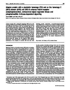
Molecular Cell 856
Figure 2. Modular Domain Organization of SH2-Containing Proteins Classification and domain composition of the 110 nonredundant human SH2 domain-containing proteins identified in human and mouse by Pfam (Bateman et al., 2004) and SMART (http://smart.embl-heidelberg.de/). More information on SH2-containing proteins can be found at http://sh2. uchicago.edu/. More information on the individual domains portrayed can be found at http://www.mshri.on.ca/pawson/domains.html and http:// smart.embl-heidelberg.de/.
The Human and Mouse Complement of SH2 Domains 857
In addition, a significant number of SH2 proteins regulate the duration and location of PTK signaling. For example, SOCS proteins are transcriptionally induced by JAK-STAT signals and provide an inhibitory feedback both by blocking JAK tyrosine kinase activity and by promoting ubiquitylation (Ilangumaran et al., 2004). Cbl proteins induce pTyr-dependent receptor multiubiquitylation and thus create binding sites for endocytic proteins with ubiquitin interaction motifs, involved in receptor trafficking (Haglund and Dikic, 2005). SH2 proteins can also directly interact with active sites of RTKs, exemplified by the SH2 domain of the APS protein, which simultaneously homodimerizes and binds the phosphorylated activation loop of the insulin receptor (INSR), potentially stabilizing the active state of the receptor (Hu et al. 2003). Conversely, the SH2 domain of the related Grb14 protein is proposed to bind the phosphorylated INSR activation loop but has a 45 residue sequence (BPS region) located between the SH2 and PH domains that antagonizes receptor activity by acting as a pseudosubstrate (Depetris et al., 2005). Thus, SH2 proteins, in addition to acting as ‘‘on’’ or ‘‘off’’ regulators of intracellular biochemical pathways, can modify the kinetics, activity, and substrate specificity of tyrosine kinase signals. Given the large repertoire of distinct interaction and catalytic domains in the human proteome, it is evident that SH2 domains are associated with a focused subset of modules, most notably SH3, PTB, PH, kinase, phosphatase, GEF, and GAP domains (Figure 2). This may reflect the requirement that SH2 proteins initially act near the plasma membrane to engage specific signaling networks that promote cell growth, differentiation, morphology, and metabolism, although proteins such as STAT transcription factors subsequently move to other subcellular compartments. By virtue of their modular design, SH2 domains can be artificially joined to domains from which they are normally segregated, to create aberrant signaling pathways (Howard et al., 2003). Presumably, such nonphysiological combinations were selected against in the course of evolution to insulate PTKs from inducing counterproductive intracellular responses. In this analysis, we have not considered shorter peptide motifs, which can serve as ligands for interaction domains and thereby extend the binding properties of SH2 domain proteins. Multiple Alignments and the SH2 Tree Both Pfam and SMART HMMs identify SH2 sequences that are somewhat smaller than the full folded domain, revealed by corresponding structural information. The C-terminal region of the SH2 structural domain is often excluded from the sequence identified by bioinformatic means, and this can eliminate important elements, notably b strand G. Although this C-terminal region of the SH2 domain is less well conserved than either the N-terminal or central core sequences, it is important for the formation of the ligand binding pocket and for the stable folding of the intact domain. Thus, we extended the protein sequence from the Pfam- and SMART-identified SH2 domains by some 20 amino acids on both the N and C termini to obtain a sequence group for multiple sequence alignment. The 120 SH2 domain sequences were aligned with ClustalW (1.8), and this alignment
was manually refined based on structural information (Figure S1). From the raw ClustalW data, we generated an unrooted neighbor-joining tree represented as a dendrogram (Figure 3). This tree was then color coded such that the branch colors represent the functional classes of the SH2-domain-containing proteins identified in Figure 2. Those SH2 domains for which one or more structures exist are highlighted in red. Uncharacterized SH2 Domain Proteins A small number of SH2 domains have not previously been described or have been only briefly mentioned in the literature. LOC284948 is a member of the family of SH2 scaffold proteins that includes SLP-76, BLNK, and MIST/CLNK. We propose naming the LOC284948 gene product SLNK for SH2 linker protein related to BLNK, consistent with the nomenclature currently utilized in this family. SH2D5 (LOC400745) is an uncharacterized protein that has similar domain architecture to the Shc family of scaffolding proteins, containing a PTB domain followed by an SH2 domain. Despite this architectural similarity, SH2D5 bears minimal sequence resemblance to the Shc family. The Shc family of adaptor proteins does have a bona fide fourth family member that has been reported as Rai-like protein (RaLP) but is of unknown function. We propose terming this protein Shc4 (or ShcD), consistent with the current nomenclature in this family. Related to HSH2D and SH2D2A are two additional SH2 proteins, which the HUGO-HGNC has named SH2D4A and SH2D4B. Each member of this family of four proteins contains a single SH2 domain in the absence of other recognizable modules, and we have classified them as signal regulatory proteins, though little is known of their cellular roles. SH2D4A and SH2D4B, while highly similar to one another, are distinct from HSH2D and SH2D2A in both the sequences of their SH2 domains, as well as in the position of the SH2 domain within the primary sequence, and may therefore need to be considered as a distinct subclass. Supt6h is an archaic protein that is conserved from yeast to man (Chiang et al., 1996). It contains the most primitive example of an SH2-like sequence and the only such sequence in yeast. Unlike the majority of other SH2 proteins, Supt6h has not expanded in number in more complex organisms, and it contains a variant SH2 domain, which in our hands does not bind to pTyr-containing peptides (B.A.L. and P.D.N., unpublished data). Nonetheless, the evolutionary conservation of the Supt6h SH2 domain argues that it serves a functional role. SH2 Domain Proteins in Development and Disease SH2 domain proteins have been extensively studied for their roles in invertebrate and mammalian development, and a number have been identified as altered in hereditary or sporadic human diseases. The genes for 81 SH2containing proteins have been disrupted in mice (Table 2). Several SH2 domain proteins are critical for early to midembryogenesis, including Grb2, RasGAP, Csk, Shp2, Shc1, PLC-g1, Jak2, and Stat-3, while other proteins are first required later in development or in specific tissues postnatally. In this latter category are proteins with primary functions in more specialized cell types,
Molecular Cell 858
Figure 3. Dendrogram of the Human Complement of SH2 Domains The dendrogram, modified for ease of viewing, shows the sequence similarity between the SH2 domains indicated in Table S1. The initial branching pattern was built from a neighbor-joining tree derived from a clustalW protein sequence alignment of the domains. This was modified by reference to other alignment and tree-building methods (hmmalign and parsimony trees) and by pairwise alignment of SH2 domains. The curved layout was created manually. The overall branching pattern is more informative than that produced by any single automated method, though many branch lengths are semiquantitative. Branches of the tree are colored according to the presumed function of the protein in which each SH2 domain is embedded according to the legend provided. The protein name is indicated in red if one or more structures exist as PDB files (indicated in Table S2). An interactive version of this dendrogram is available at http://sh2.uchicago.edu/.
The Human and Mouse Complement of SH2 Domains 859
such as those of the immune system. For example, Lck, ZAP-70, Slp-76, and Gads are interconnected proteins in the signaling network downstream of the T cell antigen receptor, and their corresponding genes are required for thymic development and functional signaling in thymocytes (Arpaia et al., 1994; Molina et al., 1992; Pivniouk et al., 1998; Yoder et al., 2001). Several SH2 proteins have overlapping functions with closely related family members; Table 2 indicates 14 cases in which combined inactivation of two or more family members reveals a more severe phenotype than observed in mice with individual gene mutations. For example, individual Vav12/2, Vav22/2, or Vav32/2 mice have minimal phenotypic abnormalities (Fujikawa et al., 2003; Stein et al., 1994; Tedford et al., 2001), while the Vav1/2/3 triple knockout mice have significant defects in lymphocytic cell development (Fujikawa et al., 2003). With respect to disease, known mutations in the genes for 18 distinct SH2 domain proteins contribute to human disorders, including cancers and leukemias, developmental disorders, diabetes, and immunodeficiencies (Table 1). These can arise from either loss- or gain-of-function mutations in SH2 domains. In the former group, mutations that affect the peptide binding properties of SH2 proteins such as SH2D1A/SAP or Btk induce immunodeficiencies. An example of the latter class is provided by the tyrosine phosphatase Shp2, which has two N-terminal SH2 domains preceding the catalytic domain. Shp2 enzymatic activity is controlled through an intramolecular interaction between the Nterminal (N2) SH2 domain and the active site of the phosphatase domain, which inactivates both domains (Hof et al., 1998). The binding of tyrosine-phosphorylated peptides to the SH2 domains activates the catalytic domain and juxtaposes it to its targets, resulting in the stimulation of the ERK/MAP kinase pathway. In Noonan syndrome (NS), a developmental disorder characterized by congenital heart disease, skeletal defects, and cognitive impairment, this autoinhibitory regulation is disrupted by mutations in the gene for human Shp2 (PTPN11) (Tartaglia et al., 2001). The resulting substitutions cluster at the SH2 domain-phosphatase interface, causing a loss of autoinhibition without obvious adverse effect on catalytic or pTyr binding properties. Mutations that affect the same Shp2 residues but introduce less conservative amino acid substitutions are associated with myeloid leukemias (i.e., juvenile myelomonocytic leukemia), possibly because they elicit a more potent activation of Shp2 (Bentires-Alj et al., 2004). SH2 domains, and SH2-mediated interactions, can also be critical functional components of chimeric human oncoproteins such as Bcr-Abl (Zhang et al., 2001) and contribute to the specific action of kinase inhibitors such as imatinib/Gleevec (Nagar et al., 2003). Mutation of SH2 domain binding sites can also contribute to the oncogenicity of human RTKs, as in the case of the Met RTK in human lung cancers, which is upregulated due to loss of the Cbl SH2 binding site and consequent decreased ubiquitination (Kong-Beltran et al., 2006). Structural and Functional Diversity The first structures of SH2 domains appeared in 1992 (Booker et al., 1992; Overduin et al., 1992; Waksman et al., 1992), and subsequent analysis has revealed
both conserved and variable features of SH2 domain interactions. We located 169 structures covering 43 SH2 domains in 38 proteins (Supplemental Data), many of which were solved with peptide or synthetic ligands. In several cases, the structures encompass larger regions of the SH2-containing proteins or the SH2 domain ligand and thus provide molecular insight into the regulatory properties of SH2-mediated interactions. Table S2 summarizes the current state of SH2 structures, and SH2 domains for which structures exist are highlighted in the dendrogram (Figure 3), indicating a number of subfamilies of SH2 domains that are underrepresented in terms of structural understanding. The structural features of SH2 domains, and their interactions with both phosphopeptides and small molecules, have been extensively reviewed (Bradshaw and Waksman, 2002; Machida and Mayer, 2005), and we confine our comments to a few salient points. SH2 domains contain approximately 100 amino acids, which usually form an N-terminal a helix (aA) and a central antiparallel b sheet (strands bA–bD), followed by a smaller b sheet (bD0 , bE, bF), a second a helix (aB), and a C-terminal b strand (bG). This creates a bipartite structure in which the central b sheet separates a conserved pTyr binding pocket from a more variable binding surface that typically engages residues C-terminal to the pTyr. SH2 domains are therefore configured to bind a four to seven residue tyrosine-based peptide, dependent on phosphorylation of the tyrosine (which yields about half the free energy of binding) and the presence of C-terminal amino acids that can be accommodated by the specificity pocket. In a physiological context, these short phosphopeptide motifs are components of larger polypeptides, such as activated RTKs or phosphorylated scaffolds (Kuriyan and Cowburn, 1997). The preferred selectivity of SH2 domains for peptide ligands has been explored by in vitro approaches such as the probing of degenerate phosphopeptide libraries (Songyang et al., 1994), as summarized in Table S3. These data yield position-specific scoring matrices that can be exploited to predict potential SH2 domain binding sites (Yaffe et al., 2001) (see Scansite at http:// scansite.mit.edu) (Obenauer et al., 2003) and can be compared to experimentally identified SH2 recruitment sites (for example, see the Phospho.ELM database at http://phospho.elm.eu.org/) (Diella et al., 2004). Although SH2 domains are generally considered to be relatively uniform in their ligand binding properties, recent data indicate that they show considerable versatility, in part because they can bind phosphopeptides in several different modes (Figure 1). Most commonly, (1) the phosphopeptide binds as an extended strand that crosses the central b sheet to present at least three C-terminal residues to the more variable specificity pocket. For example, the specificity pocket of the C-terminal SH2 domain of phospholipase C (PLC)-g1 forms a hydrophobic cleft (Pascal et al., 1994), and PLC-g1 SH2 domains therefore bind motifs in which the pTyr is followed by a run of hydrophobic residues (i.e., pYIILPDP in the b-PDGF receptor). Even in this simple mode, there are complexities, since the PLC-g1 C-terminal SH2 domain also binds tightly (Kd = 70 nM) to a doubly phosphorylated peptide found in the activated Syk tyrosine kinase (pYESPpYAD). In this latter complex,
Molecular Cell 860
the PLC-g1 SH2 domain undergoes a significant conformational change to create a secondary pTyr binding site in the specificity pocket (Groesch et al., 2006). (2) Some SH2 domains bind longer phosphopeptides in an extended conformation, through the recognition of both N- and C-terminal residues (relative to the pTyr), as in the case of the SH2D1A SH2 domain, which is unique in being able to bind a nonphosphorylated tyrosinebased peptide with relatively high affinity (Li et al., 1999; Poy et al., 1999). The SOCS3 SH2 domain has an extended N-terminal region that forms a hydrophobic pocket for Val at the peptide 22 position and thus binds tightly to a pTyr peptide from the IL-6 receptor (Kd = 150 nM) (Babon et al., 2006). In contrast, (3) pTyr-X-Asn peptides bound to the Grb2 SH2 domain traverse the b sheet but are forced into a b turn by a bulky Trp residue at position EF1 of the SH2 domain (Rahuel et al., 1996). The phosphorylated INSR activation loop, which engages the APS SH2 domain (4), is structurally constrained by virtue of its integration with the kinase domain. One of the exposed IRK pTyr residues (pTyr-1158) occupies the conventional pTyr binding pocket of the APS SH2 domain, but the activation loop then makes a sharp turn to run parallel to the b sheet and positions pTyr1162 at the +4 position for interaction with two Lys residues in the bD strand (Hu et al., 2003). SH2 domains can also mediate idiosyncratic proteinprotein interactions through binding surfaces that are distinct from the conventional phosphopeptide recognition region (Figure 4). As a consequence, an individual SH2 domain can potentially bind multiple partners. Some SH2 domains, such as that of SH2D1A, have secondary binding surfaces that engage specific SH3 domains and can thereby act as self-contained adaptors that link tyrosine-phosphorylated ligands to SH3 domain proteins. In T cells, SH2D1A engages the SLAM receptor through the conventional ligand binding surface but recruits the SH3 domain of the Fyn tyrosine kinase through a distinct basic surface centered around Arg78 of the SH2 domain. This stimulates Fyn kinase activity and targets it to phosphorylate tyrosine residues in the tail of the receptor, which consequently recruit SH2 proteins that constrain T cell activation (Latour et al., 2001, 2003). Similarly, the Crk SH2 domain can bind the Abl SH3 domain through a proline-rich loop (Figure 4B). In a further example of noncanonical interactions, the SH2 domains of Grb10, Grb14, APS, and likely Grb7 and SH2-B form noncovalent homodimers through a conserved dimer interface in the aB helix (Figure 4) (Depetris et al., 2005; Hu et al., 2003; Stein et al., 2003) while simultaneously engaging the phosphorylated activation loop of a RTK. The covalent joining of interaction domains can potentially yield new specificities and affinities. Thus, the tandem SH2 domains of PI3K p85, ZAP-70, Syk, Shp2, and phospholipase C-g1 each bind with substantially enhanced affinity to specific bisphosphorylated tyrosine-based motifs, corresponding to their appropriate biological partners (Ottinger et al., 1998). In a related fashion, the Cbl SH2 domain is embedded in a larger structural module that also contains a four-helix bundle and an EF hand and engages residues both N and C terminal to the pTyr of a phosphopeptide ligand. Indeed, peptide residues in the 25 and 26 positions contact
the four-helix bundle of the extended Cbl SH2 domain (Hu and Hubbard, 2005). Phosphotyrosine-Independent SH2 Domain Interactions As noted above, the Supt6h SH2 domain likely lacks pTyr binding activity. This may apply to a number of other SH2 domains, such as those predicted on the basis of sequence in JAK tyrosine kinases. One JAK family member, TYK2, has a His in place of the critical ArgbB5 that coordinates the phosphate group of pTyr. Although the related SH2 domains in human JAK1, JAK2, and JAK3 all have an Arg at bB5, they are divergent in the conserved N-terminal region of the SH2 encompassing bA, aA, bB, and bC, and substitution of ArgbB5 in the JAK1 SH2 domain does not alter JAK1 localization or function (Radtke et al., 2005). However, an intact SH2 is required for JAK1 to bind to cytokine receptors, suggesting that JAK SH2 domains may lack conventional pTyr binding properties but are nonetheless structurally important for receptor recognition. Indeed, modeling of JAK2 suggests that the SH2 domain may contact the N-terminal FERM domain (Giordanetto and Kroemer, 2002). Similarly, the SH2 domains of Rin2 (HisbB5) and SH2D5 (TrpbB5) also lack ArgbB5 and may also not bind pTyr ligands, though little is presently known about the function of the SH2 domain in either protein. Some SH2 domains may also bind nonpeptide ligands. The SH2 domains of phosphatidylinositol (PI) 30 -kinase can bind PI(3,4,5)P3, which interferes with recognition of pTyr-containing peptides and may thereby provide a feedback inhibition of SH2-phosphoprotein interactions (Rameh et al., 1995). Similarly, the Src SH2 domain can also bind PIP3 (Rameh et al., 1995), as well as sulfogalactose (Lingwood et al., 2005), in addition to phosphopeptides. These results are consistent with the notion that modules now prevalent in the human proteome, such as SH2 domains, may have been selected during the course of evolution for their ability to acquire new binding modes. Interestingly, the three-dimensional SH2 fold appears to be more ancient than the conventional SH2 domain itself. The E. coli biotin ligase, BirA, contains a structural region analogous to SH2 domains, though it shares no apparent sequence homology to known SH2 domains (Russell and Barton, 1993). The SH2-like region of BirA binds phosphate, likely in the form of ATP, and biotin on a binding face corresponding to that used by SH2 domains to bind phosphopeptides. While the lack of sequence similarity makes an evolutionary link uncertain, this does hint at the diverse functional applications of the SH2 fold. Conclusions Our analysis of SH2 domain proteins is complementary to existing proteome-wide information regarding protein-tyrosine kinases (Manning et al., 2002) and tyrosine phosphatases (Alonso et al., 2004; Andersen et al., 2004). These data sets provide an inventory of specific gene/ protein families involved in generating and interpreting pTyr signals and are therefore useful for a systems-level understanding of signal transduction. A number of experimental and bioinformatics approaches have the potential to illuminate these issues. By understanding
The Human and Mouse Complement of SH2 Domains 861
Figure 4. Variety and Complexity in SH2 Domain Structures Structural ribbon diagrams of SH2 domains and associated peptide ligands or binding partners. (A) Structure of the phosphotyrosine binding region of Cbl with the SH2 domain indicated in dark blue, the EF hand in green, and the four-helix bundle in cyan and a peptide corresponding to a portion of ZAP-70 (GpYTPEPA) represented as a ball and stick model painted orange modified from PDB 2CBL (Meng et al., 1999). (B) The SH2 domain of the Crk adaptor protein in a ternary complex with a Crk pY221 phosphopeptide (pYAQPS) via the Crk SH2 phosphotyrosine binding pocket and the SH3 domain of Abl via an interaction between the proline-rich loop between bD and bE of the Crk SH2 and the Abl SH3 domain modified from PDB 1JU5 (Donaldson et al., 2002). (C) The atypical structure of the SH2 domain of STAT-1 (blue) and the STAT-1 linker region (green). The tyrosine-phosphorylated (cyan) C-terminal segment (orange) promotes STAT-1 dimerization. Modified from PDB 1BF5 (Chen et al., 1998). (D) The dimerization of APS SH2 domains through contacts in aB. Individual APS SH2 domains are indicated in cyan and blue. Modified from PDB 1RPY (Hu et al., 2003).
the substrate specificity of kinases and the phosphopeptide binding properties of SH2 domains, it is possible to predict potentially relevant signaling complexes (Johnson and Hunter, 2005; Joughin et al., 2005; Obenauer and Yaffe, 2004). In addition, mass spectrometry-based approaches allow a more comprehensive analysis of actual pTyr sites, as well as quantitative investigation of tyrosine phosphorylation events and SH2-mediated interactions (Blagoev et al., 2003, 2004). Similarly, chip-based techniques, in which extensive sets of domains or phosphopeptides are arrayed and probed with potential binding partners, facilitate a broad analysis of the affinities with which SH2 domains bind
specific pTyr sites on activated RTKs or scaffolds (Jones et al., 2006; Stoevesandt et al., 2005). Taken with expression data and functional analysis, these varied approaches provide a means to explore the complex responses of normal and disease cells to pTyr-SH2 signals and for profiling cancer cells (Nollau and Mayer, 2001). The SH2 domain is one of around 100 families of interaction domains, each of which can be found in tens or hundreds of copies in human proteins; these therefore represent a prevalent feature of the human proteome. Generating comprehensive data regarding the biochemical properties of these domains, and the organization and functions of the proteins in which
Molecular Cell 862
they reside, will provide an important level of information for understanding cellular behavior. Experimental Procedures Method of Data Retrieval and SH2 Identification The Pfam HMM and SMART HMM domain descriptions (Bateman et al., 2004; Bateman and Haft, 2002; Letunic et al., 2004; Schultz et al., 1998) were used to search the protein sequence data available from Uniprot, Ensembl, and NCBI databases (Apweiler et al., 2004; Bairoch et al., 2005; Hubbard et al., 2005). This was enhanced with a search of translated cDNA and genomic sequence data (including predicted ORFs) from the Pawson lab in-house database (COBRA) using RPS-BLAST searches using the SMART, Pfam, and CDD profiles for the SH2 domain (Marchler-Bauer et al., 2003; Pandit et al., 2004). This complete set contained a significant degree of sequence duplication and redundancy. The overpopulated domain set was filtered for identity and for proteins identified as having identical genetic loci or representing sequence polymorphisms/sequencing errors of the same protein. Duplications, splice variants, and pseudogenes were manually removed following detailed inspection and comparisons of sequence similarity. This resulted in a nonredundant set of 120 SH2 domains contained in 110 distinct proteins (Table S1). The specific genetic locus responsible for encoding each SH2 protein was determined by BLAST analysis of SH2 domain cDNA against the human genomic sequences. Entrez Gene identifiers were determined and a list of alternate symbols and names compiled from these sources as well as from available literature (Maglott et al., 2005). Mouse SH2 domains were determined independently in a similar manner, and homologs between mouse and human SH2 domains were determined as closest relatives by BLAST, supported by information from UniGene (Wheeler et al., 2005). While mice also have 120 SH2 domains in 110 proteins, the set is not entirely overlapping, as SH2D1C (ERT) is present in mouse but is a pseudogene in human while the human gene encoding SH2D3A appears to be absent in the rodent lineage, though it is conserved in primates. Database Release Dates and Versions UniProtKB Release 6, February 7, 2006; NCBI data, February 7, 2006; PDB files, February 7, 2006; PFAM definition of the SH2 domain, HMMER2.0 [2.3.2]; ACC:PF00017.12, Saturday, October 22, 2005, 15:17:03. Identification of SH2 Domain Structures A combination of BLAST and HMM was used to interrogate sequence extracted from the PDB files for SH2 domains using the Nash lab in-house database to establish a list of SH2 domain structures. This was supported by extensive searches of the literature and manual updating of novel structures. Supplemental Data Supplemental data include one figure and three tables and can be found with this article online at http://www.molecule.org/cgi/ content/full/22/6/851/DC1/. Acknowledgments We gratefully acknowledge the helpful discussions and assistance of Dr. David Austin, Dr. Jerry Gish, and Dr. Karen Colwill. This work was supported by funds provided by The University of Chicago Cancer Research Center (PN) and grants from The Cancer Research Foundation (PN) and Genome Canada (TP). P.D.N. was a Senior Research Fellow of the Canadian Institutes for Health Research (CIHR) at the outset of this work; B.A.L. is supported by a Committee on Cancer Biology training grant from the NIH. T.P. is a CIHR distinguished investigator. The companion website (http://sh2.uchicago. edu/) was constructed by K.J. and Conrad Lee with assistance from B.A.L. and Eshana Shah. Received: February 28, 2006 Revised: May 19, 2006 Accepted: June 2, 2006 Published: June 22, 2006
References Alonso, A., Sasin, J., Bottini, N., Friedberg, I., Friedberg, I., Osterman, A., Godzik, A., Hunter, T., Dixon, J., and Mustelin, T. (2004). Protein tyrosine phosphatases in the human genome. Cell 117, 699–711. Andersen, J.N., Jansen, P.G., Echwald, S.M., Mortensen, O.H., Fukada, T., Del Vecchio, R., Tonks, N.K., and Moller, N.P. (2004). A genomic perspective on protein tyrosine phosphatases: gene structure, pseudogenes, and genetic disease linkage. FASEB J. 18, 8–30. Apweiler, R., Bairoch, A., Wu, C.H., Barker, W.C., Boeckmann, B., Ferro, S., Gasteiger, E., Huang, H., Lopez, R., Magrane, M., et al. (2004). UniProt: the Universal Protein knowledgebase. Nucleic Acids Res. 32, D115–D119. Arnould, C., Philippe, C., Bourdon, V., Gr goire, M.J., Berger, R., and Jonveaux, P. (1999). The signal transducer and activator of transcription STAT5b gene is a new partner of retinoic acid receptor alpha in acute promyelocytic-like leukaemia. Hum. Mol. Genet. 8, 1741–1749. Arpaia, E., Shahar, M., Dadi, H., Cohen, A., and Roifman, C.M. (1994). Defective T cell receptor signaling and CD8+ thymic selection in humans lacking zap-70 kinase. Cell 76, 947–958. Arrandale, J.M., Gore-Willse, A., Rocks, S., Ren, J.M., Zhu, J., Davis, A., Livingston, J.N., and Rabin, D.U. (1996). Insulin signaling in mice expressing reduced levels of Syp. J. Biol. Chem. 271, 21353–21358. Babon, J.J., McManus, E.J., Yao, S., DeSouza, D.P., Mielke, L.A., Sprigg, N.S., Willson, T.A., Hilton, D.J., Nicola, N.A., Baca, M., et al. (2006). The structure of SOCS3 reveals the basis of the extended SH2 domain function and identifies an unstructured insertion that regulates stability. Mol. Cell 22, 205–216. Bairoch, A., Apweiler, R., Wu, C.H., Barker, W.C., Boeckmann, B., Ferro, S., Gasteiger, E., Huang, H., Lopez, R., Magrane, M., et al. (2005). The Universal Protein Resource (UniProt). Nucleic Acids Res. 33, D154–D159. Bateman, A., and Haft, D.H. (2002). HMM-based databases in InterPro. Brief. Bioinform. 3, 236–245. Bateman, A., Coin, L., Durbin, R., Finn, R.D., Hollich, V., GriffithsJones, S., Khanna, A., Marshall, M., Moxon, S., Sonnhammer, E.L., et al. (2004). The Pfam protein families database. Nucleic Acids Res. 32(Database issue), D138–D141. Benes, C.H., Wu, N., Elia, A.E., Dharia, T., Cantley, L.C., and Soltoff, S.P. (2005). The C2 domain of PKCdelta is a phosphotyrosine binding domain. Cell 121, 271–280. Bentires-Alj, M., Paez, J.G., David, F.S., Keilhack, H., Halmos, B., Naoki, K., Maris, J.M., Richardson, A., Bardelli, A., Sugarbaker, D.J., et al. (2004). Activating mutations of the noonan syndrome-associated SHP2/PTPN11 gene in human solid tumors and adult acute myelogenous leukemia. Cancer Res. 64, 8816–8820. Bladt, F., Aippersbach, E., Gelkop, S., Strasser, G.A., Nash, P., Tafuri, A., Gertler, F.B., and Pawson, T. (2003). The murine Nck SH2/ SH3 adaptors are important for the development of mesoderm-derived embryonic structures and for regulating the cellular actin network. Mol. Cell. Biol. 23, 4586–4597. Blagoev, B., Kratchmarova, I., Ong, S.E., Nielsen, M., Foster, L.J., and Mann, M. (2003). A proteomics strategy to elucidate functional protein-protein interactions applied to EGF signaling. Nat. Biotechnol. 21, 315–318. Blagoev, B., Ong, S.E., Kratchmarova, I., and Mann, M. (2004). Temporal analysis of phosphotyrosine-dependent signaling networks by quantitative proteomics. Nat. Biotechnol. 22, 1139–1145. Booker, G.W., Breeze, A.L., Downing, A.K., Panayotou, G., Gout, I., Waterfield, M.D., and Campbell, I.D. (1992). Structure of an SH2 domain of the p85 alpha subunit of phosphatidylinositol-3-OH kinase. Nature 358, 684–687. Bradshaw, J.M., and Waksman, G. (2002). Molecular recognition by SH2 domains. Adv. Protein Chem. 61, 161–210. Brender, C., Columbus, R., Metcalf, D., Handman, E., Starr, R., Huntington, N., Tarlinton, D., Odum, N., Nicholson, S.E., Nicola, N.A., et al. (2004). SOCS5 is expressed in primary B and T lymphoid cells
The Human and Mouse Complement of SH2 Domains 863
but is dispensable for lymphocyte production and function. Mol. Cell. Biol. 24, 6094–6103. Buday, L., Wunderlich, L., and Tamas, P. (2002). The Nck family of adapter proteins: regulators of actin cytoskeleton. Cell. Signal. 14, 723–731. Burnett, R.C., David, J.C., Harden, A.M., Le Beau, M.M., Rowley, J.D., and Diaz, M.O. (1991). The LCK gene is involved in the t(1;7)(p34;q34) in the T-cell acute lymphoblastic leukemia derived cell line, HSB-2. Genes Chromosomes Cancer 3, 461–467. Cardoso, C., Leventer, R.J., Ward, H.L., Toyo-Oka, K., Chung, J., Gross, A., Martin, C.L., Allanson, J., Pilz, D.T., Olney, A.H., et al. (2003). Refinement of a 400-kb critical region allows genotypic differentiation between isolated lissencephaly, Miller-Dieker syndrome, and other phenotypes secondary to deletions of 17p13.3. Am. J. Hum. Genet. 72, 918–930. Cazzaniga, G., Tosi, S., Aloisi, A., Giudici, G., Daniotti, M., Pioltelli, P., Kearney, L., and Biondi, A. (1999). The tyrosine kinase abl-related gene ARG is fused to ETV6 in an AML-M4Eo patient with a t(1;12)(q25;p13): molecular cloning of both reciprocal transcripts. Blood 94, 4370–4373. Chan, A.C., Kadlecek, T.A., Elder, M.E., Filipovich, A.H., Kuo, W.L., Iwashima, M., Parslow, T.G., and Weiss, A. (1994). ZAP-70 deficiency in an autosomal recessive form of severe combined immunodeficiency. Science 264, 1599–1601. Chandrasekharan, S., Qiu, T.H., Alkharouf, N., Brantley, K., Mitchell, J.B., and Liu, E.T. (2002). Characterization of mice deficient in the Src family nonreceptor tyrosine kinase Frk/rak. Mol. Cell. Biol. 22, 5235– 5247. Charalambous, M., Smith, F.M., Bennett, W.R., Crew, T.E., Mackenzie, F., and Ward, A. (2003). Disruption of the imprinted Grb10 gene leads to disproportionate overgrowth by an Igf2-independent mechanism. Proc. Natl. Acad. Sci. USA 100, 8292–8297. Chen, X., Vinkemeier, U., Zhao, Y., Jeruzalmi, D., Darnell, J.E., Jr., and Kuriyan, J. (1998). Crystal structure of a tyrosine phosphorylated STAT-1 dimer bound to DNA. Cell 93, 827–839. Cheng, A.M., Rowley, B., Pao, W., Hayday, A., Bolen, J.B., and Pawson, T. (1995). Syk tyrosine kinase required for mouse viability and B-cell development. Nature 378, 303–306.
Crotty, S., Kersh, E.N., Cannons, J., Schwartzberg, P.L., and Ahmed, R. (2003). SAP is required for generating long-term humoral immunity. Nature 421, 282–287. Czar, M.J., Kersh, E.N., Mijares, L.A., Lanier, G., Lewis, J., Yap, G., Chen, A., Sher, A., Duckett, C.S., Ahmed, R., and Schwartzberg, P.L. (2001). Altered lymphocyte responses and cytokine production in mice deficient in the X-linked lymphoproliferative disease gene SH2D1A/DSHP/SAP. Proc. Natl. Acad. Sci. USA 98, 7449–7454. de Klein, A., van Kessel, A.G., Grosveld, G., Bartram, C.R., Hagemeijer, A., Bootsma, D., Spurr, N.K., Heisterkamp, N., Groffen, J., and Stephenson, J.R. (1982). A cellular oncogene is translocated to the Philadelphia chromosome in chronic myelocytic leukaemia. Nature 300, 765–767. Depetris, R.S., Hu, J., Gimpelevich, I., Holt, L.J., Daly, R.J., and Hubbard, S.R. (2005). Structural basis for inhibition of the insulin receptor by the adaptor protein grb14. Mol. Cell 20, 325–333. Dhaka, A., Costa, R.M., Hu, H., Irvin, D.K., Patel, A., Kornblum, H.I., Silva, A.J., O’Dell, T.J., and Colicelli, J. (2003). The RAS effector RIN1 modulates the formation of aversive memories. J. Neurosci. 23, 748–757. Diella, F., Cameron, S., Gemund, C., Linding, R., Via, A., Kuster, B., Sicheritz-Ponten, T., Blom, N., and Gibson, T.J. (2004). Phospho.ELM: a database of experimentally verified phosphorylation sites in eukaryotic proteins. BMC Bioinformatics 5, 79. Digilio, M.C., Conti, E., Sarkozy, A., Mingarelli, R., Dottorini, T., Marino, B., Pizzuti, A., and Dallapiccola, B. (2002). Grouping of multiplelentigines/LEOPARD and Noonan syndromes on the PTPN11 gene. Am. J. Hum. Genet. 71, 389–394. Donaldson, L.W., Gish, G., Pawson, T., Kay, L.E., and Forman-Kay, J.D. (2002). Structure of a regulatory complex involving the Abl SH3 domain, the Crk SH2 domain, and a Crk-derived phosphopeptide. Proc. Natl. Acad. Sci. USA 99, 14053–14058. Duan, C., Yang, H., White, M.F., and Rui, L. (2004). Disruption of the SH2-B gene causes age-dependent insulin resistance and glucose intolerance. Mol. Cell. Biol. 24, 7435–7443. Dupuis, S., Dargemont, C., Fieschi, C., Thomassin, N., Rosenzweig, S., Harris, J., Holland, S.M., Schreiber, R.D., and Casanova, J.L. (2001). Impairment of mycobacterial but not viral immunity by a germline human STAT1 mutation. Science 293, 300–303.
Cheng, A.M., Negishi, I., Anderson, S.J., Chan, A.C., Bolen, J., Loh, D.Y., and Pawson, T. (1997). The Syk and ZAP-70 SH2-containing tyrosine kinases are implicated in pre-T cell receptor signaling. Proc. Natl. Acad. Sci. USA 94, 9797–9801.
Eerola, I., Boon, L.M., Mulliken, J.B., Burrows, P.E., Dompmartin, A., Watanabe, S., Vanwijck, R., and Vikkula, M. (2003). Capillary malformation-arteriovenous malformation, a new clinical and genetic disorder caused by RASA1 mutations. Am. J. Hum. Genet. 73, 1240– 1249.
Cheng, A.M., Saxton, T.M., Sakai, R., Kulkarni, S., Mbamalu, G., Vogel, W., Tortorice, C.G., Cardiff, R.D., Cross, J.C., Muller, W.J., and Pawson, T. (1998). Mammalian Grb2 regulates multiple steps in embryonic development and malignant transformation. Cell 95, 793– 803.
Eichinger, L., Pachebat, J.A., Glockner, G., Rajandream, M.A., Sucgang, R., Berriman, M., Song, J., Olsen, R., Szafranski, K., Xu, Q., et al. (2005). The genome of the social amoeba Dictyostelium discoideum. Nature 435, 43–57.
Chervitz, S.A., Aravind, L., Sherlock, G., Ball, C.A., Koonin, E.V., Dwight, S.S., Harris, M.A., Dolinski, K., Mohr, S., Smith, T., et al. (1998). Comparison of the complete protein sets of worm and yeast: orthology and divergence. Science 282, 2022–2028.
Elchebly, M., Payette, P., Michaliszyn, E., Cromlish, W., Collins, S., Loy, A.L., Normandin, D., Cheng, A., Himms-Hagen, J., Chan, C.C., et al. (1999). Increased insulin sensitivity and obesity resistance in mice lacking the protein tyrosine phosphatase-1B gene. Science 283, 1544–1548.
Chiang, P.W., Wang, S., Smithivas, P., Song, W.J., Ramamoorthy, S., Hillman, J., Puett, S., Van Keuren, M.L., Crombez, E., Kumar, A., et al. (1996). Identification and analysis of the human and murine putative chromatin structure regulator SUPT6H and Supt6h. Genomics 34, 328–333.
Elder, M.E., Lin, D., Clever, J., Chan, A.C., Hope, T.J., Weiss, A., and Parslow, T.G. (1994). Human severe combined immunodeficiency due to a defect in ZAP-70, a T cell tyrosine kinase. Science 264, 1596–1599.
Chiang, Y.J., Kole, H.K., Brown, K., Naramura, M., Fukuhara, S., Hu, R.J., Jang, I.K., Gutkind, J.S., Shevach, E., and Gu, H. (2000). Cbl-b regulates the CD28 dependence of T-cell activation. Nature 403, 216–220. Cooney, G.J., Lyons, R.J., Crew, A.J., Jensen, T.E., Molero, J.C., Mitchell, C.J., Biden, T.J., Ormandy, C.J., James, D.E., and Daly, R.J. (2004). Improved glucose homeostasis and enhanced insulin signalling in Grb14-deficient mice. EMBO J. 23, 582–593. Craig, A.W., Zirngibl, R., Williams, K., Cole, L.A., and Greer, P.A. (2001). Mice devoid of fer protein-tyrosine kinase activity are viable and fertile but display reduced cortactin phosphorylation. Mol. Cell. Biol. 21, 603–613.
Ellmeier, W., Jung, S., Sunshine, M.J., Hatam, F., Xu, Y., Baltimore, D., Mano, H., and Littman, D.R. (2000). Severe B cell deficiency in mice lacking the tec kinase family members Tec and Btk. J. Exp. Med. 192, 1611–1624. Feller, S.M. (2001). Crk family adaptors-signalling complex formation and biological roles. Oncogene 20, 6348–6371. Flemming, A., Brummer, T., Reth, M., and Jumaa, H. (2003). The adaptor protein SLP-65 acts as a tumor suppressor that limits preB cell expansion. Nat. Immunol. 4, 38–43. Fournier, E., Isakoff, S.J., Ko, K., Cardinale, C.J., Inghirami, G.G., Li, Z., Curotto de Lafaille, M.A., and Skolnik, E.Y. (2003). The B cell SH2/ PH domain-containing adaptor Bam32/DAPP1 is required for T cellindependent II antigen responses. Curr. Biol. 13, 1858–1866.
Molecular Cell 864
Friedman, E., Gejman, P.V., Martin, G.A., and McCormick, F. (1993). Nonsense mutations in the C-terminal SH2 region of the GTPase activating protein (GAP) gene in human tumours. Nat. Genet. 5, 242– 247.
Hibbs, M.L., Tarlinton, D.M., Armes, J., Grail, D., Hodgson, G., Maglitto, R., Stacker, S.A., and Dunn, A.R. (1995). Multiple defects in the immune system of Lyn-deficient mice, culminating in autoimmune disease. Cell 83, 301–311.
Fruman, D.A., Snapper, S.B., Yballe, C.M., Davidson, L., Yu, J.Y., Alt, F.W., and Cantley, L.C. (1999). Impaired B cell development and proliferation in absence of phosphoinositide 3-kinase p85alpha. Science 283, 393–397.
Hof, P., Pluskey, S., Dhe-Paganon, S., Eck, M.J., and Shoelson, S.E. (1998). Crystal structure of the tyrosine phosphatase SHP-2. Cell 92, 441–450.
Fu, J.F., Hsu, J.J., Tang, T.C., and Shih, L.Y. (2003). Identification of CBL, a proto-oncogene at 11q23.3, as a novel MLL fusion partner in a patient with de novo acute myeloid leukemia. Genes Chromosomes Cancer 37, 214–219.
Howard, P.L., Chia, M.C., Del Rizzo, S., Liu, F.F., and Pawson, T. (2003). Redirecting tyrosine kinase signaling to an apoptotic caspase pathway through chimeric adaptor proteins. Proc. Natl. Acad. Sci. USA 100, 11267–11272. Hu, J., and Hubbard, S.R. (2005). Structural characterization of a novel Cbl phosphotyrosine recognition motif in the APS family of adapter proteins. J. Biol. Chem. 280, 18943–18949.
Fujikawa, K., Miletic, A.V., Alt, F.W., Faccio, R., Brown, T., Hoog, J., Fredericks, J., Nishi, S., Mildiner, S., Moores, S.L., et al. (2003). Vav1/ 2/3-null mice define an essential role for Vav family proteins in lymphocyte development and activation but a differential requirement in MAPK signaling in T and B cells. J. Exp. Med. 198, 1595–1608.
Hu, J., Liu, J., Ghirlando, R., Saltiel, A.R., and Hubbard, S.R. (2003). Structural basis for recruitment of the adaptor protein APS to the activated insulin receptor. Mol. Cell 12, 1379–1389.
Giordanetto, F., and Kroemer, R.T. (2002). Prediction of the structure of human Janus kinase 2 (JAK2) comprising JAK homology domains 1 through 7. Protein Eng. 15, 727–737.
Huang, H., Jiao, Y., Xu, R., and Gao, Y. (2004). Construction of a nonredundant human SH2 domain database. Genomics Proteomics Bioinformatics 2, 119–122.
Goldberg, J.M., Manning, G., Liu, A., Fey, P., Pilcher, K.E., Xu, Y., and Smith, J.L. (2006). The dictyostelium kinome—analysis of the protein kinases from a simple model organism. PLoS Genet. 2, e38 10.1371/ journal.pgen.0020038.
Hubbard, T., Andrews, D., Caccamo, M., Cameron, G., Chen, Y., Clamp, M., Clarke, L., Coates, G., Cox, T., Cunningham, F., et al. (2005). Ensembl 2005. Nucleic Acids Res. 33, D447–D453.
Goldman, F.D., Ballas, Z.K., Schutte, B.C., Kemp, J., Hollenback, C., Noraz, N., and Taylor, N. (1998). Defective expression of p56lck in an infant with severe combined immunodeficiency. J. Clin. Invest. 102, 421–429.
Ilangumaran, S., Ramanathan, S., and Rottapel, R. (2004). Regulation of the immune system by SOCS family adaptor proteins. Semin. Immunol. 16, 351–365.
Hunter, T. (2000). Signaling—2000 and beyond. Cell 100, 113–127.
Griffiths, E.K., Sanchez, O., Mill, P., Krawczyk, C., Hojilla, C.V., Rubin, E., Nau, M.M., Khokha, R., Lipkowitz, S., Hui, C.C., and Penninger, J.M. (2003). Cbl-3-deficient mice exhibit normal epithelial development. Mol. Cell. Biol. 23, 7708–7718.
Imaizumi, T., Araki, K., Miura, K., Araki, M., Suzuki, M., Terasaki, H., and Yamamura, K. (1999). Mutant mice lacking Crk-II caused by the gene trap insertional mutagenesis: Crk-II is not essential for embryonic development. Biochem. Biophys. Res. Commun. 266, 569–574.
Groesch, T.D., Zhou, F., Mattila, S., Geahlen, R.L., and Post, C.B. (2006). Structural basis for the requirement of two phosphotyrosine residues in signaling mediated by Syk tyrosine kinase. J. Mol. Biol. 356, 1222–1236.
Imamoto, A., and Soriano, P. (1993). Disruption of the csk gene, encoding a negative regulator of Src family tyrosine kinases, leads to neural tube defects and embryonic lethality in mice. Cell 73, 1117– 1124.
Gu, H., and Neel, B.G. (2003). The ‘‘Gab’’ in signal transduction. Trends Cell Biol. 13, 122–130.
Irby, R.B., Mao, W., Coppola, D., Kang, J., Loubeau, J.M., Trudeau, W., Karl, R., Fujita, D.J., Jove, R., and Yeatman, T.J. (1999). Activating SRC mutation in a subset of advanced human colon cancers. Nat. Genet. 21, 187–190.
Guris, D.L., Fantes, J., Tara, D., Druker, B.J., and Imamoto, A. (2001). Mice lacking the homologue of the human 22q11.2 gene CRKL phenocopy neurocristopathies of DiGeorge syndrome. Nat. Genet. 27, 293–298. Hackenmiller, R., Kim, J., Feldman, R.A., and Simon, M.C. (2000). Abnormal Stat activation, hematopoietic homeostasis, and innate immunity in c-fes2/2 mice. Immunity 13, 397–407. Haglund, K., and Dikic, I. (2005). Ubiquitylation and cell signaling. EMBO J. 24, 3353–3359. Hamaguchi, I., Yamaguchi, N., Suda, J., Iwama, A., Hirao, A., Hashiyama, M., Aizawa, S., and Suda, T. (1996). Analysis of CSK homologous kinase (CHK/HYL) in hematopoiesis by utilizing gene knockout mice. Biochem. Biophys. Res. Commun. 224, 172–179. Hamosh, A., Scott, A.F., Amberger, J.S., Bocchini, C.A., and McKusick, V.A. (2005). Online Mendelian Inheritance in Man (OMIM), a knowledgebase of human genes and genetic disorders. Nucleic Acids Res. 33, D514–D517. Han, A., Saijo, K., Mecklenbrauker, I., Tarakhovsky, A., and Nussenzweig, M.C. (2003). Bam32 links the B cell receptor to ERK and JNK and mediates B cell proliferation but not survival. Immunity 19, 621–632. Heisterkamp, N., Groffen, J., and Stephenson, J.R. (1983). The human v-abl cellular homologue. J. Mol. Appl. Genet. 2, 57–68. Helgason, C.D., Damen, J.E., Rosten, P., Grewal, R., Sorensen, P., Chappel, S.M., Borowski, A., Jirik, F., Krystal, G., and Humphries, R.K. (1998). Targeted disruption of SHIP leads to hemopoietic perturbations, lung pathology, and a shortened life span. Genes Dev. 12, 1610–1620. Henkemeyer, M., Rossi, D.J., Holmyard, D.P., Puri, M.C., Mbamalu, G., Harpal, K., Shih, T.S., Jacks, T., and Pawson, T. (1995). Vascular system defects and neuronal apoptosis in mice lacking ras GTPaseactivating protein. Nature 377, 695–701.
Iseki, M., Kubo, C., Kwon, S.M., Yamaguchi, A., Kataoka, Y., Yoshida, N., Takatsu, K., and Takaki, S. (2004). Increased numbers of B-1 cells and enhanced responses against TI-2 antigen in mice lacking APS, an adaptor molecule containing PH and SH2 domains. Mol. Cell. Biol. 24, 2243–2250. James, C., Ugo, V., Le Couedic, J.P., Staerk, J., Delhommeau, F., Lacout, C., Garcon, L., Raslova, H., Berger, R., Bennaceur-Griscelli, A., et al. (2005). A unique clonal JAK2 mutation leading to constitutive signalling causes polycythaemia vera. Nature 434, 1144–1148. Ji, Q.S., Winnier, G.E., Niswender, K.D., Horstman, D., Wisdom, R., Magnuson, M.A., and Carpenter, G. (1997). Essential role of the tyrosine kinase substrate phospholipase C-gamma1 in mammalian growth and development. Proc. Natl. Acad. Sci. USA 94, 2999–3003. Johnson, S.A., and Hunter, T. (2005). Kinomics: methods for deciphering the kinome. Nat. Methods 2, 17–25. Jones, R.B., Gordus, A., Krall, J.A., and MacBeath, G. (2006). A quantitative protein interaction network for the ErbB receptors using protein microarrays. Nature 439, 168–174. Joughin, B.A., Tidor, B., and Yaffe, M.B. (2005). A computational method for the analysis and prediction of protein:phosphopeptidebinding sites. Protein Sci. 14, 131–139. Jumaa, H., Bossaller, L., Portugal, K., Storch, B., Lotz, M., Flemming, A., Schrappe, M., Postila, V., Riikonen, P., Pelkonen, J., et al. (2003). Deficiency of the adaptor SLP-65 in pre-B-cell acute lymphoblastic leukaemia. Nature 423, 452–456. Kaplan, M.H., Sun, Y.L., Hoey, T., and Grusby, M.J. (1996). Impaired IL-12 responses and enhanced development of Th2 cells in Stat4deficient mice. Nature 382, 174–177. Karaghiosoff, M., Neubauer, H., Lassnig, C., Kovarik, P., Schindler, H., Pircher, H., McCoy, B., Bogdan, C., Decker, T., Brem, G., et al.
The Human and Mouse Complement of SH2 Domains 865
(2000). Partial impairment of cytokine responses in Tyk2-deficient mice. Immunity 13, 549–560.
duction by SAP, the X-linked lymphoproliferative gene product. Nat. Immunol. 2, 681–690.
Katzmann, D.J., Sarkar, S., Chu, T., Audhya, A., and Emr, S.D. (2004). Multivesicular body sorting: ubiquitin ligase Rsp5 is required for the modification and sorting of carboxypeptidase S. Mol. Biol. Cell 15, 468–480.
Latour, S., Roncagalli, R., Chen, R., Bakinowski, M., Shi, X., Schwartzberg, P.L., Davidson, D., and Veillette, A. (2003). Binding of SAP SH2 domain to FynT SH3 domain reveals a novel mechanism of receptor signalling in immune regulation. Nat. Cell Biol. 5, 149– 154.
Kawata, T., Shevchenko, A., Fukuzawa, M., Jermyn, K.A., Totty, N.F., Zhukovskaya, N.V., Sterling, A.E., Mann, M., and Williams, J.G. (1997). SH2 signaling in a lower eukaryote: a STAT protein that regulates stalk cell differentiation in dictyostelium. Cell 89, 909–916. Kerner, J.D., Appleby, M.W., Mohr, R.N., Chien, S., Rawlings, D.J., Maliszewski, C.R., Witte, O.N., and Perlmutter, R.M. (1995). Impaired expansion of mouse B cell progenitors lacking Btk. Immunity 3, 301–312. Khan, W.N., Alt, F.W., Gerstein, R.M., Malynn, B.A., Larsson, I., Rathbun, G., Davidson, L., Muller, S., Kantor, A.B., Herzenberg, L.A., et al. (1995). Defective B cell development and function in Btk-deficient mice. Immunity 3, 283–299.
Letunic, I., Copley, R.R., Schmidt, S., Ciccarelli, F.D., Doerks, T., Schultz, J., Ponting, C.P., and Bork, P. (2004). SMART 4.0: towards genomic data integration. Nucleic Acids Res. 32(Database issue), D142–D144. Li, S.C., Gish, G., Yang, D., Coffey, A.J., Forman-Kay, J.D., Ernberg, I., Kay, L.E., and Pawson, T. (1999). Novel mode of ligand binding by the SH2 domain of the human XLP disease gene product SAP/ SH2D1A. Curr. Biol. 9, 1355–1362. Li, M., Ren, D., Iseki, M., Takaki, S., and Rui, L. (2006). Differential role of SH2-B and APS in regulating energy and glucose homeostasis. Endocrinology 147, 2163–2170.
King, N., Hittinger, C.T., and Carroll, S.B. (2003). Evolution of key cell signaling and adhesion protein families predates animal origins. Science 301, 361–363.
Liao, X.C., and Littman, D.R. (1995). Altered T cell receptor signaling and disrupted T cell development in mice lacking Itk. Immunity 3, 757–769.
Kofoed, E.M., Hwa, V., Little, B., Woods, K.A., Buckway, C.K., Tsubaki, J., Pratt, K.L., Bezrodnik, L., Jasper, H., Tepper, A., et al. (2003). Growth hormone insensitivity associated with a STAT5b mutation. N. Engl. J. Med. 349, 1139–1147.
Lingwood, C., Mylvaganam, M., Minhas, F., Binnington, B., Branch, D.R., and Pomes, R. (2005). The sulfogalactose moiety of sulfoglycosphingolipids serves as a mimic of tyrosine phosphate in many recognition processes. Prediction and demonstration of Src homology 2 domain/sulfogalactose binding. J. Biol. Chem. 280, 12542– 12547.
Kohmura, N., Yagi, T., Tomooka, Y., Oyanagi, M., Kominami, R., Takeda, N., Chiba, J., Ikawa, Y., and Aizawa, S. (1994). A novel nonreceptor tyrosine kinase, Srm: cloning and targeted disruption. Mol. Cell. Biol. 14, 6915–6925. Koleske, A.J., Gifford, A.M., Scott, M.L., Nee, M., Bronson, R.T., Miczek, K.A., and Baltimore, D. (1998). Essential roles for the Abl and Arg tyrosine kinases in neurulation. Neuron 21, 1259–1272. Kong-Beltran, M., Seshagiri, S., Zha, J., Zhu, W., Bhawe, K., Mendoza, N., Holcomb, T., Pujara, K., Stinson, J., Fu, L., et al. (2006). Somatic mutations lead to an oncogenic deletion of met in lung cancer. Cancer Res. 66, 283–289. Kralovics, R., Passamonti, F., Buser, A.S., Teo, S.S., Tiedt, R., Passweg, J.R., Tichelli, A., Cazzola, M., and Skoda, R.C. (2005). A gain-offunction mutation of JAK2 in myeloproliferative disorders. N. Engl. J. Med. 352, 1779–1790. Krebs, D.L., Uren, R.T., Metcalf, D., Rakar, S., Zhang, J.G., Starr, R., De Souza, D.P., Hanzinikolas, K., Eyles, J., Connolly, L.M., et al. (2002). SOCS-6 binds to insulin receptor substrate 4, and mice lacking the SOCS-6 gene exhibit mild growth retardation. Mol. Cell. Biol. 22, 4567–4578. Krebs, D.L., Metcalf, D., Merson, T.D., Voss, A.K., Thomas, T., Zhang, J.G., Rakar, S., O’Bryan, M.K., Willson, T.A., Viney, E.M., et al. (2004). Development of hydrocephalus in mice lacking SOCS7. Proc. Natl. Acad. Sci. USA 101, 15446–15451. Kuno, Y., Abe, A., Emi, N., Iida, M., Yokozawa, T., Towatari, M., Tanimoto, M., and Saito, H. (2001). Constitutive kinase activation of the TEL-Syk fusion gene in myelodysplastic syndrome with t(9;12)(q22;p12). Blood 97, 1050–1055. Kuriyan, J., and Cowburn, D. (1997). Modular peptide recognition domains in eukaryotic signaling. Annu. Rev. Biophys. Biomol. Struct. 26, 259–288. Lacronique, V., Boureux, A., Valle, V.D., Poirel, H., Quang, C.T., Mauchauffe, M., Berthou, C., Lessard, M., Berger, R., Ghysdael, J., and Bernard, O.A. (1997). A TEL-JAK2 fusion protein with constitutive kinase activity in human leukemia. Science 278, 1309–1312. Lai, K.M., and Pawson, T. (2000). The ShcA phosphotyrosine docking protein sensitizes cardiovascular signaling in the mouse embryo. Genes Dev. 14, 1132–1145. Lander, E.S., Linton, L.M., Birren, B., Nusbaum, C., Zody, M.C., Baldwin, J., Devon, K., Dewar, K., Doyle, M., FitzHugh, W., et al. (2001). Initial sequencing and analysis of the human genome. Nature 409, 860–921. Latour, S., Gish, G., Helgason, C.D., Humphries, R.K., Pawson, T., and Veillette, A. (2001). Regulation of SLAM-mediated signal trans-
Liu, X., Robinson, G.W., Wagner, K.U., Garrett, L., Wynshaw-Boris, A., and Hennighausen, L. (1997). Stat5a is mandatory for adult mammary gland development and lactogenesis. Genes Dev. 11, 179–186. Lo, S.H., Yu, Q.C., Degenstein, L., Chen, L.B., and Fuchs, E. (1997). Progressive kidney degeneration in mice lacking tensin. J. Cell Biol. 136, 1349–1361. Lock, L.S., Royal, I., Naujokas, M.A., and Park, M. (2000). Identification of an atypical Grb2 carboxyl-terminal SH3 domain binding site in Gab docking proteins reveals Grb2-dependent and -independent recruitment of Gab1 to receptor tyrosine kinases. J. Biol. Chem. 275, 31536–31545. Lowell, C.A., Soriano, P., and Varmus, H.E. (1994). Functional overlap in the src gene family: inactivation of hck and fgr impairs natural immunity. Genes Dev. 8, 387–398. Lowell, C.A., Niwa, M., Soriano, P., and Varmus, H.E. (1996). Deficiency of the Hck and Src tyrosine kinases results in extreme levels of extramedullary hematopoiesis. Blood 87, 1780–1792. Machida, K., and Mayer, B.J. (2005). The SH2 domain: versatile signaling module and pharmaceutical target. Biochim. Biophys. Acta 1747, 1–25. Maglott, D., Ostell, J., Pruitt, K.D., and Tatusova, T. (2005). Entrez Gene: gene-centered information at NCBI. Nucleic Acids Res. 33, D54–D58. Manning, G., Whyte, D.B., Martinez, R., Hunter, T., and Sudarsanam, S. (2002). The protein kinase complement of the human genome. Science 298, 1912–1934. Marchler-Bauer, A., Anderson, J.B., DeWeese-Scott, C., Fedorova, N.D., Geer, L.Y., He, S., Hurwitz, D.I., Jackson, J.D., Jacobs, A.R., Lanczycki, C.J., et al. (2003). CDD: a curated Entrez database of conserved domain alignments. Nucleic Acids Res. 31, 383–387. Marine, J.C., McKay, C., Wang, D., Topham, D.J., Parganas, E., Nakajima, H., Pendeville, H., Yasukawa, H., Sasaki, A., Yoshimura, A., and Ihle, J.N. (1999). SOCS3 is essential in the regulation of fetal liver erythropoiesis. Cell 98, 617–627. Marion, E., Kaisaki, P.J., Pouillon, V., Gueydan, C., Levy, J.C., Bodson, A., Krzentowski, G., Daubresse, J.C., Mockel, J., Behrends, J., et al. (2002). The gene INPPL1, encoding the lipid phosphatase SHIP2, is a candidate for type 2 diabetes in rat and man. Diabetes 51, 2012–2017. Meng, W., Sawasdikosol, S., Burakoff, S.J., and Eck, M.J. (1999). Structure of the amino-terminal domain of Cbl complexed to its binding site on ZAP-70 kinase. Nature 398, 84–90.
Molecular Cell 866
Meraz, M.A., White, J.M., Sheehan, K.C., Bach, E.A., Rodig, S.J., Dighe, A.S., Kaplan, D.H., Riley, J.K., Greenlund, A.C., Campbell, D., et al. (1996). Targeted disruption of the Stat1 gene in mice reveals unexpected physiologic specificity in the JAK-STAT signaling pathway. Cell 84, 431–442. Metcalf, D., Greenhalgh, C.J., Viney, E., Willson, T.A., Starr, R., Nicola, N.A., Hilton, D.J., and Alexander, W.S. (2000). Gigantism in mice lacking suppressor of cytokine signalling-2. Nature 405, 1069–1073. Minami, A., Iseki, M., Kishi, K., Wang, M., Ogura, M., Furukawa, N., Hayashi, S., Yamada, M., Obata, T., Takeshita, Y., et al. (2003). Increased insulin sensitivity and hypoinsulinemia in APS knockout mice. Diabetes 52, 2657–2665. Minegishi, Y., Rohrer, J., Coustan-Smith, E., Lederman, H.M., Pappu, R., Campana, D., Chan, A.C., and Conley, M.E. (1999). An essential role for BLNK in human B cell development. Science 286, 1954–1957. Minoguchi, M., Minoguchi, S., Aki, D., Joo, A., Yamamoto, T., Yumioka, T., Matsuda, T., and Yoshimura, A. (2003). STAP-2/BKS, an adaptor/docking protein, modulates STAT3 activation in acutephase response through its YXXQ motif. J. Biol. Chem. 278, 11182–11189. Molina, T.J., Kishihara, K., Siderovski, D.P., van Ewijk, W., Narendran, A., Timms, E., Wakeham, A., Paige, C.J., Hartmann, K.U., Veillette, A., et al. (1992). Profound block in thymocyte development in mice lacking p56lck. Nature 357, 161–164. Moniakis, J., Funamoto, S., Fukuzawa, M., Meisenhelder, J., Araki, T., Abe, T., Meili, R., Hunter, T., Williams, J., and Firtel, R.A. (2001). An SH2-domain-containing kinase negatively regulates the phosphatidylinositol-3 kinase pathway. Genes Dev. 15, 687–698. Nada, S., Yagi, T., Takeda, H., Tokunaga, T., Nakagawa, H., Ikawa, Y., Okada, M., and Aizawa, S. (1993). Constitutive activation of Src family kinases in mouse embryos that lack Csk. Cell 73, 1125–1135. Nagar, B., Hantschel, O., Young, M.A., Scheffzek, K., Veach, D., Bornmann, W., Clarkson, B., Superti-Furga, G., and Kuriyan, J. (2003). Structural basis for the autoinhibition of c-Abl tyrosine kinase. Cell 112, 859–871. Naramura, M., Kole, H.K., Hu, R.J., and Gu, H. (1998). Altered thymic positive selection and intracellular signals in Cbl-deficient mice. Proc. Natl. Acad. Sci. USA 95, 15547–15552. Naramura, M., Jang, I.K., Kole, H., Huang, F., Haines, D., and Gu, H. (2002). c-Cbl and Cbl-b regulate T cell responsiveness by promoting ligand-induced TCR down-modulation. Nat. Immunol. 3, 1192–1199. Negishi, I., Motoyama, N., Nakayama, K., Nakayama, K., Senju, S., Hatakeyama, S., Zhang, Q., Chan, A.C., and Loh, D.Y. (1995). Essential role for ZAP-70 in both positive and negative selection of thymocytes. Nature 376, 435–438. Neubauer, H., Cumano, A., Muller, M., Wu, H., Huffstadt, U., and Pfeffer, K. (1998). Jak2 deficiency defines an essential developmental checkpoint in definitive hematopoiesis. Cell 93, 397–409. Nishizumi, H., Taniuchi, I., Yamanashi, Y., Kitamura, D., Ilic, D., Mori, S., Watanabe, T., and Yamamoto, T. (1995). Impaired proliferation of peripheral B cells and indication of autoimmune disease in lyn-deficient mice. Immunity 3, 549–560. Nollau, P., and Mayer, B.J. (2001). Profiling the global tyrosine phosphorylation state by Src homology 2 domain binding. Proc. Natl. Acad. Sci. USA 98, 13531–13536. Nosaka, T., van Deursen, J.M., Tripp, R.A., Thierfelder, W.E., Witthuhn, B.A., McMickle, A.P., Doherty, P.C., Grosveld, G.C., and Ihle, J.N. (1995). Defective lymphoid development in mice lacking Jak3. Science 270, 800–802. Obenauer, J.C., and Yaffe, M.B. (2004). Computational prediction of protein-protein interactions. Methods Mol. Biol. 261, 445–468. Obenauer, J.C., Cantley, L.C., and Yaffe, M.B. (2003). Scansite 2.0: Proteome-wide prediction of cell signaling interactions using short sequence motifs. Nucleic Acids Res. 31, 3635–3641. Ohtsuka, S., Takaki, S., Iseki, M., Miyoshi, K., Nakagata, N., Kataoka, Y., Yoshida, N., Takatsu, K., and Yoshimura, A. (2002). SH2-B is required for both male and female reproduction. Mol. Cell. Biol. 22, 3066–3077.
Ottinger, E.A., Botfield, M.C., and Shoelson, S.E. (1998). Tandem SH2 domains confer high specificity in tyrosine kinase signaling. J. Biol. Chem. 273, 729–735. Overduin, M., Rios, C.B., Mayer, B.J., Baltimore, D., and Cowburn, D. (1992). Three-dimensional solution structure of the src homology 2 domain of c-abl. Cell 70, 697–704. Pandit, S.B., Bhadra, R., Gowri, V.S., Balaji, S., Anand, B., and Srinivasan, N. (2004). SUPFAM: a database of sequence superfamilies of protein domains. BMC Bioinformatics 5, 28. Parganas, E., Wang, D., Stravopodis, D., Topham, D.J., Marine, J.C., Teglund, S., Vanin, E.F., Bodner, S., Colamonici, O.R., van Deursen, J.M., et al. (1998). Jak2 is essential for signaling through a variety of cytokine receptors. Cell 93, 385–395. Park, C., Li, S., Cha, E., and Schindler, C. (2000). Immune response in Stat2 knockout mice. Immunity 13, 795–804. Pascal, S.M., Singer, A.U., Gish, G., Yamazaki, T., Shoelson, S.E., Pawson, T., Kay, L.E., and Forman-Kay, J.D. (1994). Nuclear magnetic resonance structure of an SH2 domain of phospholipase Cgamma 1 complexed with a high affinity binding peptide. Cell 77, 461–472. Pawson, T., and Nash, P. (2000). Protein-protein interactions define specificity in signal transduction. Genes Dev. 14, 1027–1047. Pawson, T., Gish, G.D., and Nash, P. (2001). SH2 domains, interaction modules and cellular wiring. Trends Cell Biol. 11, 504–511. Pawson, T., Gish, G.D., and Nash, P. (2004). The SH2 domain: a prototype for protein interaction domains. In Modular Protein Domains, G. Cesareni, ed. (Weinheim, Germany: Wiley VCH Press), pp. 5–36. Peeters, P., Raynaud, S.D., Cools, J., Wlodarska, I., Grosgeorge, J., Philip, P., Monpoux, F., Van Rompaey, L., Baens, M., Van den Berghe, H., and Marynen, P. (1997). Fusion of TEL, the ETS-variant gene 6 (ETV6), to the receptor-associated kinase JAK2 as a result of t(9;12) in a lymphoid and t(9;15;12) in a myeloid leukemia. Blood 90, 2535–2540. Peterson, A.C., Marks, R.E., Fields, P.E., Imamoto, A., and Gajewski, T.F. (2003). T cell development and function in CrkL-deficient mice. Eur. J. Immunol. 33, 2687–2695. Pivniouk, V., Tsitsikov, E., Swinton, P., Rathbun, G., Alt, F.W., and Geha, R.S. (1998). Impaired viability and profound block in thymocyte development in mice lacking the adaptor protein SLP-76. Cell 94, 229–238. Poy, F., Yaffe, M.B., Sayos, J., Saxena, K., Morra, M., Sumegi, J., Cantley, L.C., Terhorst, C., and Eck, M.J. (1999). Crystal structures of the XLP protein SAP reveal a class of SH2 domains with extended, phosphotyrosine-independent sequence recognition. Mol. Cell 4, 555–561. Radtke, S., Haan, S., Jorissen, A., Hermanns, H.M., Diefenbach, S., Smyczek, T., Schmitz-Vandeleur, H., Heinrich, P.C., Behrmann, I., and Haan, C. (2005). The Jak1 SH2 domain does not fulfill a classical SH2 function in Jak/STAT signaling but plays a structural role for receptor interaction and up-regulation of receptor surface expression. J. Biol. Chem. 280, 25760–25768. Rahuel, J., Gay, B., Erdmann, D., Strauss, A., Garcia-Echeverria, C., Furet, P., Caravatti, G., Fretz, H., Schoepfer, J., and Grutter, M.G. (1996). Structural basis for specificity of Grb2-SH2 revealed by a novel ligand binding mode. Nat. Struct. Biol. 3, 586–589. Rajagopal, K., Sommers, C.L., Decker, D.C., Mitchell, E.O., Korthauer, U., Sperling, A.I., Kozak, C.A., Love, P.E., and Bluestone, J.A. (1999). RIBP, a novel Rlk/Txk- and itk-binding adaptor protein that regulates T cell activation. J. Exp. Med. 190, 1657–1668. Rajantie, I., Ekman, N., Iljin, K., Arighi, E., Gunji, Y., Kaukonen, J., Palotie, A., Dewerchin, M., Carmeliet, P., and Alitalo, K. (2001). Bmx tyrosine kinase has a redundant function downstream of angiopoietin and vascular endothelial growth factor receptors in arterial endothelium. Mol. Cell. Biol. 21, 4647–4655. Rameh, L.E., Chen, C.S., and Cantley, L.C. (1995). Phosphatidylinositol (3,4,5)P3 interacts with SH2 domains and modulates PI 3-kinase association with tyrosine-phosphorylated poteins. Cell 83, 821–830. Rawlings, D.J., Saffran, D.C., Tsukada, S., Largaespada, D.A., Grimaldi, J.C., Cohen, L., Mohr, R.N., Bazan, J.F., Howard, M.,
The Human and Mouse Complement of SH2 Domains 867
Copeland, N.G., et al. (1993). Mutation of unique region of Bruton’s tyrosine kinase in immunodeficient XID mice. Science 261, 358–361. Rivera, G.M., Briceno, C.A., Takeshima, F., Snapper, S.B., and Mayer, B.J. (2004). Inducible clustering of membrane-targeted SH3 domains of the adaptor protein Nck triggers localized actin polymerization. Curr. Biol. 14, 11–22. Roberts, A.W., Robb, L., Rakar, S., Hartley, L., Cluse, L., Nicola, N.A., Metcalf, D., Hilton, D.J., and Alexander, W.S. (2001). Placental defects and embryonic lethality in mice lacking suppressor of cytokine signaling 3. Proc. Natl. Acad. Sci. USA 98, 9324–9329. Rodig, S.J., Meraz, M.A., White, J.M., Lampe, P.A., Riley, J.K., Arthur, C.D., King, K.L., Sheehan, K.C., Yin, L., Pennica, D., et al. (1998). Disruption of the Jak1 gene demonstrates obligatory and nonredundant roles of the Jaks in cytokine-induced biologic responses. Cell 93, 373–383. Roncagalli, R., Taylor, J.E., Zhang, S., Shi, X., Chen, R., Cruz-Munoz, M.E., Yin, L., Latour, S., and Veillette, A. (2005). Negative regulation of natural killer cell function by EAT-2, a SAP-related adaptor. Nat. Immunol. 6, 1002–1010. Rozakis-Adcock, M., Fernley, R., Wade, J., Pawson, T., and Bowtell, D. (1993). The SH2 and SH3 domains of mammalian Grb2 couple the EGF receptor to the Ras activator mSos1. Nature 363, 83–85. Russell, R.B., and Barton, G.J. (1993). An SH2-SH3 domain hybrid. Nature 364, 765. Russell, S.M., Tayebi, N., Nakajima, H., Riedy, M.C., Roberts, J.L., Aman, M.J., Migone, T.S., Noguchi, M., Markert, M.L., Buckley, R.H., et al. (1995). Mutation of Jak3 in a patient with SCID: essential role of Jak3 in lymphoid development. Science 270, 797–800. Sakai, R., Henderson, J.T., O’Bryan, J.P., Elia, A.J., Saxton, T.M., and Pawson, T. (2000). The mammalian ShcB and ShcC phosphotyrosine docking proteins function in the maturation of sensory and sympathetic neurons. Neuron 28, 819–833. Savage, P.D., Shapiro, M., Langdon, W.Y., Geurts van Kessel, A.D., Seuanez, H.N., Akao, Y., Croce, C., Morse, H.C., 3rd, and Kersey, J.H. (1991). Relationship of the human protooncogene CBL2 on 11q23 to the t(4;11), t(11;22), and t(11;14) breakpoints. Cytogenet. Cell Genet. 56, 112–115.
et al. (2000). Tyk2 plays a restricted role in IFN alpha signaling, although it is required for IL-12-mediated T cell function. Immunity 13, 561–571. Shultz, L.D., Schweitzer, P.A., Rajan, T.V., Yi, T., Ihle, J.N., Matthews, R.J., Thomas, M.L., and Beier, D.R. (1993). Mutations at the murine motheaten locus are within the hematopoietic cell protein-tyrosine phosphatase (Hcph) gene. Cell 73, 1445–1454. Sigurdsson, S., Nordmark, G., Goring, H.H., Lindroos, K., Wiman, A.C., Sturfelt, G., Jonsen, A., Rantapaa-Dahlqvist, S., Moller, B., Kere, J., et al. (2005). Polymorphisms in the tyrosine kinase 2 and interferon regulatory factor 5 genes are associated with systemic lupus erythematosus. Am. J. Hum. Genet. 76, 528–537. Sleeman, M.W., Wortley, K.E., Lai, K.M., Gowen, L.C., Kintner, J., Kline, W.O., Garcia, K., Stitt, T.N., Yancopoulos, G.D., Wiegand, S.J., and Glass, D.J. (2005). Absence of the lipid phosphatase SHIP2 confers resistance to dietary obesity. Nat. Med. 11, 199–205. Songyang, Z., Shoelson, S.E., McGlade, J., Olivier, P., Pawson, T., Bustelo, X.R., Barbacid, M., Sabe, H., Hanafusa, H., Yi, T., et al. (1994). Specific motifs recognized by the SH2 domains of Csk, 3BP2, fps/fes, GRB-2, HCP, SHC, Syk, and Vav. Mol. Cell. Biol. 14, 2777–2785. Soriano, P., Montgomery, C., Geske, R., and Bradley, A. (1991). Targeted disruption of the c-src proto-oncogene leads to osteopetrosis in mice. Cell 64, 693–702. Sosinowski, T., Killeen, N., and Weiss, A. (2001). The Src-like adaptor protein downregulates the T cell receptor on CD4+CD8+ thymocytes and regulates positive selection. Immunity 15, 457–466. Starr, R., Metcalf, D., Elefanty, A.G., Brysha, M., Willson, T.A., Nicola, N.A., Hilton, D.J., and Alexander, W.S. (1998). Liver degeneration and lymphoid deficiencies in mice lacking suppressor of cytokine signaling-1. Proc. Natl. Acad. Sci. USA 95, 14395–14399. Stein, P.L., Vogel, H., and Soriano, P. (1994). Combined deficiencies of Src, Fyn, and Yes tyrosine kinases in mutant mice. Genes Dev. 8, 1999–2007. Stein, E.G., Ghirlando, R., and Hubbard, S.R. (2003). Structural basis for dimerization of the Grb10 Src homology 2 domain. Implications for ligand specificity. J. Biol. Chem. 278, 13257–13264.
Saxton, T.M., Henkemeyer, M., Gasca, S., Shen, R., Rossi, D.J., Shalaby, F., Feng, G.S., and Pawson, T. (1997). Abnormal mesoderm patterning in mouse embryos mutant for the SH2 tyrosine phosphatase Shp-2. EMBO J. 16, 2352–2364.
Stoevesandt, O., Elbs, M., Kohler, K., Lellouch, A.C., Fischer, R., Andre, T., and Brock, R. (2005). Peptide microarrays for the detection of molecular interactions in cellular signal transduction. Proteomics 5, 2010–2017.
Sayos, J., Wu, C., Morra, M., Wang, N., Zhang, X., Allen, D., van Schaik, S., Notarangelo, L., Geha, R., Roncarolo, M.G., et al. (1998). The X-linked lymphoproliferative-disease gene product SAP regulates signals induced through the co-receptor SLAM. Nature 395, 462–469.
Streubel, B., Vinatzer, U., Willheim, M., Raderer, M., and Chott, A. (2005). Novel t(5;9)(q33;q22) fuses ITK to SYK in unspecified peripheral T-cell lymphoma. Leukemia 20, 313–318.
Schaeffer, E.M., Debnath, J., Yap, G., McVicar, D., Liao, X.C., Littman, D.R., Sher, A., Varmus, H.E., Lenardo, M.J., and Schwartzberg, P.L. (1999). Requirement for Tec kinases Rlk and Itk in T cell receptor signaling and immunity. Science 284, 638–641. Schlessinger, J., and Lemmon, M.A. (2003). SH2 and PTB domains in tyrosine kinase signaling. Sci. STKE 2003, RE12. Schultz, J., Milpetz, F., Bork, P., and Ponting, C.P. (1998). SMART, a simple modular architecture research tool: identification of signaling domains. Proc. Natl. Acad. Sci. USA 95, 5857–5864. Schwartzberg, P.L., Stall, A.M., Hardin, J.D., Bowdish, K.S., Humaran, T., Boast, S., Harbison, M.L., Robertson, E.J., and Goff, S.P. (1991). Mice homozygous for the ablm1 mutation show poor viability and depletion of selected B and T cell populations. Cell 65, 1165–1175. Shen, R., Ouyang, Y.B., Qu, C.K., Alonso, A., Sperzel, L., Mustelin, T., Kaplan, M.H., and Feng, G.S. (2002). Grap negatively regulates T-cell receptor-elicited lymphocyte proliferation and interleukin-2 induction. Mol. Cell. Biol. 22, 3230–3236.
Suzuki, H., Terauchi, Y., Fujiwara, M., Aizawa, S., Yazaki, Y., Kadowaki, T., and Koyasu, S. (1999). Xid-like immunodeficiency in mice with disruption of the p85alpha subunit of phosphoinositide 3kinase. Science 283, 390–392. Takaki, S., Sauer, K., Iritani, B.M., Chien, S., Ebihara, Y., Tsuji, K., Takatsu, K., and Perlmutter, R.M. (2000). Control of B cell production by the adaptor protein lnk. Definition of a conserved family of signal-modulating proteins. Immunity 13, 599–609. Takeda, K., Noguchi, K., Shi, W., Tanaka, T., Matsumoto, M., Yoshida, N., Kishimoto, T., and Akira, S. (1997). Targeted disruption of the mouse Stat3 gene leads to early embryonic lethality. Proc. Natl. Acad. Sci. USA 94, 3801–3804. Takenawa, T., Miki, H., and Matuoka, K. (1998). Signaling through Grb2/Ash-control of the Ras pathway and cytoskeleton. Curr. Top. Microbiol. Immunol. 228, 325–342. Tartaglia, M., Mehler, E.L., Goldberg, R., Zampino, G., Brunner, H.G., Kremer, H., van der Burgt, I., Crosby, A.H., Ion, A., Jeffery, S., et al. (2001). Mutations in PTPN11, encoding the protein tyrosine phosphatase SHP-2, cause Noonan syndrome. Nat. Genet. 29, 465–468.
Shimoda, K., van Deursen, J., Sangster, M.Y., Sarawar, S.R., Carson, R.T., Tripp, R.A., Chu, C., Quelle, F.W., Nosaka, T., Vignali, D.A., et al. (1996). Lack of IL-4-induced Th2 response and IgE class switching in mice with disrupted Stat6 gene. Nature 380, 630–633.
Tartaglia, M., Niemeyer, C.M., Fragale, A., Song, X., Buechner, J., Jung, A., Hahlen, K., Hasle, H., Licht, J.D., and Gelb, B.D. (2003). Somatic mutations in PTPN11 in juvenile myelomonocytic leukemia, myelodysplastic syndromes and acute myeloid leukemia. Nat. Genet. 34, 148–150.
Shimoda, K., Kato, K., Aoki, K., Matsuda, T., Miyamoto, A., Shibamori, M., Yamashita, M., Numata, A., Takase, K., Kobayashi, S.,
Tedford, K., Nitschke, L., Girkontaite, I., Charlesworth, A., Chan, G., Sakk, V., Barbacid, M., and Fischer, K.D. (2001). Compensation
Molecular Cell 868
between Vav-1 and Vav-2 in B cell development and antigen receptor signaling. Nat. Immunol. 2, 548–555.
Yaffe, M.B. (2002). Phosphotyrosine-binding domains in signal transduction. Nat. Rev. Mol. Cell Biol. 3, 177–186.
Teglund, S., McKay, C., Schuetz, E., van Deursen, J.M., Stravopodis, D., Wang, D., Brown, M., Bodner, S., Grosveld, G., and Ihle, J.N. (1998). Stat5a and Stat5b proteins have essential and nonessential, or redundant, roles in cytokine responses. Cell 93, 841–850.
Yaffe, M.B., Leparc, G.G., Lai, J., Obata, T., Volinia, S., and Cantley, L.C. (2001). A motif-based profile scanning approach for genomewide prediction of signaling pathways. Nat. Biotechnol. 19, 348–353.
Texido, G., Su, I.H., Mecklenbrauker, I., Saijo, K., Malek, S.N., Desiderio, S., Rajewsky, K., and Tarakhovsky, A. (2000). The B-cell-specific Src-family kinase Blk is dispensable for B-cell development and activation. Mol. Cell. Biol. 20, 1227–1233. Thomis, D.C., Gurniak, C.B., Tivol, E., Sharpe, A.H., and Berg, L.J. (1995). Defects in B lymphocyte maturation and T lymphocyte activation in mice lacking Jak3. Science 270, 794–797. Turner, M., Mee, P.J., Costello, P.S., Williams, O., Price, A.A., Duddy, L.P., Furlong, M.T., Geahlen, R.L., and Tybulewicz, V.L. (1995). Perinatal lethality and blocked B-cell development in mice lacking the tyrosine kinase Syk. Nature 378, 298–302. Tybulewicz, V.L., Crawford, C.E., Jackson, P.K., Bronson, R.T., and Mulligan, R.C. (1991). Neonatal lethality and lymphopenia in mice with a homozygous disruption of the c-abl proto-oncogene. Cell 65, 1153–1163. Ueki, Y., Tiziani, V., Santanna, C., Fukai, N., Maulik, C., Garfinkle, J., Ninomiya, C., doAmaral, C., Peters, H., Habal, M., et al. (2001). Mutations in the gene encoding c-Abl-binding protein SH3BP2 cause cherubism. Nat. Genet. 28, 125–126. Ueki, K., Yballe, C.M., Brachmann, S.M., Vicent, D., Watt, J.M., Kahn, C.R., and Cantley, L.C. (2002). Increased insulin sensitivity in mice lacking p85beta subunit of phosphoinositide 3-kinase. Proc. Natl. Acad. Sci. USA 99, 419–424. Uhlik, M.T., Temple, B., Bencharit, S., Kimple, A.J., Siderovski, D.P., and Johnson, G.L. (2005). Structural and evolutionary division of phosphotyrosine binding (PTB) domains. J. Mol. Biol. 345, 1–20. Utting, O., Sedgmen, B.J., Watts, T.H., Shi, X., Rottapel, R., Iulianella, A., Lohnes, D., and Veillette, A. (2004). Immune functions in mice lacking Clnk, an SLP-76-related adaptor expressed in a subset of immune cells. Mol. Cell. Biol. 24, 6067–6075. van Oers, N.S., Lowin-Kropf, B., Finlay, D., Connolly, K., and Weiss, A. (1996). alpha beta T cell development is abolished in mice lacking both Lck and Fyn protein tyrosine kinases. Immunity 5, 429–436. Van Zant, G., and Shultz, L. (1989). Hematologic abnormalities of the immunodeficient mouse mutant, viable motheaten (mev). Exp. Hematol. 17, 81–87. Velazquez, L., Cheng, A.M., Fleming, H.E., Furlonger, C., Vesely, S., Bernstein, A., Paige, C.J., and Pawson, T. (2002). Cytokine signaling and hematopoietic homeostasis are disrupted in Lnk-deficient mice. J. Exp. Med. 195, 1599–1611. Venter, J.C., Adams, M.D., Myers, E.W., Li, P.W., Mural, R.J., Sutton, G.G., Smith, H.O., Yandell, M., Evans, C.A., Holt, R.A., et al. (2001). The sequence of the human genome. Science 291, 1304–1351. Waksman, G., Kominos, D., Robertson, S.C., Pant, N., Baltimore, D., Birge, R.B., Cowburn, D., Hanafusa, H., Mayer, B.J., Overduin, M., et al. (1992). Crystal structure of the phosphotyrosine recognition domain SH2 of v-src complexed with tyrosine-phosphorylated peptides. Nature 358, 646–653. Waksman, G., Shoelson, S.E., Pant, N., Cowburn, D., and Kuriyan, J. (1993). Binding of a high affinity phosphotyrosyl peptide to the Src SH2 domain: crystal structures of the complexed and peptide-free forms. Cell 72, 779–790. Wang, D., Feng, J., Wen, R., Marine, J.C., Sangster, M.Y., Parganas, E., Hoffmeyer, A., Jackson, C.W., Cleveland, J.L., Murray, P.J., and Ihle, J.N. (2000). Phospholipase Cgamma2 is essential in the functions of B cell and several Fc receptors. Immunity 13, 25–35. Wheeler, D.L., Barrett, T., Benson, D.A., Bryant, S.H., Canese, K., Church, D.M., DiCuccio, M., Edgar, R., Federhen, S., Helmberg, W., et al. (2005). Database resources of the National Center for Biotechnology Information. Nucleic Acids Res. 33, D39–D45. Wu, C., Nguyen, K.B., Pien, G.C., Wang, N., Gullo, C., Howie, D., Sosa, M.R., Edwards, M.J., Borrow, P., Satoskar, A.R., et al. (2001). SAP controls T cell responses to virus and terminal differentiation of TH2 cells. Nat. Immunol. 2, 410–414.
Yagi, T., Aizawa, S., Tokunaga, T., Shigetani, Y., Takeda, N., and Ikawa, Y. (1993). A role for Fyn tyrosine kinase in the suckling behaviour of neonatal mice. Nature 366, 742–745. Yang, W., Klaman, L.D., Chen, B., Araki, T., Harada, H., Thomas, S.M., George, E.L., and Neel, B.G. (2006). An Shp2/SFK/Ras/Erk signaling pathway controls trophoblast stem cell survival. Dev. Cell 10, 317–327. Yoder, J., Pham, C., Iizuka, Y.M., Kanagawa, O., Liu, S.K., McGlade, J., and Cheng, A.M. (2001). Requirement for the SLP-76 adaptor GADS in T cell development. Science 291, 1987–1991. Zhang, X., Wong, R., Hao, S.X., Pear, W.S., and Ren, R. (2001). The SH2 domain of bcr-Abl is not required to induce a murine myeloproliferative disease; however, SH2 signaling influences disease latency and phenotype. Blood 97, 277–287. Zhang, R., Tsai, F.Y., and Orkin, S.H. (1994). Hematopoietic development of vav2/2 mouse embryonic stem cells. Proc. Natl. Acad. Sci. USA 91, 12755–12759.