letters to nature
.................................................................
Retrospective and prospective coding for predicted reward in the sensory thalamus Yutaka Komura*², Ryoi Tamura*, Teruko Uwano*, Hisao Nishijo*, Kimitaka Kaga² & Taketoshi Ono* * Department of Physiology, Faculty of Medicine, Toyama Medical and Pharmaceutical University, Toyama 930-0194, Japan ² Department of Otorhinolaryngology, University of Tokyo School of Medicine, Tokyo 113-0033, Japan ..............................................................................................................................................
Reward is important for shaping goal-directed behaviour1±4. After stimulus±reward associative learning, an organism can assess the motivational value of the incoming stimuli on the basis of past experience (retrospective processing), and predict forthcoming rewarding events (prospective processing)1±5. The traditional role of the sensory thalamus is to relay current sensory information to cortex. Here we ®nd that non-primary thalamic neurons respond to reward-related events in two ways. The early, phasic responses occurred shortly after the onset of the stimuli and depended on the sensory modality. Their magnitudes resisted extinction and correlated with the learning experience. The late responses a
Cue
gradually increased during the cue and delay periods, and peaked just before delivery of the reward. These responses were independent of sensory modality and were modulated by the value and timing of the reward. These observations provide new evidence that single thalamic neurons can code for the acquired signi®cance of sensory stimuli in the early responses (retrospective coding) and predict upcoming reward value in the late responses (prospective coding). The motivational control of goal-directed action requires the extraction of reward information from various environmental stimuli1±4. On the basis of both clinical and experimental evidence, motivation and reward are associated with the mesolimbic system, striatum and frontal cortex2±4,6±15. In these structures, no specialized peripheral receptors exist for rewards2. How and where does the external sensory information meet the internal motivation in the brain? The posterior thalamus, projecting to the amygdala and temporal cortex16,17, seems to have a potential role in aversive information processing18±20. To explore thalamic functions in processing rewarding information, we recorded the activity of single neurons from the posterior thalamic region of well trained rats performing a delayed stimulus±reward association task. The task required discrimination between cue stimuli that did and did not predict rewards. Auditory or visual cue stimuli were presented for 2 s and, after a 1-s delay, a spout automatically protruded close to the mouth of the rat. If the rat licked the spout within 2 s after the delay period, the rat could obtain sucrose solution or intracranial c
Reward
Delay
Cue
Reward
Delay
Auditory Tone 1 (ICSS) Tone 2 (sucrose) Tone 3 (neutral) Tone 1 (extinction)
100 Spikes s–1
Spikes s–1
100
50
0 –1
0
b
1 Cue
2 Time (s)
3
4
0 –1
5
0
d
Reward
Delay
50
1 Cue
2 Time (s)
3
Delay
4
5
Reward
Visual 100 Spikes s–1
100 Spikes s–1
Light 1 (ICSS) Light 2 (neutral) Light 1 (extinction)
50
0 –1
0
1
2 Time (s)
3
4
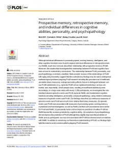
Figure 1 Rastergrams and spike-density functions (SDFs) of the neuronal activity to each cue stimulus (top panels, auditory cues; bottom panels, visual cues). Each dot below a raster line indicates one lick. a, b, Dual response to an auditory cue of a representative 546
5
50
0 –1
0
1
2 Time (s)
3
4
5
neuron in the posterior intralaminar nucleus. c, d, The similar response of a representative neuron in the lateral posterior nucleus, except that the cue was a visual stimulus.
© 2001 Macmillan Magazines Ltd
NATURE | VOL 412 | 2 AUGUST 2001 | www.nature.com
letters to nature
a
b
200
Cue
Reward
Reaction time (ms) 0
Water Sucrose (50 µl) Sucrose (100 µl) ICSS (70 µA) ICSS (140 µA) Extinction
Spikes s–1
150
100
100
200
No response
50
0 –1
0
1
2 Time (s)
3
4
F4,33 = 45.2, P , 0.001, with post hoc Bonferroni test, P , 0.001), but the magnitude of the early response did not. Moreover, as the value of the reward increased, the latency to ®rst lick, that is reaction time, shortened (one-way ANOVA, F4,33 = 52.3, P , 0.001, with post hoc Bonferroni test, P , 0.001) (Fig. 2b). We tested a total of 56 neurons (35 auditory and 21 visual) with two response components in the same way. In 43 neurons (28 auditory and 15 visual), the magnitude of the late response was modulated signi®cantly by reward value, and the reward value was negatively correlated with the reaction time of the initial lick. Next, we examined the effects of reward delay time on the activity of these neurons. The delay between the end of the cue and the onset of reward was set as 0, 1 or 2 s. If the reward delay was reduced from 1 to 0 s (Fig. 2c) or increased from 1 to 2 s (Fig. 2d), a peak of the late response shifted to the left from approximately 3 to 2 s or the right from approximately 3 to 4 s, respectively, after the onset of the cue. During the ®rst seven trials after the delay period was altered from 1 to 0 or 2 s, the late component displayed two peaks, which were not necessarily accompanied by licking. In contrast to the late component, reward timing did not modulate the peak of the early component (F2,21 = 0.33, not signi®cant). We found similar adaptations to reward timing in the peak of the late responses of all 41 neurons tested (25 auditory and 16 visual). Third, we assessed the plasticity of the cells during extinction of learning and relearning of stimulus±reward association (Fig. 3). After eight pre-extinction trials (block 1), the rat continued to lick the spout during the ®rst eight extinction trials (block 2), and then ceased licking the spout in the following trials (blocks 3±9). During these extinction trials, the magnitude of the early responses decreased slowly to reach a stable level by block 7, which was larger than the response to the neutral stimuli. The magnitude of the late response, on the other hand, decreased rapidly and did not differ from spontaneous activity by block 3. For the next six blocks (4±9), the neural activity did not change signi®cantly. In the next a
5
b
Spikes s–1
150
Spikes s–1
c Reward
Cue
Delay = 1 s Delay = 0 s (trial 2) Delay = 0 s (trial 5) Delay = 0 s (trial 7–13)
100
No licking
150 100 50
1
(8 trials)
2
0
1 2
(8 trials)
3 4 5 6 7 8 9 10 11 12 Trial block number
50 3 0 –1
0
1
2 Time (s)
3
4
5
4
6
6
rnin
lea
(7 trials)
g
Delay = 1 s Delay = 2 s (trial 2) Delay = 2 s (trial 5) Delay = 2 s (trial 7–13)
100
(7 trials)
Re
5
tion
Reward
Cue
7
12
(7 trials)
8
50
9
1
2 Time (s)
3
4
5
6
Figure 2 Effects of reward value and timing on the activity of the neuron with two response components, shown in Fig. 1a. The cue was tone 1. a, SDFs generated by different reward values. b, Histograms of reaction time (the interval between protrusion of the spout and the onset of licking). c, SDFs from the 2nd and 5th trials after changing the reward delay from 1 to 0 s. Trial 7±13 is the average SDF from trials 7±13 after changing the delay. d, SDFs from the 2nd and 5th trials after changing the delay from 1 to 2 s. Trial 7±13 is the average SDF from trials 7±13 after changing the delay. NATURE | VOL 412 | 2 AUGUST 2001 | www.nature.com
(1 trial)
10
(7 trials) Cue
0
(7 trials)
11
(7 trials)
–1 0 –1
Reward
(8 trials)
inc
150
Early Late
(8 trials)
Ext
d
Spikes s–1
200
Spikes s–1
self-stimulation (ICSS) in the medial forebrain bundle as a reward. Figure 1 shows two representative examples of responsive neurons in the posterior thalamic region. These neurons exhibited two temporally separated response components to auditory (Fig. 1a, b) or visual (Fig. 1c, d) stimuli that predicted a reward: the early component at the onset of stimulus presentation and the late component before the reward delivery. The early component was suppressed and the late component disappeared if the stimulus did not predict a reward. We isolated and tested a total of 593 neurons in the posterior thalamic region. Of these cells, 377 responded to one or more of the conditioned stimuli in this task. Of these responsive neurons, 185 showed responses with the two response components shown in Fig. 1. We next looked at the type of information that is conveyed in the early and late components of the responses. First, we investigated the effects of a change in value of the reward on the activity of the neurons with two response components (Fig. 2a). The same cue stimulus was associated with ®ve different rewards: water (50 ml); two different amounts of sucrose solution (50 and 100 ml); and two different intensities of ICSS (70 and 140 mA). Reward value was assumed to increase in the order21: water, ,50 ml sucrose solution and ,100 ml sucrose solution for the natural rewards; and 70 mA ICSS and ,140 mA ICSS for the arti®cial rewards. For the neuron in Fig. 2a, as reward value increased, the magnitude of the late response component also increased (one-way analysis of variance (ANOVA)
0
1
(1 trial)
Reward
3 2 Time (s)
4
Cue
5
–1
0
1
Reward
3 2 Time (s)
4
5
Figure 3 The plasticity of responses to tone 1 of the neuron shown in Fig. 1a during extinction and relearning. The shaded areas indicate trials with reward delivery. a, SDFs across blocks of trials. Each block consists of 7 or 8 trials, except for blocks 10 and 11 (single trial). Arrows indicate trials during which the rat licked the spout after the delay period. Scale bar, 50 spikes s-1. b, Change in the magnitude of the early and late responses. Each asterisk shows that the response magnitudes were signi®cantly different than those of each previous block (one-way ANOVA F11,64 = 64.9, P , 0.001, with post hoc Bonferroni test, all P , 0.001). The dotted line indicates the ®ring level of the early response to a neutral auditory stimulus.
© 2001 Macmillan Magazines Ltd
547
letters to nature trial (block 10), an ICSS reward was delivered after the cue, regardless of the rat's behavioural response, to reinstitute the association between the stimulus and the reward. The rat began to lick the spout again (block 11), and the magnitude of both the early and late responses recovered rapidly to pre-extinction levels. The magnitude of the late response correlated well with the behavioural responses: both rapidly decreased and increased during extinction and relearning, respectively. On the other hand, the magnitude of the early component slowly decreased during extinction, but rapidly recovered during relearning. Extinction does not erase the memory of conditioning22, as the behavioural response returns readily with relearning. The asymmetrical change in the early response during extinction and relearning indicated that this component was in¯uenced by the memory of a previously acquired stimulus±reward association. Even after behavioural extinction, the early response to the conditioned stimuli (n = 185 neurons) was signi®cantly greater than that to the neutral stimuli (paired t-test, P , 0.001). Furthermore the responses to the highest and the lowest frequency cue tones, which were associated with rewards, were greater than to the intermediate,
a Primary sensory cortex
Secondary sensory cortex
Perirhinal cortex
Reward circuit
LP
DLG
Frontal cortex
Thalamus SG MGD
Amygdala Striatum
SN VTA
LHA
MGM
MGV
PIL Goal-directed behaviour Non-primary pathway
Primary pathway Auditory Visual
Auditory/visual
b DLG (n = 43)
RI Early (auditory) Early (visual) Early (audiovisual) Late
1.0 0.5 0 –1.0
–0.5
0
MGV (n = 59)
0.5
1.0
MI
LP (n = 35) SG (n = 35)
0.5 0 –1.0
–0.5
0.5
1.0
0.5
1.0
MI
0.5
0.5 0 –0.5
0
MGD (n = 28) RI MGM (n = 40) 1.0 PIL (n = 47)
RI
1.0
–1.0
RI 1.0
0
0.5
1.0
MI
0 –1.0
–0.5
0
MI
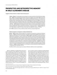
Figure 4 Functional topography. a, Parallel pathways through the auditory and visual thalamic nuclei (recording sites). In the auditory system, the primary pathway ascends from the ventral part of the medial geniculate nucleus (MGV) to the primary auditory cortex. The non-primary pathway ascends from the dorsal or medial part of the medial geniculate nucleus (MGD and MGM, respectively), suprageniculate nucleus (SG) and posterior intralaminar nucleus (PIL) to the secondary auditory cortex. In the visual system, the primary pathway is from the dorsal lateral geniculate nucleus (DLG) to the primary visual cortex. The extrageniculate or non-primary pathway is from the lateral posterior nucleus (LP) to the secondary visual cortex. SN, substantia nigra; VTA, ventral tegmental area; LHA, lateral hypothalamic area. b, The effects of the sensory speci®city and reward association in each nucleus. The indices (MI and RI) of the neuron shown in Fig. 1a and b were 0.98 and 0.22, respectively, for the early component and -0.06 and 1.00, respectively, for the late component. MI and RI values of the neuron shown in Fig. 1c and d were -1.01 and 0.23, respectively, for the early component and -0.01 and 1.00, respectively, for the late component. 548
non-rewarded tones (one-way ANOVA, F2,405 = 988.8, P , 0.001, with post hoc Bonferroni test, P , 0.001) and equivalent to one another (P = 0.11). The results are different from previous studies23,24 on the sensory properties of the posterior thalamus of naive rats that demonstrated that neurons are more sensitive to higher frequency tones within the same frequency range that was used in our study. Therefore, differences of the early component documented here probably result from the experiences of reward contingencies rather than the physical properties of the stimuli. Finally we investigated the functional anatomy of these responses. Thalamocortical connections are categorized into two parallel primary and non-primary pathways, and the posterior thalamic region contributes to both of these (Fig. 4a)16,17,25,26. All of the 185 neurons with two response components (Fig. 1) were located in the non-primary thalamic nuclei. Of these, the neurons recorded in the dorsal or medial part of the medial geniculate nucleus and the posterior intralaminar nucleus coded predominantly auditory stimuli in the early phase. The neurons in the lateral posterior nucleus and suprageniculate nucleus coded visual stimuli. In contrast, the cells in the primary thalamic region showed a clear modality speci®city without reward dependency: the ventral medial geniculate nucleus neurons and the dorsal lateral geniculate neurons responded only to the auditory and visual stimuli, respectively, and only during the cue presentation. The magnitude of the responses of these cells did not change after the manipulation of reward contingency. Figure 4b summarizes the relationships between sensory speci®city and reward dependency for the responsive neurons in the sample. How does the dual temporal coding in the posterior thalamus work together with processing reward information in other brain areas2±4,6±15? The functional distribution shown in Fig. 4b is consistent with the fact that only the non-primary thalamus sends efferents directly to components of the reward circuit, such as the amygdala, striatum and perirhinal cortex (Fig. 4a)16,17. Because the magnitude of the early components varied with the conditioning experience (Figs 1 and 3), the posterior thalamus might serve as a ®lter for the forebrain that favours behaviourally signi®cant stimuli over the others by amplifying response magnitude. This hypothesis also corresponds with a theoretical model and with human studies that stipulate the thalamus, especially the primate pulvinar (homologue of the rat lateral posterior nucleus26), selectively enhances attended sensory signals20,27. On the other hand, the climbing activity that characterizes the late responses may code prospectively for the anticipated reward. These responses probably arise from interaction with the other brain areas contributing to reward expectation2±4,8,9,11,12,14,15, via the perirhinal cortex. This is because the perirhinal cortex seems to be important in attaching motivational signi®cance to stimuli28, and among the constituents of the presumed reward circuits, only the perirhinal cortex is known to send the direct inputs to the non-primary thalamus16. In this way, the posterior thalamus could communicate with the reward circuits, through the dual temporal coding for mnemonic and anticipatory process. M
Methods Animal care The experimental procedures were similar to those used previously9,11, and the experiments were conducted in accordance with the US National Institutes of Health Guide for the Care and Use of Laboratory Animals, and with Guideline for the Care and Use of Laboratory Animals in Toyama Medical and Pharmaceutical University.
Animal preparation Twelve male Wistar rats (270±330 g) were anaesthetized (sodium pentobarbital 40 mg per kg, intraperitoneal injection) and had a cranioplastic cap attached to the skull. This made it possible to ®x the animal's head painlessly in the stereotaxic device. At the same time, two concentric bipolar electrodes for ICSS were implanted in the medial forebrain bundle (anterior, -3.8 from bregma; lateral, 6 1.4 mm; ventral, 8.4 mm or anterior, -5.2 mm; lateral, 6 0.8 mm; ventral, 8.2 mm) according to the atlas of Paxinos and Watson29. A hole
© 2001 Macmillan Magazines Ltd
NATURE | VOL 412 | 2 AUGUST 2001 | www.nature.com
letters to nature (3.0±5.0 mm diameter) for chronic recording was drilled through the cranioplastic cap and the underlying skull.
Behavioural task The rats were required to lick a spout to obtain rewards of 0.3 M sucrose solution or ICSS (0.5-s train of 100-Hz, 0.3-ms capacitor-coupled negative square wave pulses). We monitored licking behaviour with a photoelectric sensor. The threshold level for ICSS to maintain the licking behaviour in the operant task was determined in behavioural tests before recording. In this study, the thresholds ranged from 65 to 110 mA. The licking frequency depended on the method of reward delivery, that is, low for ICSS and high for a liquid reward. This was probably because ICSS was delivered only once at the ®rst lick even if the rat licked several times, whereas liquid was delivered continuously during a 2-s reward period, although the property of the reward, natural or arti®cial, could also be involved in this difference. In the cued operant task, a 1,200 Hz tone (tone 1) and a white light on the left side (light 1) signalled availability of the ICSS, and a 4,300 Hz tone (tone 2) signalled availability of sucrose solution. A 2,800 Hz tone (tone 3) and a white light on the right side (light 2) were neutral, that is, not associated with reward. A speaker positioned 10 cm anterior to the rats delivered tones that were 85 dB at each ear and two white light lamps, 458 lateral and 7 cm from each eye, delivered the visual stimuli.
Electrophysiology Before the unit recording session, we searched for auditory neural responses by clapping and, thus, localized the margins of the medial geniculate body. On the basis of the coordinates derived from this localizing procedure, a glass-insulated tungsten microelectrode (Z = 1.0±1.5 MQ at 1,000 Hz) was inserted stereotaxically stepwise by a micromanipulator into various parts of the posterior thalamus. Extracellular discharges of single neurons were recorded using conventional recording procedure.
Histology At the end of recording session, several small electrolytic lesions (20 mA for 20 s) were made stereotaxically around the recording sites with a glass-insulated tungsten microelectrode. The brains were sectioned coronally (30 mm) and stained with cresyl violet. All marking and stimulation sites were then veri®ed microscopically. The positions of the recorded neurons were mapped onto the appropriate tissue sections, using stereotaxic coordinates, and the sections were compared with the atlas of Paxinos and Watson29.
Data analysis Spike trains were smoothed by convolution with a gaussian kernel (j = 20 ms) to obtain spike-density functions (SDFs)30. Spontaneous activity was de®ned as the mean discharge rate during the 1,000 ms preceding cue onset. The early response was de®ned as the number of spike counts in the ®rst 100 ms after cue onset. The late response was de®ned as the number of spike counts in the 500 ms period from 2,500 to 3,000 ms after cue onset. A neuron was classi®ed as responsive to any cue stimuli if the difference in neural activity in three periods (spontaneous activity, early response and late response) was signi®cant (one-way ANOVA, P , 0.01). A modality coupling index (MI) and a reward coupling index (RI) were computed for each response component: MI = (Faud(ICSS) + Faud(ext) - Fvis(ICSS) - Fvis(ext))/Fsum; RI = (F aud(ICSS) - Faud(ext) + F vis(ICSS) - F vis(ext))/F sum, where Fsum = Faud(ICSS) + Faud(ext) + Fvis(ICSS) + Fvis(ext). Faud(ICSS) or F vis(ICSS) is the average ®ring rate to auditory stimulus or visual stimulus, respectively, associated with reward minus spontaneous ®ring rate in each neuron. Faud(ext) or Fvis(ext) is the average ®ring rate to auditory stimulus or visual stimulus, respectively, in the extinction test minus spontaneous ®ring rate in each neuron. MI represents sensory modality selectivity of the neural responses and RI represents reward dependency. For example, if the value of MI was 1.0 or -1.0, this indicates a purely auditory-speci®c response or a visual one, respectively. An RI value of 1.0 or 0 indicates that a neural response is totally dependent on or independent of reward contingency, respectively. In principle, memory and anticipation are mutually dependent. However, the temporal response patterns (Fig. 1) and the effects of manipulating reward parameters (Figs 2 and 3) suggested that the early and late components conveyed distinguishable information. Therefore we designated the early component as related to memory (retrospective coding) and the late one as related to anticipation (prospective coding). Received 26 January; accepted 13 June 2001. 1. Dickinson, A. & Balleine, B. Motivational control of goal-directed action. Anim. Learn. Behav. 22, 1±18 (1994). 2. Schultz, W. Multiple reward signals in the brain. Nature Rev. Neurosci. 1, 199±207 (2000). 3. Schultz, W., Tremblay, L. & Hollerman, J. R. Reward processing in primate orbitofrontal cortex and basal ganglia. Cereb. Cortex 10, 272±283 (2000). 4. Rolls, E. T. The Brain and Emotion. (Oxford Univ. Press, New York, 1999). 5. Rainer, G., Rao, S. C. & Miller, E. K. Prospective coding for objects in primate prefrontal cortex. J. Neurosci. 19, 5493±5505 (1999). 6. Fibiger, H. C. & Phillips, A. G. in Handbook of PhysiologyÐThe Nervous System Vol. IV (ed. Bloom, F. E.) 647±675 (Williams and Wilkins, Baltimore, 1986). 7. Wise, R. A. & Rompre, P. P. Brain dopamine and reward. Annu. Rev. Psychol. 40, 191±225 (1989). 8. Mirenowicz, J. & Schultz, W. Preferential activation of midbrain dopamine neurons by appetitive rather than aversive stimuli. Nature 379, 449±451 (1996). 9. Nakamura, K., Ono, T. & Tamura, R. Central sites involved in the lateral hypothalamus conditioned neural responses to acoustic cues in the rat. J. Neurophysiol. 58, 1123±1148 (1987). 10. Robbins, T. W. & Everitt, B. J. Neurobehavioral mechanism of reward and motivation. Curr. Opin. Neurobiol. 6, 228±236 (1996).
NATURE | VOL 412 | 2 AUGUST 2001 | www.nature.com
11. Ono, T., Nishijo, H. & Uwano, T. Amygdala role in conditioned associative learning. Prog. Neurobiol. 46, 401±422 (1995). 12. Schoenbaum, G., Chiba, A. A. & Gallagher, M. Orbitofrontal cortex and basolateral amygdala encode expected outcomes during learning. Nature Neurosci. 1, 155±159 (1998). 13. Bechara, A., Damasio, H., Tranel, D. & Anderson, S. W. Dissociation of working memory from decision making within the human prefrontal cortex. J. Neurosci. 18, 428±437 (1998). 14. Watanabe, M. Reward expectancy in primate prefrontal neurons. Nature 382, 629±632 (1996). 15. Kawagoe, R., Takikawa, Y. & Hikosaka, O. Expectation of reward modulates cognitive signals in the basal ganglia. Nature Neurosci. 1, 411±416 (1998). 16. LeDoux, J. E., Farb, C. R., & Romanski, L. M. Overlapping projections to the amygdala and striatum from auditory processing areas of the thalamus and cortex. Neurosci. Lett. 134, 139±144 (1991). 17. Linke, R., De Lima, A. D., Schwegler, H. & Pare, H.-C. Direct synaptic connections of axons from superior colliculus with identi®ed thalamo-amygdaloid projection neurons in the rat: possible substrates of a subcortical visual pathway to the amygdala. J. Comp. Neurol. 403, 158±170 (1999). 18. Weinberger, N. M. Learning-induced changes of auditory receptive ®elds. Curr. Opin. Neurobiol. 3, 570±577 (1993). 19. LeDoux, J. E. Emotion circuits in the brain. Ann. Rev. Neurosci. 23, 155±184 (2000). 20. Dolan, R. J. in The New Cognitive Neurosciences 2nd edn (ed. Gazzaniga, M. S.) 1115±1131 (MIT Press, Cambridge, Massachusetts, 2000). 21. Gallistel, C. R., Leon, M., Waraczynski, M. & Hanau, M. S. Effect of current on the maximum possible reward. Behav. Neurosci. 105, 901±912 (1991). 22. Bouton, M. E. Context, ambiguity, and classical conditioning. Curr. Direct. Psychol. Sci. 3, 49±53 (1994). 23. Bordi, F. & LeDoux, J. E. Response properties of single units in areas of rat auditory thalamus that project to the amygdala. I. Acoustic discharge patterns and frequency receptive ®elds. Exp. Brain Res. 98, 261±274 (1994). 24. Edeline, J. M., Manunta, Y., Nodal, F. R. & Bajo, V. M. Do auditory responses recorded from awake animals re¯ect the anatomical parcellation of the auditory thalamus? Hear. Res. 131, 135±152 (1999). 25. Winer, J. A. & Morest, D. K. The medial division of the medial geniculate body of the cat: implications for thalamic organization. J. Neurosci. 3, 2629±2651 (1983). 26. Sefton, A. J. & Dreher, B. in The Rat Nervous System 2nd edn (ed. Paxinos, G.) 833±898 (Academic, San Diego, 1995). 27. LaBerge, D. Thalamic and cortical mechanisms of attention suggested by recent positron emission tomographic experiments. J. Cogn. Neurosci. 2, 358±372 (1990). 28. Liu, Z., Murray, E. A. & Richmond, B. J. Learning motivational signi®cance of visual cues for reward schedules requires rhinal cortex. Nature Neurosci. 3, 1307±1315 (2000). 29. Paxinos, G. & Watson, C. The Rat Brain in Stereotaxic Coordinates 4th edn (Academic, San Diego, 1998). 30. Richmond, B. J. & Optican, L. M. Temporal encoding of two-dimensional patterns by single units in primate inferior temporal cortex. II. Quanti®cation of response waveform. J. Neurophysiol. 57, 147± 161 (1987).
Acknowledgements We thank R. Norgren (invited by Gofo Life Sciences International Fund) for helpful comments on this manuscript, and T. Kitamura for technical assistance. This work was partly supported by Grants-in-Aid for Scienti®c Research from the Japanese Ministry of Education, Science and Culture. Correspondence and requests for materials should be addressed to T.O. (e-mail:
[email protected]).
................................................................. Practising orientation identi®cation improves orientation coding in V1 neurons Aniek Schoups*, Ru®n Vogels*, Ning Qian² & Guy Orban* * Laboratorium voor Neuro-en Psychofysiologie, K.U. Leuven Medical School, B-3000 Leuven, Belgium ² Center for Neurobiology and Behavior, and Department of Physiology and Cellular Biophysics, Columbia University, New York 10032, USA ..............................................................................................................................................
The adult brain shows remarkable plasticity, as demonstrated by the improvement in ®ne sensorial discriminations after intensive practice. The behavioural aspects of such perceptual learning are well documented, especially in the visual system1±8. Speci®city for stimulus attributes clearly implicates an early cortical site, where receptive ®elds retain ®ne selectivity for these attributes; however, the neuronal correlates of a simple visual discrimination task remained unidenti®ed. Here we report electrophysiological correlates in the primary visual cortex (V1) of monkeys for learning orientation identi®cation. We link the behavioural improvement
© 2001 Macmillan Magazines Ltd
549