Segmentation-Based Multilayer Diagnosis Lossless Medical Image Compression Xin Bai1, Jesse S. Jin1, 2, Dagan Feng1, 3 1 School of Information Technologies The University of Sydney, NSW 2006, Australia 2 School of Computer Science and Engineering University of New South Wales, NSW 2052, Australia 3 Centre for Multimedia Signal Processing, Dept. of EIE Hong Kong Polytechnic University, Hong Kong
[email protected]
Abstract Hospital and clinical environments are moving towards computerisation, digitisation and centralisation, resulting in prohibitive amounts of digital medical image data. Compression techniques are, therefore, essential in archival and communication of medical image. Although lossy compression yields much higher compression rates, the medical community has relied on lossless compression for legal and clinical reasons. In this paper, we propose a segmentation-based multilayer (SML) coding scheme for lossless medical image compression. A fully automatic unseeded region growing (URG) segmentation approach is used for extracting diagnostically important regions, i.e., the regions of interest (ROI), for multilayer lossless ROI compression with the efficient Barrows-Wheeler coding (BWC) and wavelet-based JPEG2000 coding. Our proposed SML compression scheme can provide efficient compression for various medical imaging data and offer potential advantages in content-based medical image retrieval and semantic progressive transmission in telemedicine.. Keywords: Medical image coding, lossless compression, segmentation, region-based image processing.
1
Introduction
Digital medical imaging, which produces digital pictures of human body, provides powerful tools for diagnosis, treatment and surgery, hence becomes a major aspect of modern healthcare delivery. It is a fast growing area with the richest source of information and variety of modalities such as X-ray transmission imaging (X-ray), magnetic resonance imaging (MRI), computerised tomography (CT), ultrasound images, positron emission tomography (PET), X-ray mammography (MG), and many others. The use of digital medical imaging, however, is accompanied by substantial large amount of image data.
Copyright © 2004, Australian Computer Society, Inc. This paper appeared at the Pan-Sydney Area Workshop on Visual Information Processing (VIP2003), Sydney. Conferences in Research and Practice in Information Technology, Vol. 36. M. Piccardi, T. Hintz, X. He, M. L. Huang, D. D. Feng, J. Jin, Eds. Reproduction for academic, not-for profit purposes permitted provided this text is included.
Even though rapid progress has been made in mass storage density and computer network performance, the volume of digital imagery produced by hospitals has been increasing as well. The demand for transmission bandwidth and storage space in the digital radiology environment continues to outstrip the capabilities of available technologies (Huang 1999, Wong, Zaremba, Gooden and Huang 1995). Therefore, there remains a strong demand for digital medical image compression. During the past two decades, various image compression techniques have been proposed (Egger, Fleury, Ebrahimi and Kunt 1999, Kuduvalli and Rangayyan 1992, Taubman and Marcellin 2001), and can be classified mainly into lossless and lossy methods in terms of their ability to reconstruct from the compressed images. Lossless compression techniques (Akune, Yonekawa, Ishimitsu, Takeuchi, Dio and MacMahon 1991, Chande, Das and Ganesan 1998, Das and Burgett 1993, Gruter, Egger, Vesin and Kunt 2000, Ramabadran and Chen 1992, Roos, Viergever, Dijke and Peters 1988) ensure complete data fidelity but with low compression ratios, while the lossy techniques (Chen and Ramabadran 1994, Chiu, Vaisey and Atkins 2001, Effros 2000) aim to achieve much higher compression ratios by allowing some acceptable degradation in the recovered image. However, the medical professionals have been reluctant to adopt lossy compression since any information loss or error caused by compression process could affect clinical diagnostic decisions and legal challenge could be raised. The medical community has therefore instead relied on lossless compression methods, though efficient compression techniques are highly needed for medical applications. The true goal is to maximize compression while maintaining clinical relevance. A possible solution to the above dilemma is to offer hybrid compression schemes. A general theme is to provide lossless compression for diagnostically important regions (i.e., region-of-interest, or ROI), while allowing lossy compression for regions other than ROI (i.e., nROI, or unimportant regions). Such hybrid compression can also be referred to regionally lossless coding or diagnosis lossless compression. Some researchers have investigated and proposed various hybrid medical image compression techniques (Bokturk, Tomasi, Girod and Beaulieu 2001, Bruckmann and Uhl 2000, Chen, Zhang and Parker 1994, Chen, Zhang, Luo and Parker 1995, Liu, Xiong, Wu,
Compressor (Multilayer RO I / Residu al C oding) ROI Index Coding
Preprocessor (Multilayer Segm ent ation)
Segme nted ROI Set (PRL+NRL)
Barrows-Wheeler Coding (BWC)
Unseeded Region Growing (URG) Algorithm Original Medical Image
PRL Residual Lossles s Coding
Resid ual ROI Set Optima l Residual Extraction
NRL Residual Lossy- Loss less Coding
… … … …
… … … …
Multilayer Residual Coding
Backgr ound Image Set (BRL)
Diag nosis Lossless Comp ressed Image
Fully Lossless Comp ressed Image
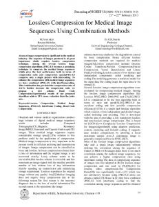
Figure 1: Flow diagram for proposed SML compression scheme Wang and Castleman 2002, Strom and Cosman 1997, Wong, Guan and Hong 1995). The proposed compression methods differ according to the different ROI delineation / segmentation approaches (i.e., manual or automatic), the different segmentation goals / layouts (i.e., a rough background-foreground distinction or a more focused segmentation), and the different coding approaches for ROIs and nROIs (i.e., subband, DCT, vector quantization, wavelets, etc.). Chen et al (1994) made use of ROIs using subband analysis and synthesis of volumetric datasets using wavelets. A structure-preserving adaptive quantization method was reported in (Chande, Das and Ganesan 1998) for further improving ROI compression. A region-coding method using predictive pruned treestructured vector quantization was proposed in (Poggi and Olshen 1995), with CT chest segment using thresholding, connectivity, and boundary smoothing. In (Wong, Guan and Hong 1995), a self-organizing neural network was first used to separate the breast area from the mammogram background, and an optimized JPEG coding method was applied to breast ROIs. In (Vlaicu, Lungu, Crisan and Persa 1995), a ROI-DCT algorithm that uses more DCT coefficients in ROI was proposed. A ROIbased compression method with two multi-resolution coding schemes (wavelet zerotree coding and the Stransform) was reported in (Strom and Cosman 1997). In (Shen and Rangayyan 1997), a hybrid compression method based on an adaptive scanning pattern and JBIG coding was proposed for high-resolution radiographic images. Recently, a motion compensated ROI coding for colon CT images was investigated in (Bokturk, Tomasi, Girod and Beaulieu 2001) and a modified SPIHT ROI coding was reported in (Liu, Xiong, Wu, Wang and Castleman 2002) for chromosome images. Most of the above region-based compression techniques seem to give good performance only for some specific types of medical images. When applied to the practice of medicine, they could meet some difficulties. The research in (Chen, Zhang and Parker 1994, Chen, Zhang, Luo and Parker 1995) required manual ROI definition which is operator dependent and thus prone to reproducibility errors and it is also time consuming. The method in (Wong, Guan and Hong 1995) simply provided foreground / background segmentation and could be only suitable for mammogram which consists of a large uninteresting background. On the other hand, the
techniques in (Bokturk, Tomasi, Girod and Beaulieu 2001, Liu, Xiong, Wu, Wang and Castleman 2002) supported automatic segmentation, however their region coding approaches are sophisticated and again only for specific types of data such as chromosome images. The region coding method in (Shen and Rangayyan 1997) was simple and efficient, but its segmentation only group spatially connected pixels lying within a small dynamic grey level range, and cannot provide object identification or analysis of clinical features. Such scheme may not tailor for interactive / semantic progressive transmission and ROI-based storage and retrieval. In this paper, we propose a segmentation-based multilayer (SML) diagnosis lossless compression scheme for medical image. The original medical image is first automatically segmented into a multilayer form and following by ROI index lossless coding and multilayer residual coding (lossless or lossy to lossless depend on different clinical studies). Our hybrid compression scheme can be widely used in various medical imaging applications, and bring an efficient way for rapid image transmission, long-term archiving and content-based medical image retrieval. The paper continues as follows: Section 2 describes proposed hybrid compression scheme. Some preliminary results are reported in Section 3, and lastly conclusion and discussion of possible future work in Section 4.
2
Proposed SML Compression Scheme
This section describes our proposed segmentation-based multilayer (SML) diagnosis lossless compression scheme. The overall flow of the compression scheme is shown in Figure 1.
2.1
Multilayer Segmentation
In this stage, the multilayer segmentation is treated as a SML pre-processor which includes initial automatic region segmentation based on an unseeded region growing (URG) algorithm, and optimal residual extraction. In region-based coding, the original image is segmented into various spatial regions based on their intensity or greyscale characteristics, and represented as a segmented index map (SIM). In our proposed scheme, any segmented region may belong to one of the following layers based on its diagnosis importance level:
•
•
•
Primary ROI Layer (PRL): all diagnostically important regions. They may vary depend on different clinical studies; Normal ROI Layer (NRL): unimportant regions which surround the PRL regions. They may help the clinician to easily observe and locate PRL within the original image, and evaluate possible interactions with surrounding organs; Background Region Layer (BRL): regions other than PRL and NRL regions. They mainly locate outside human body / organs and without any diagnostic value.
Here, we have: PRL + NRL + BRL = SIM
(1)
Unseeded Region Growing (URG)
Region-based segmentation methods postulate that neighboring pixels within the same region have similar intensity values. The general procedure is to compare a pixel with its immediate surrounding neighbours. If a criterion of homogeneity is satisfied, the pixel can be classified into the same class as one or more of its neighbours. The choice of homogeneity criterion is therefore critical to the success of the segmentation. Seeded region growing (SRG) method (Adama and Bischof 1994) is a well known region-based segmentation approach that segments intensity images into regions based on a marker set (seeds). The selection of seeds determines what is a feature of interest in the image and what is irrelevant. As the name implies, region growing is a procedure that groups pixels into larger regions. Border pixels are added to regions in an order that depends on the similarity between the pixel and the marked region, thus the segmentation result is highly dependent on the choice of seeds. A new segmentation method known as “unseeded region growing” (URG) was developed recently (Lin, Jin and Talbot 2000, Lin 2000) which is similar to SRG except that no explicit seed selection is necessary: it does not rely on fine-tuning homogeneity parameters, nor does it require manual inputs / seeds. The seeds can be generated by the segmentation procedure automatically. Therefore, the URG can achieve fully automatic segmentation which is robust, easy to use and can incorporate high level knowledge of the image composition through the choice of region threshold that can be conceptualized as the contrast between different regions. Formally, the URG segmentation process initializes with region A1 containing a single image pixel, and the running state of the segmentation process consist of a set of identified regions, A1, A2, …, An. Let T be the set of all unallocated pixels which borders at least one of these regions:
(2)
i =1
where N(x) are immediate neighboring pixels of point x. Further, we define a difference measure
δ ( x, Ai ) = g ( x ) − mean y∈A [ g ( y )] i
(3)
where g(x) denotes the image value at point x, and i is an index of the region such that N(x) intersect Ai. The growing process involves selecting a point z ∈ T and region Aj where j ∈ [1, n] such that
δ ( z, A j ) = min {δ ( x, Ak } x∈T ,k∈[1,n ]
Note that some NRL regions could become PRL regions if the image is used for different clinical studies.
2.1.1
n
T = {x ∉ U Ai ∧ ∃k : N ( x ) I Ak ≠ Θ}
(4)
If δ(z, Aj) is less than the predefined threshold t, then the pixel is added to Aj. Otherwise, we must choose the most substantially similar region A such that
A = arg min Ak {δ ( z, Ak }
(5)
If δ(z, A) < t, we can assign the pixel to A. If neither of these two conditions above apply, then it is apparent that the pixel is significantly different from all the regions found so far, so a new region, An+1 would be identified and initialized with point z. In all three cases, the statistic of the assigned region must be updated once the pixel has been added to the region. The URG segmentation procedure is inherently iterative, and the above process is repeated until all pixels have been allocated to a region. For convenience, the initial starting point has been chosen to be the first image pixel, but preliminary investigations have suggested that the starting position does not have a significant influence on the segmentation result.
2.1.2
Optimal Residual Extraction
After applying the URG algorithm for initial segmentation, we can obtain a segmented ROI set (i.e., ROI index image) which includes PRL and NRL regions, and a background image set (i.e., BRL regions). Since the ROI region needs to be lossless-compressed, its residual image set, i.e., the difference image between the ROI index image and the original image (except the BRL), must also be extracted for later coding. Such residual coding could result in low compression ratios because of the large amounts of code needed for encoding the difference (all the non-zero pixels in the residual image). Minimizing the number of non-zero pixels (or maximizing the number of zero pixels) in the residual image could therefore achieve better residual coding. In general, after the region segmentation, a segmented index map is derived by filling the individual region with the average value of original pixels inside this region. It may not result in maximum zero pixels in the residual image. In our proposed scheme, instead of using the average value, we pick up the modal value (i.e., the mostfrequently-appeared value) in the region as its index value. It provides an optimal residual extraction and provide better residual coding.
2.2
Multilayer ROI / Residual Coding
The compressor used in our proposed scheme consists of two components: the ROI index coding based on Barrows-Wheeler lossless coding (BWC) and the multilayer residual coding which includes PRL lossless coding and NRL lossy to lossless coding, as shown in Figure 1.
2.2.1
Barrows-Wheeler Coding (BWC)
The Burrows-Wheeler Transform (BWT) is a reversible sequence transformation that is becoming increasingly popular for lossless data compression (Effros 2000). The initial idea of BWT is to apply a reversible transformation to a block of text to form a new block that contains the same characters, but is easier to compress by simple compression algorithms. The transformation tends to group characters together so that the probability of finding a character close to another instance of the same character is increased substantially. Text of this kind can easily be compressed with fast locally adaptive algorithms, such as move-to-front (MTF) (Bentley, Sleator, Tarjan and Wei 1986) encoding in combination with Huffman or arithmetic coding (Burrows and Wheeler 1994). The transformation operates by lexicographically sorting the symbols in the data block, using its context as the sort key. The contexts are compared for sorting by working from right to left, and comparisons go as far back as necessary to order any two contexts. However, most of the time, only a few symbols are needed to distinguish between contexts. The transformed data is simply the symbols in the order of their sorted contexts, so the permuted data block contains a special permutation of the original data. The MTF transformation is further applied on the transformed data. In MTF transform, a symbol is deleted from its current position and moved to the front of the list after it is used. This attempts to ensure that frequently used symbols appear near the front of the list. If a symbol has been recently used then it will be near the front of the list and therefore have a short decimal encoding (Bentley, Sleator, Tarjan and Wei 1986). The advantage of using MTF transform is to increase uneven distribution of symbols, hence it results more efficient entropy coding. Detailed algorithms description can be found in (Burrows and Wheeler 1994).
be compressed in lossy manner for rapid transmission, or in lossless mode for long-term archiving; the rest BRL regions are not encoded in our diagnosis compression scheme but can be simply marked / displayed as entire black by the decoder. Since the wavelet-based JPEG2000 standard (Taubman and Marcellin 2001) is currently the basis for the best general-purpose lossy and lossless compression of still images, in this paper, we adopt it for both PRL and NRL residual coding. However, for different medical imaging applications, their residual images may appear with different statistic patterns. Simply applying JPEG2000 to these various residual images may not be optimal for coding. We are now conducting the research on adaptive coding techniques which can provide more accurate and efficient compression for different medical imaging modalities.
3
Preliminary Results
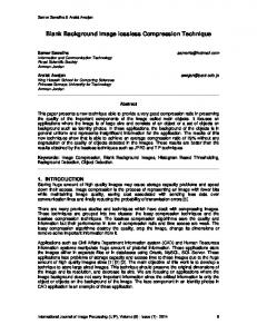
We first use a set of MRI brain images to test our SML pre-processor. The original image plane with tumour regions (i.e., the bright-grey mass shown in the up-right hand side of the brain) is shown in Figure 2(a). After the URG segmentation, we obtain the segmented ROI index image in Figure 2(b) with the PRL (tumour regions) and the NRL regions, and the background image (BRL) in Figure 2(c). Since this MRI image is for clinical tumour study, we mark all the tumour regions as PRL, and discard the BRL regions. If we apply the image for other pathology studies, for example, thalamus study, then related centre regions need to be marked as PRL. The residual ROI image is also derived using our optimal residual extraction method, as shown in Figure 2(d).
(a)
(b)
(c)
(d)
The BWT-based coding (BWC) technique (also called “the block-sorting compression”) works well on both text and non-text inputs. It provides fast compression and especially achieves much higher compression ratios for bi-level (binary) images and segmented images, because of the reduced symbol alphabet and repeated patterns.
2.2.2
Multilayer Residual Coding
The aim of our work is to develop a diagnosis lossless compression scheme which can deliver different levels of reconstruction quality (lossless or lossy) in different regions of the image, according to their diagnosis importance level, to maximize compression performance. The residuals of the pixels inside the PRL regions are coded in lossless mode; the residuals of NRL regions can
Figure 2: (a) The original MRI brain image with 256 by 256 and 8 bits of grey levels. (b) The segmented ROI index image (PRL+NRL). (c) The background image (BRL). (d) The residual ROI image.
In order to compare the compression performance, we conduct experiments based on two sets of images (MRI brain image and CT chest image), using the SLIC technique (Shen and Rangayyan 1997), JPEG2000 coding and our proposed SML scheme. The comparison results (with encoded bytes and compression ratios, i.e., CR) on MRI brain image and CT chest image are listed in Tables 1 and 2, respectively. Our SML technique has provided higher compression ratios than the other methods. In these two examples, we assume that all the brain regions (i.e., PRL and NRL regions) are diagnosis important regions and thus compressed in lossless manner for longterm digital archiving. Therefore the overall compression results listed are not with high performance. In the case of rapid transmission in teleradiology, we may only provide the tumour region at original quality by applying lossless coding on PRL, but the remainder of the image sustains degradation with lossy coding on NRL. In the example shown in Figure 2, the diagnosis important region (i.e., the circular tumour region) marked as PRL occupies only 5% of the pixels in the entire image. It therefore results a dramatically higher compression ratio and provides much better coding performance. Bytes CR
Original
SLIC
JPEG2k
SML
65,594
49,277
39,586
29,758
1:1
1.33:1
1.66:1
2.20:1
Table 1: Comparison of the proposed segmentationbased multilayer diagnosis lossless coding scheme with other methods on MRI brain image (256x256x8bits)
Bytes CR
Original
SLIC
JPEG2k
SML
262,202
128,758
73,452
48,709
1:1
2.04:1
3.57:1
5.38:1
Table 2: Comparison of the proposed segmentationbased multilayer diagnosis lossless coding scheme with other methods on CT chest image (512x512x8bits)
4
Conclusions
In region-based lossless compression techniques, the residual ROI coding is a very important step which determines the final compression performance. In our proposed SML coding scheme, although the residual ROI image is optimally extracted using modal value, it is then only coded by the universal approach – JPEG2000, instead of any tailored residual coding methods for medical imaging domain. A possible and promising improvement of compression could be the introduction of different codebooks for various medical imaging modalities and / or various clinical studies by using certain training sets and exploiting the prior known information as much as possible. Our proposed segmentation-based multilayer diagnosis lossless coding scheme could potentially open up interesting new avenues for content-based medical image retrieval. In our SML scheme, the image data are coded in
such a way that ROI shape and texture features are preextracted and directly stored in the ROI index map and multilayer residual set, respectively. It means that the SML compression scheme delivers basic information on the image content in the same process, and features from each ROI region can be easily extracted for indexing and fast retrieval. Progressive transmission is a highly desirable feature for many image compression applications, especially telemedicine. In our proposed SML scheme, the information in the bitstream is arranged in order of diagnosis importance. More important information, such as all PRL regions appear at the beginning of the bitstream, while less important information (i.e., NRL regions) appears towards the end. Acoarse version of the image can therefore be recovered by decoding the initial portion of the bitstream, and the image can be refined by progressive decoding process until perfect reconstruction is achieved. Furthermore, our SML scheme provides unique semantic progressive transmission which can facilitate the interactive selection of ROI.
5
Acknowledgements
This research is supported by the ARC and UGC grants.
6
References
Adams, R. and Bischof, L. (1994): Seeded region growing. IEEE Trans. Pattern Anal. Mach. Intelligence 16(6): 641-647. Akune, J., Yonekawa, H., Ishimitsu, Y., Takeuchi, H., Doi, K. and MacMahon, H. (1991): Development of a data compression module for digital radiography. Med. Biolog. Eng. Comput. 29(Suppl). Bentley, J.L., Sleator, D., Tarjan, R. and Wei, V. (1986): A locally adaptive data compression scheme. Communications of the ACM 29(4):320-330. Bokturk, S., Tomasi, C., Girod, B. and Beaulieu, C. (2001): Medical image compression based on region of interest with application to colon CT images. Proc. 23rd Annual International Conference of the IEEE on medical and Biomedical Engineering, Istanbul, TGurdey. Bruckmann, A. and Uhl, A. (2000): Selective medical image compression techniques for telemedical and archiving applications Computers in Biology and Medicine, 30:153-169. Burrows, M. and Wheeler, D.J. (1994): A block-sorting lossless data compression algorithm. SRC research report 124. Digital systems research center. Chande, S.B., Das, M. and Ganesan, S. (1998): An algorithm driven architecture for a lossless image compression scheme based on multiplicative autoregressive models. Proc. 1998 Midwest Symposium on Circuits and systems, 1998:395-398. Chen, C.W., Zhang, Y.Q. and Parker, K.J. (1994): Subband analysis and synthesis of volumetric medical images using wavelet. Visual Communications and
Image Processing’94, Chicago, IL, USA, 2308(3):15441555, SPIE, Bellingham, WA.
segmentation and improved prediction. IEEE Trans. Image Processing 4(6):734-742.
Chen, C.W., Zhang, Y.Q., Luo, J. and Parker, K.J. (1995): Medical image compression with structurepreserving adaptive quantization. Visual Communications and Image Processing’95, Taipei, Taiwan, 2501(2):983-994, SPIE, Bellingham, WA.
Ramabadran, T.V. and Chen, K. (1992): The use of contextual information in the reversible compression of medical images. IEEE Trans. Med. Imag. 11(2):185195.
Chen, K. and Ramabadran, R (1994): Near lossless compression of medical images through entropy-coded DPCM. IEEE Trans. Med. Imag. 13(3):538-548. Chen, Z.D., Chang, R.F. and Kuo, W.J. (1999): Adaptive predictive multiplicative autoregressive model for medical image compression IEEE Trans. Med. Imag. 18(2):181-184. Chiu, E., Vaisey, J. and Atkins, M. (2001): Waveletbased space-frequency compression of ultrasound images IEEE Trans. Info. Tech. Biomedicine 5(4)300310. Das, M. and Burgett, S. (1993): Lossless compression of medical images using two-dimensional multiplicative autoregressive models. IEEE Trans. Med. Imag. 12(4):721-726. Effros, M. (2000): PPM performance with BWT complexity: a new method for lossless data compression. Data Compression Conference 2000, 203212. Egger, O., Fleury, P., Ebrahimi, T. and Kunt, M. (1999): High-performance compression of visual information – a tutorial review – Part I: Still Pictures. Proceedings of the IEEE 87(6):976-1011. Gruter, R., Egger, O., Vesin, J. and Kunt, M. (2000): Rank-order polynomial subband decomposition for medical image compression. IEEE Trans. Med. Imag. 19(10):1044-1052. Huang, H.K. (1999): PACS Basic principles and applications. Wiley-Liss. Kuduvalli, G.R. and Rangayyan, R.M. (1992): Performance analysis of reversible image compression techniques for high-resolution digital teleradiology. IEEE Trans. Med. Imag. 11(3): 430-445. Lin, Z., Jin, J.S. and Talbot, H. (2000): Unseeded region growing for 3D image segmentation. Visualisation 2000. Conferences in Research and Practice in Information Technology, Sydney, Australia, 2:31-37, ACS. Lin, Z. (2000): 3D magnetic resonance image segmentation and medical image compression. Thesis. University of New South Wales, Sydney. Liu, Z., Xiong, Z., Wu, Q., Wang, Y. and Castleman, K. (2002): Cascaded differential and wavelet compression of chromosome images. IEEE Trans. Biomedical Eng. 49(4):372-383. Poggi, G. and Olshen, R.A. (1995): Pruned treestructured vector quantization of medical images with
Ramaswamy, A. and Mikhael, W. (1996): A mixed transform approach for efficient compression of medical images. IEEE Trans. Med. Imag. 15(3): 343-352. Roos, P., Viergever, M.A., Dijke, M.C.A. and Peters, J.H. (1988): Reversible intraframe compression of medical images. IEEE Trans. Med. Imag. 7(4):328-336. Shen, L. and Rangayyan, R. M. (1997): A segmentationbased lossless image coding method for high-resolution medical image compression. IEEE Trans. Med. Imag. 16(3):301-307. Strom, J. and Cosman, P. (1997): Medical image compression with lossless regions of interest. Signal Processing 59(1997):155-171. Taubman, D. and Marcellin, M. (2001): Image compression fundamentals, standards and practice. Norwell, MA: Kuluwer. Vlaicu, A., Lungu, S., Crisan, N. and Persa, S. (1995): New compression techniques for storage and transmission of 2-D and 3-D medical images. In Advanced Image and Video Communications and Storage Technologies, Amsterdam, Netherlands, 2451:370-377, SPIE. Wong, H.S., Guan, L. and Hong, H. (1995): Compression of digital mammogram database using a near-lossless scheme. Proc. ICIP-95, Washington DC, USA, II:2124, IEEE Computer Society Press, Sliver Spring. Wong, S., Zaremba, L., Gooden, D. and Huang, H.K. (1995): Radiologic image compression--A Review. Proc. IEEE 83(2):194-219.