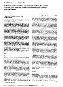
IL, intermediate layer; P, Purkinje cell. Asterisk and P in d-d are at corresponding positions. Bars (a-c") 50 ~m; (d-d") 10 lam. The Journal of Cell Biology, Volume ...
Sequential Expression and Differential Function of Multiple Adhesion Molecules during the Formation of CerebeUar Cortical Layers C h e n g - M i n g C h u o n g , K a t h r y n L. Crossin, a n d G e r a l d M. E d e l m a n Laboratory of Developmental and Molecular Biology, The Rockefeller University, New York 10021
Abstract. We have correlated the times of appearance of the neural cell adhesion molecule (N-CAM), the neuron-glia cell adhesion molecule (Ng-CAM), and the extracellular matrix protein, cytotactin, during the development of the chicken cerebellar cortex, and have shown that these molecules make different functional contributions to granule cell migration. Immunofluorescent staining showed distinct spatiotemporal expression sequences for each adhesion molecule. N-CAM was present at all times in all layers. However, the large cytoplasmic domain polypeptide of N-CAM was always absent from the external granular layer and was enriched in the molecular layer as development proceeded. Ng-CAM began to be expressed in the premigratory granule cells just before migration and later disappeared from cell bodies but remained on parallel fibers. Cytotactin, which is synthesized by glia and not by neurons, appeared first in a speckled pattern within the external granular layer and later appeared in a continuous pattern along the Bergmann glia; it was also enriched in the molecular layer. After we established their order of appearance, we tested the separate functions of these adhesion molecules in
granule cell migration by adding specific antibodies against each molecule to cerebellar explant cultures that had been labeled with tritiated thymidine and then measuring the differential distribution of labeled cells in the forming layers. Anti-N-CAM showed marginal effects. In contrast, anti-Ng-CAM arrested most cells in the external granular layer, while anti-cytotactin arrested most cells in the molecular layer. Time course analyses combined with sequential addition of different antibodies in different orders showed that anti-NgCAM had a major effect in the early period (first 36 h in culture) and a lesser effect in the second part of the culture period, while anti-cytotactin had essentially no effect at the earlier time but had major effects at a later period (18-72 h in culture). The two major stages of cerebellar granule cell migration thus appear to be differentially affected by distinct adhesion molecules of different cellular origins, binding mechanisms, and overall distributions. The results indicated that local cell surface modulation of adhesion molecules of different specificities at defined stages and sites is essential to the formation of cerebellar cortical layers.
Prominent feature during the development of the nervous system is the orderly formation of horizontal cellular and fibrous layers such as those found in the cerebeUar and the cerebral cortexes (Cowan, 1982). This morphogenetic process depends heavily on ordered cell migration in a radial direction, which depends in turn upon complex interactions among neurons and glia (Rakic, 1971). It has recently been suggested that the spatiotemporal regulation of adhesion among cells and substrates mediated by various adhesion molecules plays a regulatory role in determining the borders of histological layers in both neural and nonneural tissues, particularly by affecting cell movement (Edelman, 1985). To test this hypothesis in the central nervous system, it is useful to compare the functions of a set of adhesion molecules of different specificities, binding mechanisms, and cellular origins. By application of in vitro binding assays, two
neuronal cell adhesion molecules, the neural cell adhesion molecule (N-CAM) ~ and the neuron-glia cell adhesion molecule (Ng-CAM), have been identified, isolated, and structurally analyzed (for review see Edelman, 1986). Each of these structurally and functionally different neuronal membrane glycoproteins has a distinct binding specificity and binding mechanism. Both can mediate neuron-neuron and neuron-glia interactions, but each contributes differentially to different functions in various regions of the nervous system, depending upon dynamic modulation of its local expression (Hoffman et al., 1986). A substrate adhesion molecule, cytotactin, that is synthesized by glia but not by neurons
© The Rockefeller University Press, 0021-9525/87/02/331/12 $1.00 The Journal of Cell Biology, Volume 104, February 1987 331-342
1. Abbreviations used in this paper: EGL, external granular layer; IGL, internal granular layer; ld, large cytoplasmic domain of N-CAM; ML, molecular layer; N-CAM, neural cell adhesion molecule; Ng-CAM, neuron-glia adhesion molecule; PLZ, proliferative zone; PMZ, premigratory zone.
331
Figure 1. (a-a") E9 transverse sections; (b-b") El2 sagittal sections; (c-c") El8 sagittal sections; (a-c) cytotactin; (a'-c') Ng-CAM; (a"-c") N-CAM; (b") N-CAM ld. b" and b" and c and c' are pairs of double stained sections. Rectangular regions in b' are shown in Fig. 2, a and b; rectangular regions in c' are shown in Fig. 3 b. (Broken line) Pia mater. DN, deep nuclei; FZ, fibrous layer; IL, intermediate layer;
The Journal of Cell Biology, Volume 104, 1987
332
and mediates neuron-glia binding in vitro has also been identified (Grumet et al., 1985). Cytotactin is a general extracellular matrix protein that differs from other substrate adhesion molecules in its map of distribution in both neural and nonneural tissues during development (Crossin et al., 1986). Binding assays have indicated that the mechanisms of neuron-glia binding mediated by Ng-CAM and cytotactin are additive and thus may act separately (Grumet et al., 1985). To examine the regulatory roles played by these different adhesion molecules in cell migration and cortical layer formarion, we have chosen the histogenesis of cerebellar cortex for further study, both because it has been suggested that granule cell migration involves neuron-glia interactions (Rakic, 1971), and because the sequence of formation of histological layers is well characterized in the rodent (Altman, 1972; Altman and Bayer, 1978; Fujita, 1969) and chicken (Hanaway, 1967; Mugnaini and Forstronen, 1967; Fujita, 1969; Daniloff et al., 1986). A differential distribution of neuronal cell adhesion molecules across cerebellar layers has been suggested in several studies. Ng-CAM, which is identical to L1 and NILE (nerve growth factor-induced large external) glycoprotein (Faissner et al., 1984; Grnmet et al., 1984; Friedlander et al., 1986), is known to be present on neurons in the premigratory zone (PMZ) of the external granular layer (EGL) and in the molecular layer (ML). Cytotactin is present in a radial pattern similar to that of the Bergmann glial fibers and is particularly prominent in the ML (Grumet et al., 1985). N-CAM is found on all cells at various stages, but the large domain (ld) chain of N-CAM (Hemperly et al., 1986) is seen only in certain neurite layers (Pollerberg et al., 1985; Murray et al., 1986b). Although these studies suggest a differential distribution of these molecules, a detailed spatiotemporal analysis correlating both their expression and their function in modulating cell migration was not performed. The purpose of the present study was to attempt such an analysis. We found that the expression of each of these adhesion molecules followed a specific sequence that was correlated with the emergence of new histological layers and the establishment of borders between them. We then tested differential functional roles by examining the effects of specific antibodies against each molecule on granule cell migration in tritiated thymidine-labeled cultures of cerebellar explants (Moonen et al., 1982; Lindner et al., 1983; Hoffman et al., 1986). Although anti-N-CAM antibodies had only minimal effects on migration, both anti-Ng-CAM and anticytotactin each markedly inhibited cell migration in a characteristic fashion. Antibodies to Ng-CAM arrested cells in the EGL and to a lesser extent in the ML; antibodies to cytotactin had little effect on cells of the EGL, and mainly arrested cells in the ML.
Materials and Methods Animals White leghorn chicken embryos (H and R Poultry Farm, Cochecton, NY) were staged according to Hamburger and Hamilton (1951).
Antibodies Polyclonal and monoclonal antibodies were prepared as described in each of the following: N-CAM (Brackenbury et al., 1977), Id chain of N-CAM (Murray et al., 1986b), Ng-CAM (Grumet and Edelrnan, 1984), and cytotactin (Grumet et al., 1985). Fab' fragments and Ig were prepared as described by Brackenbury et al. (1977) and dialyzed against media.
lmmunofluorescence Indirect immunotluorescence was performed as previously described (Chuong and Edelman, 1985). Briefly, cerebella were fixed with 2.5% paraformaldehyde/0.02% glutaraldehyde/100 mM phosphate buffer for 30 rain, impregnated with 30% sucrose, and sectioned (10 ~tm) in a cryostat. Section planes are indicated in each figure legend. The sections were incubated with primary antibodies (30 gg/ml) followed by appropriate secondary antibodies (Miles Lab, Naperville, IL). The results were observed through a Zeiss Universal microscope equipped with HI RS fluorescence optics.
Granule Cell Migration Assay Chicken cerebellar explants were cultured using methods modified from published procedures (Mconen et al., 1982; Liudner et al., 1983; Hoffman et al., 1986). Cerebellar cortexes were dissected from embryonic day 15 (E15) chicken brains and cut into 0.5 x 1 x 2-mm pieces. Only medial tissues 1 mm on either side of the midline were used. The explants were labeled in medium (DME/0.1% ITS premix [Collaborative Research, Inc., Waltham, MA]/0.02 mM L-Glutamine) containing 1 gCi/ml of [3H]thymidine (specific activity, 6.7 Ci/mmol, New England Nuclear, Boston, MA) for 60 min. After washing, four explants were cultured in 1.5 ml of minimum essential medium with Earle's salt (Gibco, Grand Island, NY), supplemented with NaHCO3 (2.2 g/i), Hepes (20 mM), dextrose (2.6 g/liter), glutamine (2 raM), and fetal calf serum (10%). The cultures were gassed with 95% 02/5% CO2 mixture, tightly sealed in tissue culture flasks (50 ml size, Costar, Cambridge, MA), and shaken at 50 rpm at 3"/°C. At 36 h the explants were fed with fresh medium. In some cases, they were washed three times, and fresh medium containing different antibodies was added. At the end of each experiment, explants were fixed at designated times with 1% glutaraldehyde/2.5% paraformaldehyde/100 mM phosphate buffer (pH 7.4) overnight, dehydrated through an alcohol series, and infiltrated with paraffin (Tissue Prep 2, Fisher Scientific Co., Pittsburgh, PA). The cerebellar blocks were sectioned sagittally in 5-1tin sections in a rotatory microtome (Reichert-Jung 2030; Reicbert Scientific Instruments, Buffalo, NY). Slides were deparaflinized, dipped in NTB 2 emulsion (Eastman Kodak Co., Rochester, NY), and developed (Lane, 1979) 1-3 wk later. These slides were counterstained with hematoxylin and eosin to allow differentiation of the cerebellar layers. The extent of cell migration was measured by counting all of the tritiated thymidine-labeled cells under a 40x objective lens in the EGL, ML, and internal granule cell layer (IGL) in a rectangular area spanning from the pia mater to the IGL (~200 x 100 gm). A cell was scored as thymidine-labeled if it contained more than 10 silver grains. 5-10 areas (each containing *100 labeled ceils) from the four explants were scored in each experiment. Tritiated thymidine-positive cells were counted and the percentage of cells in each cell layer was then calculated.
Results Sequential Expression of Adhesion Molecules during the Formation of CerebeUarCortical Layers For orientation, we first describe cerebellar histogenesis showing the distributions of cell adhesion molecules and cytotactin in low magnification micrographs (Fig. 1). We then discuss the detailed localization of these molecules during the period of layer formation in the cerebellar cortex: first, the segregation of the EGL into the proliferative zone (PLZ) and the PMZ (Fig. 2), and then the formation of the
V, 4th ventricle; VL, ventricular layer. In b', to the right of the intermediate layer are the PMZ, then the PLZ. In c', to the right of the fiber tract are the IGL, ML, PMZ, and PLZ, in this order. Bar, 100 gm.
Chuong et al. Cell Adhesion Molecules in Cerebellar Cell Migration
333
Figure 2. (a-a ~ and b-b") El2 sagittal sections are high magnification micrographs of the rectangular regions of Fig. 1 b; (c and c') El4 transverse sections; (d and d') El6 transverse sections; (a-c) cytotactin; (a'-d') Ng-CAM; (a ~ and b") N-CAM; (a '~) N-CAM ld; (d) phase micrograph corresponding to d'. a ~ and a" and c and c' are double staining pairs, in which arrows point to the corresponding cells. Arrow in d' points to the cells that begin to show Ng-CAM within the PMZ. (Broken line) Pia mater. IL, intermediate layer; VL, ventricular layer. Bar, 50 ~tm. ML and the IGL resulting from the migration of the external granule cells (Fig. 3). After this immunochemical description of in vivo expression sequences, we present results from in vitro experiments in which layer formation was perturbed by antibodies to Ng-CAM or to cytotactin. In the chicken, the cerebellar cortex develops a horizontally layered arrangement between E9 and about El8 (Fig. 1), corresponding to postnatal day 7 to 14 in the rodent (Fujita, 1969). At approximately E9, a band of tissue across the midline swells to form the cerebellar anlage (Romanoff, 1960;
Altman and Bayer, 1978). The intermediate layer of the cerebellar swelling was positive for N-CAM, Ng-CAM, and cytotactin (Fig. 1, a-a"). Around Ell-E13, the E G L forms on the surface of the cerebellar anlage as a result of migration of cells from the rhombic lip (Altman and Bayer, 1978). The newly formed EGL was weakly positive for N-CAM and also stained at low levels for cytotactin but not for N-CAM ld chain or Ng-CAM (Fig. 1, b-b" and Fig. 2, b-b"). Subsequently, the E G L begins to segregate into the PLZ and PMZ (Altman, 1972; Fujita, 1969), and at around El4, granule
Figure 3. (a-a") El6 sagittal sections; (b-b" and c") El8 sagittal sections; (c-c') El6 tangential sections; (d-d") El6 transverse sections; (a-c, c", and d) cytotactin; (a'-c' and d") Ng-CAM; (a" and b") N-CAM ld. a and a' and b and b' are double stained pairs, in which arrows point to the corresponding cells, d-d' are double stained pairs; d" is the corresponding phase-contrast photomicrograph to d and d'. Arrows point to glial fibers in d and to external granule cells in d'. (Broken lines) Pia mater. IL, intermediate layer; P, Purkinje cell. Asterisk and P in d-d ~ are at corresponding positions. Bars (a-c") 50 ~m; (d-d") 10 lam.
The Journal of Cell Biology, Volume 104, 1987
334
Chuong et al. Cell Adhesion Molecules in Cerebellar Cell Migration
335
cells in the lower PMZ, which were strongly stained for NgCAM at this stage (Daniloff et al., 1986), send out parallel fibers that become the major components of the ML, while their cell bodies descend to form the IGL (Altman, 1972; Hanaway, 1967; Mugnaini and Forstronen, 1967). The ML stained strongly for cytotactin, Ng-CAM, N-CAM, and N-CAM ld chain, while the IGL was positive for cytotactin and N-CAM but negative for Ng-CAM (Fig. 1, c-c"). During this period, the intermediate layer becomes segregated into the deep nuclei and fibrous layers (Fig. 1). The fibrous layers expressed cytotactin and N-CAM (Fig. 1, b, b", c, and c"), but were enriched with Ng-CAM and the ld chain of N-CAM (Fig. 1, b', b", and c'). This overview indicates that changes in expression of adhesion molecules and cytotactin accompany the emergence of new layers in the developing cerebellum. Because we were interested mainly in the formation of cortical layers, we focused in detail on events in each of the emerging layers: the PLZ, PMZ, ML, and IGL. Segregation of Zones of the EGL. At El2, the cerebellum is composed of the EGL and the ventricular layer with the thick intermediate layer in between. At higher magnification (Fig. 2, a and b), we observed that both the EGL and the ventricular layer stained with anti-N-CAM antibodies (Fig. 2, a" and b") but not with antibodies specific for the ld chain of N-CAM (Fig. 2 a"), or with antibodies to Ng-CAM (Fig. 2, a' and b'); this is consistent with the absence of Ng-CAM and N-CAM ld chain from immature neurons (Daniloff et al., 1985; Murray et al., 1986b). Both layers were positive for cytotactin (Fig. 2, a and b) in a speckled pattern that reflected the extracellular nature of this substrate adhesion molecule. At approximately El4, the EGL began to segregate into the PLZ and the PMZ. In transverse sections, these two layers can be differentiated by their morphologies. The mitotic PLZ cells are round and the PMZ cells acquire a spindle shape as they extend processes corresponding to the precursors of the parallel fibers of the ML (Altman, 1972). Cytotactin staining became more organized in the EGL where its distribution corresponded to the border between the PLZ and the newly generated PMZ (Fig. 2 c). Staining appeared to be concentrated around the cell body region and coated only part of the cell body surface (cf. Fig. 2, c and c'). The PLZ cells were negative for Ng-CAM, but just before their migration, the elongated PMZ cells began to express Ng-CAM on their surface. Granule cells with the typical T-shape of newly formed parallel fibers and descending processes (Rakic, 1984) are shown in Fig. 2 c' (white arrows). As the development of the PMZ proceeded, Ng-CAM was expressed only in the lower rows of the PMZ cells before migration (Fig. 2, d and d'). Both the PLZ and PMZ were negative for the ld chain of N-CAM, but were positive for the small cytoplasmic domain (sd) of N-CAM chain (not shown). Formation of the ML and the IGL. At El6, precursor cells in the PLZ remained negative for cytotactin and Ng-CAM (Fig. 3, a and a'). After the last mitosis, cells passed through this zone, which showed a speckled staining pattern of cytotactin, to enter the PMZ, where they stay for •28 h (Altman, 1969; Fujita, 1969). Within the PMZ, the glial molecule cytotactin (Grumet et al., 1985) appeared in a discontinuous pattern along the Bergmann glial fibers (Fig. 3 a). At El8, the cytotactin staining along the glia became continuous: Overlays of micrographs of doubly stained sections showed
The Journal of Cell Biology, Volume 104, 1987
that the Ng-CAM-positive granule cells were closely packed between the cytotactin-positive radial fibers (Fig. 3, b and b'). This is better shown in high magnification micrographs (Fig. 3, d-d"). A high magnification micrograph of the EGL stained with anti-cytotactin (Fig. 3 c") shows the typical tightly knotted appearance of the Bergmann glial fiber (de Bias, 1984). As the formation of layers proceeded, the molecular layer thickened and was brightly stained for cytotactin, Ng-CAM, and the ld chain of N-CAM (Fig. 3, b-b"). Although the ld chain was restricted to the ML, N-CAM remained present on all layers (Daniloffet al., 1986). Pollerberg et al. (1985) showed that an antibody to the mouse N-CAM 180-kD chain (which appears to be the ld chain) was present also on the PMZ; this discrepancy may be due to the recognition of different epitopes or may reflect a species difference. In the IGL, the ld chain of N-CAM was low in amount or absent, but later the IGL became more positive for this polypeptide. This may be due to the appearance of increasing numbers of neurites in this region at later stages (Fig. 3, a" and b"). To further examine the detailed distributions of Ng-CAM and cytotactin in the PMZ and the ML, tangential sections were prepared (Fig. 3, c-c'). In the PMZ, the granule cells gradually became Ng-CAM-positive as they approached the ML and elongated into spindle shapes (Fig. 3 c'). Cytotactin staining appeared as dotted patterns associated with the Bergmarm glia surrounding the granule cell surfaces (Fig. 3 c, middle). As the cells approached the ML, they became more uniformly coated by cytotactin (Fig. 3 c, left) until they were completely surrounded by cytotactin inside the ML. Whether cytotactin was on the Bergmann glia membrane, on the neurites, or in the extracellular space could not be differentiated at this resolution. The distribution of Ng-CAM in the ML was very different from that of cytotactin, being mainly on the surfaces of the parallel fibers. Most of the granule cell body surfaces were negative (Pigott and Kelly, 1986). This was better seen in the newly formed ML in Fig. 2, c and c' (black arrows). The appearance of Ng-CAM just before granule cell migration began and the distribution of cytotactin along the Bergmann glial fibers suggested the possibility that these molecules might have a mutual but complementary functional involvement in cortical layer formation. To test this hypothesis, we set up assays involving specific perturbation by antibodies to each molecule.
Differential Effects of An tibodies against Adhesion Molecules on CerebeUar Granule Cell Migration Against the background provided by the distribution studies, we compared the effects on granule cell migration of antibodies to all three adhesion molecules in cultured cerebellar slices. First, the major effect of each antibody was determined, then the time course of the effect was analyzed, and finally different specific antibodies were tested in sequence as well as for the reversibility of their effects. Effects of Antibodies. After 3 d in culture in the presence of nonimmune antibodies, tritium-positive cells decreased from 86 to 12 % in the EGL, and increased from U to 73 % in the IGL (Table I), reflecting migration of labeled cells (Lindner et al., 1983). This represented the migration of 86% of the labeled cells from the EGL. Cultures treated with
336
Table L Effects of Anti-N-CAM, Anti-Ng-CAM, and Anti-cytotactin on Cerebellar Granule Cell Migration Antibody*
Day 0 Nonimmune Day 3 Nonimmune Anti-N-CAM Anti-Ng-CAM Anti-cytotactin Mab 1D8 Mab 6G10
EGL distribution*
Cells leaving EGL§
ML distribution
Ceils that entered ML leaving MLII
IGL distribution
%
%
%
%
%
86 ± 2 12 24 64 9 25 37
± ± ± ± ± ±
9 8 6 6 6 6
3±1 15 ± 8 ± 13 ± 55 ± 44± 40 ±
86 72 26 90 71 57
11±1
9 5 7 11 3 6
81 88 48 30 30 25
73 68 23 35 31 24
± ± ± ± ± ±
13 9 5 12 4 3
* For polyclonal antibodies, 3.8 mg/ml Fab' fragments were used. For monoclonal antibodies, culture supernatant was dialyzed and used. * The cultures were labeled for 1 h with [3H]thymidine and either fixed immediately (day 0 value) or incubated with the indicated antibodies. At the end of 3 d of culture, the distribution of [3H]thymidine-labeled cells (mean -I- SD) among the three cerebellar layers was calculated as described in Materials and Methods. § The percentage of cells leaving the EGL was calculated as follows: (% cells, day 0 - % cells, day 3)/(% cells, day 0). II The percentage of cells that enter the ML and subsequently leave the ML was calculated according to the formula: (% cells in IGL, day 3 - % cells in IGL, day 0)/(% cells in ML, day 0 + % cells in EGL, day 0 - % cells in EGL, day 3).
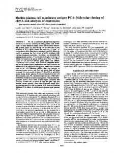
anti-N-CAM antibodies showed marginal differences from those exposed to nonimmune antibodies (72 % of cells left the EGL in the presence of anti-N-CAM) and thus these antibodies may have had a slight effect. In contrast, in the presence of anti-Ng-CAM, 64% of the cells remained in the EGL, and only 23% were in the IGL; i.e., only 26% of cells were able to migrate from the EGL. Moreover, of the cells that entered the ML, only 48 % exited, compared with 81% of control cells. When anti-cytotactin antibodies were added to the cultures, we also observed a major inhibition of migration of cells into the IGL. In contrast to the effects of anti-NgCAM, however, most of the labeled granule cells were found in the ML instead of the EGL (Fig. 4). Although 90% of cells left the EGL, only 30% of cells that entered the ML exited compared with 81% in control cultures. A similar differential distribution of cells in the ML was also seen when two different monoclonal antibodies against cytotactin were used (Table I). Exemplary histological sections and autoradiographs showing these effects are given in Fig. 4. With anti-NgCAM, the EGL remained thick and displayed many tritiumpositive cells; with anti-cytotactin, the ML became thicker and had many tritium-positive cells piled up in a radial pattern, apparently along the Bergmann glial fibers (Fig. 4 b, arrow). The control shows the normal disappearance of the EGL and the increasing thicknesses of the ML and IGL, which contains most of the labeled cells. Analyses of Time Course. To dissect the differential contribution to layer formation of Ng-CAM and cytotactin, we carded out rough kinetic analyses of the effects of their respective antibodies. The antibody experiments indicated that, with anti-Ng-CAM, blockade of migration appeared to be at the site at which cells leave the EGL to enter the ML. In contrast, with anti-cytotactin, cells entered the ML and accumulated. The accumulation of cells in the ML may have resulted from (a) enhancement of the rate of entry of cells from the EGL to the ML, (b) a general reduction of cellular migration rate in the ML, or (c) a blockade of cells entering the IGL. To distinguish among these possibilities, we examined the time course of the effects of each specific antibody as shown in Fig. 5. When cultured in the presence of nonimmune antibodies for 3 d, the percent of labeled granule cells decreased in the
Chuong et al. Cell Adhesion Molecules in Cerebellar Cell Migration
EGL at a more or less constant rate, and the increases in the IGL also occurred at a constant rate. Few cells stayed in the ML, indicating that the migration through this layer was relatively rapid as previously observed in vivo (Fujita, 1967) (Fig. 5). In the first 2-3 d in culture with anti-Ng-CAM cells left the EGL at a much slower rate and by 3 d in culture were still arrested in the EGL (Fig. 5). In contrast, with anticytotactin the loss of cells from the EGL occurred at rates similar to those found with nonimmune antibodies at all times, but there was a gradual buildup of cells in the ML at a corresponding rate, leading to an unusual accumulation of cells in the ML (Fig. 5). These experiments suggested that anti-Ng-CAM first acts at early phases of cell migration involving the transition of cells from the EGL to the ML. Although anti-cytotactin had no apparent effect at this phase, it acted at later phases either by slowing down cells in the ML or by blocking their movement from the ML to the IGL. The data do not allow clearcut discrimination between the latter two cases, but because we observed an even distribution of inhibited cells throughout the thickness of the ML rather than a piling up of cells in the lower portions of the ML (Fig. 4), we favor the possibility that there is a general slowdown of migration rates in the ML. Sequential Addition of Different Antibodies. To test this hypothesis further, we arbitrarily divided the culture period into two intervals: from day 0 to day 1.5, and from day 1.5 to day 3. We then included each of the specific antibodies during only one of the two intervals to assess the phase in which it exerted greatest effect. When anti-Ng-CAM was present only in the early interval, and the cultures were then incubated in the presence of nonimmune antibody for an additional 1.5 d, cell migration from the EGL was not significantly different from cultures incubated with antibody for all 3 d (34% compared with 43 % of cells left the EGL). This result suggested that the effect of anti-Ng-CAM is not reversible over this time period (Table II). To examine effects of anti-Ng-CAM in the later interval that could otherwise not be measured when anti-NgCAM was present from the beginning of the culture, we performed the reverse experiment: we first cultured the explants in control media for 1.5 d to allow the entry of cells into the ML, and then added anti-Ng-CAM for another 1.5 d, ex-
337
Figure 4. Effects of anti-Ng-CAM and anti-cytotactin on the migration of tritiated cerebellar granule cells. (a-c) Autoradiograms of paraffin sections of cerebellar explants that were pulse-labeled with tritiated thymidine for 1 h and cultured for 3 d. (d-f) Tracings ofa-c to highlight the silver grains and the relative thickness of the cortical layers. (g-h) Histograms of the number of tritiated cells versus distance along layers. (a, d, and g) anti-Ng-CAM; (b, e, and h) anti-cytotactin; (c, f, and 0 nonimmune control. Arrow in b points to granule cells in the ML, which are piled up along the Bergmann glia. Bar, 50 gm. amining the effects at the end of that period. Two additional observations emerged from this procedure. First, a similar percentage of cells (85 %) left the EGL as in the nonimmune control. Thus, although a percentage of the cells was still migratory between 1.5 and 3 d (Fig. 5), migration sensitive to anti-Ng-CAM was essentially completed by 1.5 d in cul-
ture. As indicated by the time course experiments, cells continue to migrate from the EGL in a linear fashion over the 3 d of culture with nonimmune antibodies. This result thus supports the interpretation that anti-Ng-CAM affects an early migration event in the EGL. Second, 63 % of cells that had entered the ML left as compared with 81% in the con-
The Journal of Cell Biology, Volume 104, 1987
338
EGL
ML
IGL
Control
A
40
~, 2o
,}._~. •
:
:
.
i
q
iO0 A n t i - N g - C A M 80
~'
60 "6 2O 0
Q
,
,
,
,
~'--~-°'i" ~ ° i
i
i
i
c_
~5 8O 6O
o
o,
a 3 o , 2 3 ; ' , Doys
~
in culture
Figure5. Time course studies of the effectsof nonimmune, anti-NgCAM and anti-~'ytotactinantibodies on cerebellar granule cell migration. Cerebellar explants were labeled for 1 h with [3H]thymidine, washed, and cultured in the presence of the indicated antibodies for 0-3 d. At each time point (abscissa), the explants were fixed, sectioned, and the number of labeled cells in each layer was calculated as described in Materials and Methods.
trois. Thus, although cells left the EGL there was still a significant accumulation of cells in the ML, suggesting that anti-Ng-CAM had additional independent effects on migration once cells had entered the ML. We then carried out similar experiments with anti-cytotactin. When anti-cytotactin was present only in the early interval, 60% of cells migrated out of the ML compared with 81% of cells in control cultures and 30% of cells when anticytotactin was present at all times. As suggested by the time course study, the major effect of anti-cytotactin occurred in the later culture period (Fig. 5). When anti-cytotactin was present only in the second half of the culture period, the same low percentage (35%) of cells left the ML as left when anti-cytotactin was present throughout the culture period (30%), which suggested that all cells migrating within the molecular layer were sensitive to the effects of anti-cytotactin. Because the major effect for anti-Ng-CAM was in the earlier period, while that for anti-cytotactin was in the later period, we reasoned that by exposing the culture to anticytotactin for the first 1.5 d and then switching to anti-NgCAM for another 1.5 d, the effects of each reagent would be reduced. The experiments showed that this was the case. The highest percentage of labeled cells was found in the IGL instead of the EGL or ML, thus migration was only moderately impaired and was affected primarily in the ML. The reverse experiment (anti-Ng-CAM followed by anti-cytotactin) showed both inhibition of cell migration from the EGL and
Table II. Differential Effects of Adhesion Molecules on Cerebellar Granule Cell Migration Analyzed by Sequential Addition of Antibodies Antibodies*
EGL distributionS:
Cells leaving EGL§
ML distribution
Cells that enter ML leaving MLII
IGL distribution
%
%
%
%
%
Day 0* NI (1-h label)
86 + 2
Day 0 - 1 . 5 / d a y 1.5-3 NI/NI Anti-Ng/anti-Ng Anti-Ng/NI NI/anti-Ng
12 57 49 13
± ± ± ±
9 5 l0 7
3 ± 1 86 34 43 85
15 11 13 28
± ± ± ±
9 2 7 4
11 + 1 81 66 68 63
73 32 38 59
± ± ± ±
13 4 5 6
Anti-CT/anti-CT Anti-CT/NI NI/anti-CT
9 ± 6 9 ± 6 7 + 2
90 90 92
55 ± 11 33 + 13 53 + 4
30 60 35
36 :t: 12 59 5 : 1 4 40 ± 4
Anti-CT/anti-Ng Anti-Ng/anti-CT
17 ± 7 41 ± 4
80 52
35 ± 8 25 ± 8
51 48
48 ± 5 34 ± 4
44 ± 11 77 ± 6 46 ± 5
49 10 47
12 ± 7 5 ± 1 21 ± 3
76 58 51
44 ± 11 18 ± 13 33 ± 6
Day 1.5¶ NI Anti-Ng Anti-CT
* The culture period was divided into two periods, day 0-1.5 and day 1.5-3. Some cultures were fixed at the end of the first period (day 1.5). For other cultures, at the end of 1.5 d, first antibodies were removed by three washes, then replaced with second antibodies. These culture conditions are indicated by the antibody used in each period and separated by a slash between the two periods. NI, nonimmune antibodies. * The cultures were labeled for 1 h with [3H]thymidine and either fixed immediately (day 0 value) or incubated with the indicated antibodies. At the end of 1.5 or 3 d of culture, the distribution of [3H]thymidine-labeled cells (mean + SD) among the three cerebellar layers was calculated as described in Materials and Methods. § The percentage of cells leaving the EGL was calculated as follows: (% cells, day 0 - % cells, day 3)/(% cells, day 0). II The percentage of cells that enter the ML and subsequently leave the ML was calculated according to the formula: (% cells in IGL, day 3 - % cells in IGL, day 0)/(% cells in ML, day 0 + % cells in EGL, day 0 - % cells in EGL, day 3). ¶ Day 1.5 values (see Fig. 5) are included for reference to the cell distribution at the middle of the culture period for each treatment.
Chuong et al. Cell Adhesion Molecules in Cerebellar Cell Migration
339
Expression Sequences
Perturbation Effects
The histogenesis of the cerebellum provides an excellent opportunity to examine the roles of different adhesion molecules in cell migration and in the formation of layers. In the
present study, we have shown by immunohistochemical means that the ld chain of N-CAM, Ng-CAM, and the extracellular matrix protein cytotactin all show a distinct and characteristic order of appearance during layer formation in the cerebellar cortex (Fig. 6). The conjugate functional studies on the effects of specific antibodies on granule cell migration in vitro showed a correlation between the main sites of appearance of Ng-CAM and cytotactin and the differential inhibition of various stages of migration (Fig. 6). Migration (and its effects on layer thickness and the location of borders) does not require just one kind of adhesion molecule but rather appears to involve several neuronal cell adhesion molecules as well as cytotactin acting differentially at different sites and stages. The characteristic order of appearance and distinct modulation mechanisms of these adhesion molecules of different specificity are consistent with the hypothesis that specific local signals resulting from sequential cellular interactions (Edelman, 1985) regulate their appearance. Previous studies using cerebellar explant cultures (Moonen et al., 1982) in which the movement of cells labeled with tritiated thymidine was followed (Lindner et al., 1983; Hoffman et al., 1986) showed that anti-Ng-CAM inhibited granule cell migration but anti-N-CAM had only marginal effects. In the present analyses, we further developed this assay by applying various antibodies in sequence to obtain dynamic information on the site and time of action of each of several different adhesion molecules. The site of major expression of Ng-CAM and cytotactin in the molecular layer was well correlated with the differential and independent effects of their respective antibodies on cell migration. The results showed that anti-Ng-CAM blocked cell movement by arresting cells in the EGL; it also showed moderate effects in blocking the movement of cells through the ML. In contrast, although anti-cytotactin had no apparent effects in blocking cells in the EGL, it exerted major effects in blocking cells in the ML (Fig. 6). Moreover, the different specific antibodies to these molecules appeared to act upon two independent mechanisms related to migration; in various combinations, their effects were additive. The experiments in which each antibody was present for only one-half of the culture period provided added support for these conclusions and also gave additional information about the timing and reversibility of the perturbation phenomena. When anti-Ng-CAM was present only in the first half of the culture period, the effects on cells leaving the EGL was similar to those where antibody was present throughout the culture period. When present only in the second half of the culture period, however, anti-Ng-CAM had its major effect in inhibiting cell migration within the ML; cell migration out of the EGL was comparable to nonimmune antibody controls. The two major stages of cerebellar granule cell migration as shown in Fig. 6 thus appear to be differentially affected by Ng-CAM and cytotactin. The first stage involves the migration of cells from the PLZ. This migration is slow (average rate ~1.8 ~tm/h [estimated from Fujita, 1967]). It may reflect a passive displacement generated by cell growth in the PLZ, or it could be the result of cell migration along the cytotactin-positive Bergmann glial fibers. This stage included a period possibly representing initial binding of granule cells to the Bergmann glia or initial process outgrowth
The Journal of Cell Biology, Volume 104, 1987
340
Time N
,.zT¢_B
Nid
N9
CT
antiNo
antiCT PLZ
PMZ
h
PMZ
ML
4 h
ML
IGL IL VL
B BBDD []
[]
IGL
IL "VL
Figure 6. Schematic representation of the formation of cerebellar layers showing the order of expression and the functions in cell migration of adhesion molecules. "Time" indicates the approximate period during which a granule cell stays in each layer, as measured in rodent by Fujita (1969). Numerals show the sequence of expression of each CAM in different layers. Bars with fine dots imply higher expression or greater perturbation effect than bars with coarse dots. The EGL is composed of the PLZ and PMZ.
an increase of cell number in the ML. This was similar to the summation of the independent results obtained with each antibody used singly in the same epoch (i.e., anti-Ng-CAM followed by nonimmune antibody plus nonimmune followed by anti-cytotactin). The lower number of cells in the ML relative to that observed when anti-cytotactin was present only in the second half of the culture period is probably due to the decreased input from the EGL caused by anti-Ng-CAM (Table II). Similar results were obtained with anti-Ng-CAM and anti-cytotactin together through the culture period (not shown). In interpreting the present data, several potential sources of error in these culture systems were considered. Differential cell death or cell proliferation in certain layers could have resulted in lower or higher counts in those layers. Although some cell death occurred in our explants (15 % of the total cells), the amount of death was similar in explants with the various antibodies, and almost all of the pyknotic nuclei observed were in the IGL. Moreover, such nuclei appeared only after 1.5 d in culture, suggesting that death resulted from central necrosis, and not through selective cell lysis by the antibodies used. Cell proliferation (gliosis and fibrosis) was rare and when it occurred could be recognized along the zone beneath the pia mater; its occurrence had no correlation with the antibodies used. Migration of granule cells could also be correlated with the thickness of the EGL, which became very thin in the presence of nonimmune antibodies (see Fig. 4) similar to that in viva. These independent observations suggested that the observed effects were specific for each antibody and were not the results of artifacts.
Discussion
of nascent parallel fibers during which binding by Ng-CAM is critical but cytotactin has a small or nonexistent role. Alternatively, because Ng-CAM is known to affect fasciculation (Stallcup and Beasley, 1985; Fischer et al., 1986; Hoffman et al., 1986) and because cell migration begins at the time that parallel fibers grow out (Altman, 1972), the effect of antibodies to Ng-CAM could be secondary to a failure of fasciculation. The second major stage involves the migration of cells through the ML into the IGL. This migration is fast, the average rate being •15 ~tm/h (estimated from Fujita, 1967; 10 grn/h was measured in vitro by Hatten et al., 1984). This movement phase involves cytotactin, and also, to a lesser extent, Ng-CAM. The involvement of both NgCAM and cytotactin in this stage favors a mechanism involving neuron-glia interaction because this stage is thought to be mainly mediated by interaction with Bergmann glial fibers (Rakic, 1971). It is likely that these processes further involve complex interplay among different molecules of neuronal and glial origins (Moonen et al., 1982; Lindner et al., 1986; Hatten et al., 1986). The fact that anti-N-CAM did not have strong effects on cell migration does not necessarily rule out an important role for this molecule in cortical layer formation. Indeed, the restricted distribution of the ld chain of N-CAM in layers mainly composed of neurites as compared with the distribution of N-CAM in all layers suggests that, through alteration of its cytoplasmic domain by RNA splicing (Murray et al., 1986a), N-CAM might be indirectly involved in cortical layer formation. Because the large domain of the ld polypeptide is intracellular, we were not able to test its contribution by using ld-specific antibodies in the present assay. However, the ld chain with its larger and structurally different cytoplasmic domain (Hemperly et al., 1986) might, for example, differentially interact with cytoskeletal or other proteins and thus influence the formation of cortical layers; its differential appearance suggests the action of a local signal affecting RNA splicing (Murray et al., 1986b). Although N-CAM is present in all layers of the cerebellum, there are known heterogeneities in which N-CAM is differentially distributed in the nervous system at different times (Chuong and Edelman, 1984) and which may involve chemical (Rothbard et al., 1982) and functional changes leading to different binding efficacy (Hoffman and Edelman, 1983). In the cell adhesion molecules examined in the present study, a variety of different modulation mechanisms act at different times and places, providing means in addition to their binding specificity for governing their cellular mode of action. As shown here, all of the molecules show prevalence modulation (Edelman, 1985, 1986). In addition, N-CAM shows cytoplasmic domain switching (Murray et al., 1986b) and embryonic to adult conversion (Rothbard et al., 1982; Edelman and Chuong, 1982; Chuong and Edelman, 1984), and both Ng-CAM (Thiery et al., 1985; Daniloff et al., 1986) and the ld chain (Pollerberg et al., 1985; Murray et al., 1986b) are polarity modulated on neurites in the molecular layer. Finally, the extracellular matrix molecule, cytotactin, clearly shows a sequence of differential expression that likely results from site-specific glial synthesis (Grumet et al., 1985; Crossin et al., 1986). Thus, there are different kinds of molecules, different specificities, and different control and modulation mechanisms contributing to the migration pattern. Although our understanding of the molecular mechanisms
Chuong et al. Cell Adhesion Molecules in Cerebellar Cell Migration
of cortical layer formation is far from complete and much remains to be learned of the actual cellular mechanisms of migration, the present studies show that the expression and function of several different adhesion molecules are correlated both in time and space with the segregation and generation of new cortical layers in the cerebellum. Moreover, the antibody perturbation experiments indicated that the separate stages of migratory events involving interactions among various cell types are affected by inhibiting the function of adhesion molecules of different cellular origins, binding mechanisms, and distributions. The results are consistent with the general hypothesis (Edelman, 1983, 1985, 1986) that local cell surface modulation of adhesion molecules of differing specificity plays a major regulatory role in the formation of histological layers. We thank Ms. Jane Steinberger and Ms. Alison Schroeder for excellent technical assistance and Dr. Leif Finkel and Dr. Joseph W. Becket for advice on presenting quantitative data. This work was supported by U.S. Public Health Service grants AM04256 and HD-09635, and a Senator Jacob Javits Center of Excellence in Neuroscience grant (NS-22789). Received for publication 21 July 1986, and in revised form 27 October 1986. References
Altman, J. 1969. Autoradiographic and histological studies of postnatal neurogenesis. Dating the time of production and onset of differentiation of cerebellar microneurons in rats. J. Comp. Neurol. 136:269-294. Airman, J. 1972. Postnatal development of the cerebellar cortex in the rat. Cytogenesis and histogenesis of the deep nuclei and the cortex of the cerebellum. The external germinal layer and the transitional molecular layer. J. Comp. Neurol. 145:353-398. Altman, J., and S. Bayer. 1978. Prenatal development of the cerebellar system in the rat. Cytogenesis and histogenesis of the deep nuclei and the cortex of the cerebellum. J. Comp. Neurol. 179:23--48. Brackenbury, R., J.-P. Thiery, U. Rutishauser, and G. M. Edelman. 1977. Adhesion among neural cells of the chick embryo. I. An immunological assay for molecules involved in cell-cell binding. J. Biol. Chem. 252:6835-6840. Chuong, C.-M., and G. M. Edelman. 1984. Alterations in neural cell adhesion molecules during development of different regions of the nervous system. J. Neurosci. 4:2354-2368. Chuong, C.-M., and G. M. Edelman. 1985. Expression of cell-adhesion molecules in embryonic induction. I. Morphogenesis of nestling feathers. J. Cell Biol. 101:1009-1026. Cowan, M. W. 1982. The development of the vertebrate central nervous system: an overview. In Development in the Nervous System. D. R. Garrod, editor. Cambridge University Press, Cambridge, England. 3-33. Crossin, K. L., S. Hoffman, M. Grumet, J.-P. Thiery, and G. M. Edelman. 1986. Site-restricted expression of cytotactin during development of the chicken embryo. J. Cell Biol. 102:1917-1930. Daniloff, J. K., C.-M. Chuong, G. Levi, and G. M. Edelman. 1986. Differential distribution of cell adhesion molecules during histogenesis of the chick nervous system. J. Neurosci. 6:739-758. de Bias, A. L. 1984. Monoclonal antibodies to specific astroglial and neuronal antigens reveal the cytoarchitecture of the Bergmann glia fibers in the cerebellum. J. Neurosci. 4:265-273. Edelman, G. M. 1983. Cell adhesion molecules. Science (Wash. DC). 219:450-457. Edelman, G. M. 1985. Expression of cell adhesion molecules during embryogenesis and regeneration. Exp. Cell Res. 161:1-16. Edelman, G. M. 1986. Cell adhesion molecules in the regulation of animal form and tissue pattern. Annu. Rev. Cell Biol. 2:81-116. Edelman, G. M., and C.-M. Chuong. 1982. Embryonic to adult conversion of neural cell adhesion molecules in normal and staggerer mice. Proc. Natl. Acad. Sci. USA. 79:7036-7040. Faissner, A., J. Kruse, J. Nieke, and M. Schachner. 1984. Expression of neural cell adhesion molecule L1 during development, in neurological mutants and in the peripheral nervous system. Dev. Brain Res. 15:69-82. Fischer, G., V. Kunemund, and M. Schachner. 1986. Neurite outgrowth patterns in cerebellar microexplant cultures are affected by antibodies to the cell surface glycoprotein L1. J. Neurosci. 6:605-612. Friedlander, D. R., M. Grumet, and G. M. Edelman. 1986. Nerve growth factor enhances expression of neuron-gila cell adhesion molecule in PC 12 cells. J. Cell Biol. 102:413-419. Fujita, S. 1967. Quantitative analysis of ceil proliferation and differentiation in the cortex of the postnatal mouse cerebellum. J. Cell Biol. 32:277-288.
341
Fujita, S. 1969. Autoradiographic studies on histogenesis of the cerebellar cortex. In Neurobiology of Cerebellar Evolution and Development. R. Llinas, editor. American Medical Association, Chicago. 743-747. Grumet, M., and G. M. Edelman. 1984. Heterntypic binding between neuronal membrane vesicles and glial cells is mediated by a specific cell adhesion molecule..L Cell Biol. 98:1746-1756. Orumet, M., S. Hoffman, C.-M. Chuong, and G. M. Edelman. 1984. Polypeptide components and binding functions of neuron-glia cell adhesion molecules. Proc. Natl. Acad. Sci. USA. 81:7989-7993. Grumet, M., S. Hoffman, K. L. Crossin, and G. M. Edelman. 1985. Cytotactin, an extracellular matrix protein of neural and non-neural tissue that mediates gila-neuron interaction. Proc. Natl. Acad. Sci. USA. 82:8075-8079. Hamburger, V., and H. Hamilton. 1951. A series of normal stages in the development in the chick embryo. J. Morphol. 88:49-92. Hanaway, J. 1967. Formation and differentiation of the external granular layer of the chick cerebellum. J. Comp. Neurol. 131:1-14. Hatten, M. E., R. K. H. Liem, and C. A. Mason. 1984. Two forms of cerebellar ceils interact differently with neurons in vitro. J. Cell Biol. 98:193-204. Hattan, M. E., C. A. Mason, and J. C. Edmondson. 1986. Glial process outgrowth and early neurnnal-astroglial interactions in vitro: a high-resolution time-lapse videomicroscopic study in the presence and absence of Fab fragments that disrupt these interactions. Soc. Neurosci. Abstr. 12:368. Hemperly, J. J., B. A. Murray, G. M. Edelman, and B. A. Cunningham. 1986. Sequence of a eDNA clone encoding the polysialic acid-rich and cytoplasmic domains of the neural cell adhesion molecule N-CAM. Proc. Natl. Acad. Sci. USA. 83:3037-3041. Hoffman, S., and G. M. Edelman. 1983. Kinetics of homophilic binding by E and A forms of the neural cell adhesion molecule. Proc. Natl. Acad. Sci. USA. 80:5762-5766. Hoffman, S., D. R. Friedlander, C.-M. Chnong, M. Grumet, and G. M. Edelma~. 1986. Differential contributions of Ng-CAM and N-CAM to cell adhesion in different neural regions. J. Cell Biol. 103:145-158. Lane, J. K. 1979. A detailed protocol for autoradiography as practiced in the laboratory ofE. G. Jones. In Neuroanatomical Techniques. Society for Neuroscience, Bethesda, MD. 27--46. Lindner, J., J. Guenther, H. Nick, G. Zinser, H. Antonicek, M. Schachner, and D. Monard. 1986. Modulation of granule cell migration by a gila-derived protein. Prac. Natl. Acad. Sci. USA. 83:4568-4571. Lindner, J., F. G. Rathjen, and M. Schachner. 1983. LI mono- and polyclonal antibodies modify cell migration in early postnatal mouse cerebellum.
The Journal of Cell Biology, Volume 104, 1987
Nature (Loud.). 305:427-429. Moonen, G., M. P. Grau-Wagemans, and I. Selak. 1982. Plasminogen activator-plasmin system and neuronal migration. Nature (Loud.). 298:753755. Mugnaini, E., and Forstronen, P. F. 1967. Ultrastructural studies on the cerebellar histogenesis. I. Differentiation of granule cells and development of glomeruli in the chick embryo. Z. Zellforsch. Mikrosk. Anat. 77:115-143. Murray, B. A., J. J. Hemperly, E. A. Prediger, G. M. Edeiman, and B. A. Cunningham. 1986a. Alternatively spliced mRNAs code for different polypeptide chains of the chicken neural cell adhesion molecule (N-CAM). J. Cell Biol. 102:189-193. Murray, B. A., G. C. Owens, E. A. Prediger, K. L. Crossin, B. A. Cunningham, and G. M. Edelman. 1986b. Ceil surface modulation of the neural cell adhesion molecule resulting from alternative mRNA splicing in a tissue-specific developmental sequence. J. Cell BioL 103:1431-1439. Pigott, R., and J. S. Kelly. 1986. Immunoeytochemieal and biochemical studies with the monoclonal antibody 69A1: similarities of the antigen with cell adhesion molecules L1, NILE and Ng-CAM. Dev. Brain Res. 29:111-122. Pollerberg, E. G., R. Sadoul, C. Goridis, and M. Schachner. 1985. Selective expression of the 180-kd component of the neural cell adhesion molecule N-CAM during development. J. Cell Biol. 101:1921-1929, Rakic, P. 1971. Neuron-glia relationship during granule cell migration in developing cerebellar cortex. A Golgi and electron microscope study in Maeaeus rhesus. J. Comp. Neurol. 141:283-312. Rakic, P, 1984. Contact regulation of neuronal migration. In The Cells in Contact: Adhesions and Junctions as Morpbogenetic Determinants. G. M. Edelman and J.-P. Thiery, editors. John Wiley and Sons, New York. 67-91. Romanoff, A. L. 1960. The Avian Embryo. Structural and Functional Development. Macmillan Publishing Co., New York. 255-262. Rothbard, J. B., R. Brackenbury, B. A. Cunningham, and G. M. Edelman. 1982. Differences in the carbohydrate structures of neural-cell adhesion molecules from adult and embryonic chicken brains. J. Biol. Chem. 257:1106411069. Stallcup, W. B., and L. L. Beasley. 1985. Involvement of the nerve growth factor-inducible large external glycoprotein (NILE) in neurite faseiculation in primary cultures of rat brain. Proc. Natl. Acad. Sci. USA. 82:1276-1280. Thiery, J.-P., A. Delouv~e, M. Grnmct, and G. M. Edelman. 1985. Initial appearance and regional distribution of the neuron-glia cell adhesion molecule in the chick embryo. J. Cell Biol. 100:442-456.
342