Demethylmenaquinones by Micrococcus varians. Depending on Cell Growth Media. Akira Hiraishi, Junta Sugiyama, and Kazuo Komagata. Institute of Applied ...
CURRENT MICROBIOLOO'~"Vol. 22 (1991), pp. 53-58
Current Microbiology © Springer-Verlag New York Inc, 1991
Simultaneous Formation of Menaquinones and Demethylmenaquinones by Micrococcus varians IAM 12146 Depending on Cell Growth Media Akira Hiraishi, Junta Sugiyama, and Kazuo Komagata Institute of Applied Microbiology, University of Tokyo, Bunkyo-ku, Tokyo 113, Japan
Abstract. Changes in the isoprenoid quinone composition of M i c r o c o c c u s v a r i a n s IAM 12146 in response to growth in different media were investigated. When the bacterium was grown in an ordinary complex medium, it produced menaquinones as the sole quinones, with a dihydrogenated menaquinone with seven isoprene units as the major component, at all growth stages. On the other hand, cells grown in a chemically defined medium containing glutamate and pyruvate as carbon sources produced both menaquinones and demethylmenaquinones. The major demethyimenaquinone homologs produced were the unsaturated and dihydrogenated types with seven isoprene units. The demethylmenaquinone/menaquinone ratio in cells varied during a batch growth in the chemically defined medium. The highest ratio was found in cells at the mid-exponential phase of growth.
Isoprenoid quinones occur as essential components of the respiratory electron transport system of bacterial plasma membranes and have attracted attention not only in their physiological aspects but from chemotaxonomic viewpoints [3, 5]. The taxonomic value of the respiratory quinones is based on the wide range of their structural variations among different taxa and their relative uniformity and stability within strains of the same taxon. In Gram-positive bacteria, the naphthoquinones, usually menaquinones, are found as the sole quinones, and the length and degree of hydrogenation of the multiprenyl side chain shown by these compounds are useful for their classification and identification. However, some observations have been made on the changes in menaquinone biosynthesis within a strain of certain Grampositive bacteria (for review, see [2]). In S t a p h y l o c o c c u s a u r e u s , the proportions of the various menaquinone homologs were found to change during the exponential phase of growth [6]. A previous study from our laboratory showed that cells of a M i c r o c o c cus luteus strain grown in an ordinary complex medium contained an unsaturated menaquinone with eight isoprene units (MK-8) as the predominant component, whereas those grown in chemically defined media formed both MK-8 and dihydrogenated menaquinone-8 (MK-8(H2)) as the major homologs [9]. In
addition, many species of Gram-negative, facultatively anaerobic bacteria have been reported to form menaquinones and demethylmenaquinones differently depending on environmental redox conditions [15, 16]. These observations require that the naphthoquinone formation by bacteria should be studied more thoroughly under various cultural conditions to find the range of variability in their quinone systems and its taxonomic implications. The present study was undertaken to investigate changes in the quinone composition of a M i c r o c o c cus varians strain in response to growth in different media. In this paper we present evidence that this bacterium formes both menaquinones and demethylmenaquinones, depending on cell growth media. Materials and Methods Bacterial strain and cultivation. Micrococcus varians IAM 12146 ( = ATCC 383) [13], which was obtained from the Culture Collection Center of the Institute of Applied Microbiology (University of Tokyo, Japan), was studied. Two liquid media were used for culturing the organism: one was the complex medium (peptone + beef extract + yeast extract) designated PBY [9], and the other was a modification of the chemically defined medium GPS broth [9]. The latter medium (designated GPS-II), which contained 20 mM glutamate and 20 mM pyruvate as the carbon sources and 15 mM (NH4)2SO 4 as the inorganic nitrogen source, was modified by replacing inosine and biotin as the growth factor
Address reprint requests to: Dr. A. Hiraishi, Division of Microbial Systematics, Institute of Applied Microbiology, University of Tokyo, Bunkyo-ku, Tokyo 113, Japan.
54
CURRENT MICROBIOLOGY Vol. 22 (1991)
MK-7(H2)
with a vitamin mixture [8]. Cells were grown aerobically in Sakaguchi's flasks, harvested, washed, and immediately subjected to quinone analysis, as previously described [9].
Extraction, purification, and analysis of quinones. Quinones were extracted from fresh wet cells with a chloroform-methanol mixture (2: 1, vol/vol) and purified by thin-layer chromatography (TLC) with Merck Kieselge160F2s 4 or Whatman PK6F precoated plates and a developing mixture of petroleum benzine-diethyl ether (9 : I, vol/vol), as reported previously [7, 10]. Quinone components were separated and identified by reverse-phase partition high-performance liquid chromatography (HPLC) with internal and external standards as previously described [7, 10, 14]. The plot of common logarithm of the retention times of unsaturated menaquinone standards against the number of isoprene units gave a straight line. This plotted line was used for calculating the equivalent number of isoprene units (ENIU) [14] for test quinone samples. The total naphthoquinone concentration was determined by ultraviolet (UV) spectroscopy, with an extinction coefficient of 19 mM• cm -~ at 249 nm [16]. The proportions of menaquinones and demethylmenaquinones in the total naphthoquinone fraction were determined by separating these components by HPLC as noted above. Mass spectrometry was performed with a JEOL DMX-110 mass spectrometer.
A
fi
MK-7(H2)
c~ CO
O4
O
O ¢Standard quinones. Menaquinone standards (MK-6 to MK-12) were prepared from M i c r o c o c c u s luteus 1AM 1056 [9] and Aureobacterium t e s t a c e u m IAM 12346 |14]. Demethylmenaquinones were prepared from a strain of Enterococcus faecalis [7].
e~
O
0~ e~
Results Upon quantitative TLC, the lipid extract from Mic r o c o c c u s varians IAM 12146 cells grown in PBY medium gave only one quinone band with an Rf of 0.6-0.7, i.e., the menaquinone fraction. HPLC experiments showed that the major component of the menaquinones showed an ENIU value of 7.40, corresponding to MK-7(H2). This finding verified the previous report of Yamada et al. [18] for M . varians menaquinones. Detailed information on the menaquinone composition of cells grown in the complex medium is described below. When the bacterium was grown in GPS-II medium, it produced another quinone component that appeared just behind the menaquinone band upon quantitative TLC. Owing to poor separation of the two quinone bands from each other, these were scraped together from the plate for the following analysis. Concurrently, we tried to scrape each quinone band as separately as possible. Thus, we obtained three quinone fractions: the upper-band (menaquinone) fraction, the lower-band fraction (unknown component), and the mixture of them. An ethanolic solution of the menaquinone fraction showed a UV absorption spectrum with absorption maxima at 243, 248, 263, 269, and 325 nm, as
C lI HI
I
I
!
i,
I
0
10
20
30
P
40
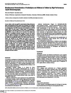
T i m e (rain) Fig. 1. HPLC elution profiles of different quinone fractions from M. varians IAM 12146 grown in the chemically defined medium (after 24 h of incubation). A, upper-band (menaquinone) fraction with MK-7(H2) predominating; B, mixture of upper-band and lower-band fractions with MK-7(H2) predominating; C, lowerband fraction with three major components (designated 1, 11, and lID. Analytical conditions: pump, Shimadzu LC-6A; column, Zorbax ODS (4.6 i.d. x 250 mm); mobile phase, methanol-isopropyl ether (4 : 1, vol/vol); flow rate, l ml/min; column temperature, 25°C.
A. Hiraishi et al.: Quinones of Micrococcus varians
~
'
'
1st batch
A
',
'
'
2nd batch
" i"
/
•
J
10 6a~I 69
,,16
o
"$
!
0
I
; 95
~,aa
z~a
~e
,10o
i,o
500
6~a~ ~lzz
~
0.1
,
8
B 0.01 62~
L
~5
le~ :aa~
i
~e~
8:
3aa
4~
~
24
~
48
~I
72 Time
J
a 96
z~ I 0 120
144
(h)
Fig. 3. Growth and menaquinone and demethylmenaquinone contents of M. varians IAM 12146 during repeated batch-culture processes with the complex ( I st batch) and the chemically defined (2nd batch) media. + , g r o w t h ; - - & - - , menaquinone content; - - A - - demethylmenaquinone content.
2::
C B2~
?%
i
__~...~.~_
0
.i I ' . . . .
~
~4B I!
,, 2oo
30o
~aa
L
5a~
Fig. 2. Mass spectra of three major components of the lowerband fraction from M. oarians IAM 12146 grown in the chemically defined medium. A, component I; B, component 11; C, component III.
expected, while the lower-band fraction had absorption maxima at 243, 248, 254 (shoulder), 263 (shoulder), and 325 nm. The results of TLC and UV spectroscopy suggested that the lower-band fraction might contain demethylmenaquinones. Comparative HPLC elution profiles of quinone components of the three fractions obtained from cells grown in the chemically defined medium are shown in Fig. 1. The menaquinone fraction gave an HPLC profile similar to that found in cells grown in PBY medium (Fig. 1A). The major component of this fraction, showing a peak with a retention time of 19.8 min, had an ENIU value of 7.40. Thus, this component could be assigned to MK-7(H2). In the lower-band fraction, the proportion of MK7(H2) was markedly decreased, and, alternatively, three major peaks with shorter retention times (8.2, 15.2, and 17.2 min) appeared (Fig. IC). The most
rapidly eluted component (designated component I) had an E N I U of 4.39, and was thus assumed to be MK-4(H2). While the detector response of component I at 248 nm did not differ significantly from that at 270 nm, the fractions eluted at 10.5 and 12.0 min, designated components II and III respectively, showed a twofold greater response in absorption at 248 and 254 nm than at 270 nm. Since this response is a characteristic feature of demethylmenaquinones [7], components II and III were suggested to be really demethylmenaquinones. The ENIU values for components II and III were 6.54 and 6.97, respectively. A previous study has shown that the ENIU of demethylmenoquinone-n (DMK-n) is expressed as (n - 1) + 0.55 + 0.02 [7]. Therefore, component II could be assigned to DMK-7. In fact, the elution time of this component corresponded to that of DMK-7 used as the standard. Component III might be assumed to be DMK-7(H2), in view of its ENIU value. As expected, the mixture of the menaquinone and lower-band fractions gave an intensive elution peak of MK-7(H2) and the other three major peaks possibly derived from MK-4(H 2) and the two demethylmenaquinone homologs (Fig. 1B). To confirm the identity of components I, II, and IlI, they were purified by HPLC and then analyzed by mass spectrometry (Fig. 2). The mass spectrum of components I showed the molecular ion peak (M +) at m/z 446 and strong fragment peaks at m/z t 87 and 225 derived from the naphthoquinone nucleus (Fig. 2A), thereby ascertaining
56
CURRENT MICROBIOLOGY Vol. 22 (1991)
Table 1. Changes in the naphthoquinone composition of M i c r o c o c c u s uarians tAM 12146 during repeated batch growth in the complex and the chemically defined media" Quinone composition (mol %) Cells grown in PBY ( I s t batch) for: Homolog MK-4(H2) MK-5(H~) MK-6(H 2) MK-7 MK-7(H.,) MK-8(H 2) DMK-7 DMK-7(H2) Others
9 h . t 1 1 78 13 . . 6
15 h .
24 h .
t 3 80 10 .
.
9 h
16 h
22 h
70 h
t 4 82 9
tb 1 I 5 73 10 2
5
7
t t I 11 63 9 8 2 5
2 t 1 8 56 7 14 5 8
5 t 3 75 6 3 3 5
. .
5
72 h
.
t 2 t 81 I1 .
Cells grown in GPS-II (2nd batch) for:
. 7
.
a For the time course of growth and quinone contents in repeated batch cultures, see Fig. 3. b Trace amount (less than t% of total content).
this to be MK-4(H2). Component II had the mass spectrum in which the molecular ion peak (M +) appeared at m/z 634 (Fig. 2B) and the base peak at m/z 211, which is 14 mass units smaller than the case of menaquinones. These results indicated that component II was DMK-7. The mass spectrum of component III was complicated compared with those of the other two (Fig. 2C). It showed two molecular ion peaks (M +) at m/z 636 and 648 and intensive fragment peaks at 211 and 225. Therefore, component III seemed to be a mixture of DMK7(H2) and MK-7. However, since the mass peaks derived from DMK-7(H 2) were considerably higher than those from MK-7, it was suggested that the demethylmenaquinone comprised a major part of this fraction. This was supported by the results of HPLC studies that component III showed the much greater response at 248 and 254 nm than at 270 rim.
In view of the results of TLC, UV spectroscopy, HPLC, and mass spectrometry, it can be concluded that M. varians IAM 12146 forms both menaquinones and demethylmenaquinones when grown in the chemically defined medium. We further investigated whether the formation of demethylmenaquinones in this bacterium depends on the phase of growth. During the period of a batch culture in PBY medium, no demethylmenaquinone formation was noted at any of the growth stages. This was the case in the subsequently repeated batch process with the complex
medium (data not shown). Figure 3 shows the changes in menaquinone and demethylmenaquinone contents in repeated batch cultures with the two different media. In the first batch culture with PBY medium, menaquinones were the sole quinone components formed, as described above, but the second batch process with GPS-II medium yielded appreciable amounts of demethylmenaquinones (0.1-1.03 /xmol/g of dry weight) in addition. The demethylmenaquinone content was highest at the mid-exponential phase of growth and decreased at the stationary growth stage. Similar results were obtained with the third transferred batch culture in the chemically defined medium (data not shown). These observations clearly show that the formation of demethyimenaquinones by M. varians IAM 12146 depends on cell growth media. The cellular concentration of menaquinones varied markedly between 1.7 and 4.8 /xmol/g of dry weight during the period of the two batch cultures. It was low at the exponential phase of growth and increased during the stationary phase, irrespective of cell growth media. Thus, the highest demethylemenaquinone/menaquinone ratio (approximately 0.2) was noted in cells at the exponential phase of growth. Changes in the homologous distribution of menaquinones and demethyimenaquinones during the period of the two repeated batch cultures are shown in Table 1. Although MK-7(H 2) was found to predominate in the whole course of the expert-
57
A. Hiraishi et al.: Quinones of Micrococcus varians
ments, there were some variations in the proportions of various naphthoquinone isoprenologs during the period of each batch process. Discussion Demethylmenaquinones have been found mainly in facultatively anaerobic Gram-negative bacteria, such as members of the families Enterobacteriaceae [16, 17] and Pasteurellaceae [11, 12]. These naphthoquinones also occur as the sole quinones in the Gram-positive bacterium E n t e r o c o c c u s f a e c a l & [1, 4]. Before this work, E . f a e c a l i s was the only species reported as demethylmenaquinone-producing Gram-positive bacteria. To our knowledge, the present study is the first to demonstrate the simultaneous occurrence of demethylmenaquinones and menaquinones in Gram-positive bacteria. The results of this study have shown that the formation of demethylmenaquinones by M. varians IAM 12146 depends on the growth in the chemically defined medium. This suggests that cell growth conditions including culture media are critical for determining the respiratory quinones for the chemotaxonomy of Gram-positive bacteria, as pointed out in our previous study [9]. In concurrent studies other Micrococcus species, such as M. luteus and M. roseus, have been found to produce no demethylmenaquinones, irrespective of cultural conditions (A, Hiraishi, J. Sugiyama, and K. Komagata, unpublished data). Therefore, if demethylmenaquinone formation is a general property of M. varians, this is of taxonomic value in the classification and identification of Gram-positive cocci. There is much need to survey demethylmenaquinones in other M . varians strains. In addition to its taxonomic interest, the question remains whether the presence of demethylmenaquinones in M. varians has any physiological implication. In the Gram-negative facultative anaerobes, menaquinones and demethylmenaquinones are formed in response to growth under different environmental conditions [15, I6] and are involved in anaerobic electron transport with different terminal acceptors such as fumarate, dimethylsulfoxide, and nitrate [15]. M. varians is an obligate aerobe for which oxygen is the only utilizable terminal electron acceptor. Our attempts to grow the bacterium under anaerobic conditions with either fumarate, dimethylsulfoxide, or nitrate as the acceptor have given negative results. Therefore, such differential roles for menaquinones and demethylmenaquinones in anaer-
obic electron transport as in the enterobacteria may not be the case in M . varians. Although we have hitherto found no full evidence for the metabolic role of demethylmenaquinones in the bacterium, further work is necessary to solve that problem.
ACKNOWLEDGMENT
We thank Dr. N. Morisaki for performing the mass spectrometry.
Literature Cited 1. Baum Rtt, Dolin MI (1965) Isolation of2-solanesyl-1,4-naphthoquinone from Streptococcusfaecalis 10 C1. J Biol Chem 240:3425-3433 2. Bentley R, Meganathan R (1982) Biosynthesis of vitamin K (menaquinone) in bacteria. Microbiol Rev 46:241-280 3. Collins MD (1985) Analysis ofisoprenoid quinones. In: Gottschalk G (ed) Methods in microbiology, vol 18. London: Academic Press, pp 329-366 4. Collins MD, Jones D (1979) The distribution of isoprenoid quinones in streptococci of serological groups D and N. J Gen Microbiol 114:27-33 5. Collins MD, Jones D (1981) Distribution of isoprenoid quinone structural types in bacteria and their taxonomic implications. Microbiol Rev 45:316-354 6. Hammond RK, White DC (1969) Formation of vitamin K~ isoprenologues by Staphylococcus aureus. J Bacteriol 100:573-578 7. Hiraishi A (1988) High-performance liquid chromatographic analysis of demethylmenaquinone and menaquinone mixtures from bacteria. J Appl Bacteriol 64:103-105 8. Hiraishi A, Kitamura H (1984) Differences in phototrophic growth on high phosphate concentrations among Rhodopseudomonas species. J Ferment Technol 62:293-296 9. Hiraishi A, Komagata K (1989) Effects of the growth medium composition on menaquinone homolog formation in Micrococcus luteus. J Gen Appl Microbiol 35:311-318 10. Hiraishi A, Hoshino Y, Kitamura H (1984) Isoprenoid quinone composition in the classification of Rhodospirillaceae. J Gen Appl Microbiol 30:197-210 11. Kroppenstedt RM, Mannheim W (1989) Lipoquinones in members of the family Pasteurellaceae. Int J Syst Bacteriol 39:304-308 12. Lester RL, White DC, Smith SL (1964) The 2-desmethyl vitamin K2's. A new group of naphthoquinones isolated from Hemophilus parainfluenzae. Biochemistry 3:949-954 13. Ogasawara-Fujita N, Sakaguchi K (1976) Classification of micrococci on the basis of deoxyribonucleic acid homology. J Gen Microbiol 94:97-106 14. Tamaoka J, Katayama-Fujimura Y, Kuraishi H (1983) Analysis of bacterial menaquinone mixtures by high performance liquid chromatography. J Appl Bacteriol 54:31-36 15. Unden G (t988) Differential roles for menaquinone and demethylmenaquinone in anaerobic electron transport of E. coli and their fnr-independent expression. Arch Microbiol 150:499-503
58
16. Whistance GR, Threlfall DR (1968) Effect of anaerobiosis on the concentrations of demethylmenaquinone, menaquinone, and ubiquinone in Escherichia freundii, Proteus mirabilis, and Aeromonas punctata. Biochem J 108:505-507 17. Whistance GR, Dillon JF, Threlfall DR (1969) The nature, intergeneric distribution and biosynthesis of isoprenoid qui-
CURRENT MICROBIOLOGY Vol. 22 (1991)
nones and phenols in Gram-negative bacteria. Biochem J 111:461-472 18. Yamada Y, Inouye G, Tahara Y, Kondo K (1976) The menaquinone system in the classification of aerobic Gram-positive cocci in the genera Micrococcus, Staphylococcus, Planococcus, and Sporosarcina. J Gen Appt Microbiol 22:227-236