platform was recorded with a cut-off time within 120 ... center of the pool, which recorded the behaviors of ..... pias) induced by X-rays in the hippocampus were.
322
ORIGINAL
Spatial learning and expression of neural cell adhesion molecule L1 in rats X-irradiated prenatally Rui Zhang, Xue-Zhi Sun*, Chun Cui, Hiromi Sakata-Haga, Kazuhiko Sawada, Changli Ye**, and Yoshihiro Fukui Department of Anatomy and Developmental Neurobiology, Institute of Health Biosciences, The University of Tokushima Graduate School, Tokushima, Japan ; *Regulatory Sciences Research Group, National Institute of Radiological Sciences, Chiba, Japan ; and **Department of Dentistry, Liaoyuan Central Hospital, Jilin, China Abstract : The present study was designed to present evidence to clarify the relationships between learning ability, neuronal cell adhesion molecule L1 expression and hippocampal structural changes in the rat model received X-irradiation at an embryonic stage (E15). Water maze task indicated that all of the irradiated rats failed to learn the task in the whole training procedure. Their latency to the platform and swimming distance were significant differences from those sham-treated controls. Histological studies showed that the hippocampal ectopias induced by X-rays in the CA1 were involved in the spatial learning impairment, in which they hampered normal processes in learning development and transmission of information. Number, size and positions of the ectopias in the dorsal parts of the hippocampus were confirmed to be related to degrees of spatial learning impairment. On the other hand, L1 expression in the hippocampus was examined with Western blot analysis. The results indicated a lower content of L1 in the irradiated rats. A decrease in L1 might be one of reasons to cause disorganization of the septohippocampal pathways. These findings suggest some mechanisms of spatial learning impairment can be attributed to the formation of the hippocampal ectopias and redaction of L1 following prenatal exposure to X-irradiation. J. Med. Invest. 54 : 322-330, August, 2007 Keywords : hippocampus, X- irradiation, L1, learning
INTRODUCTION One of the most severe consequences of prenatal irradiation exposure is damage to the development of central never system (CNS). The developing brain in the CNS is most susceptible to the irradiation. This high vulnerability of the fetal brain is a distinctive teratological characteristic commonly recognized in all mammalian species including huReceived for publication June 15, 2007 ; accepted July 12, 2007. Address correspondence and reprint requests to Dr. Xue-Zhi Sun, Regulatory Sciences Research Group, National Institute of Radiological Sciences, Anagawa 4-9-1, Inage-ku, Chiba 263-8555, Japan and Fax : +81-43-251-4853.
man beings (1-4, 7, 8), typified by a reduction in the size of the cerebral hemisphere (microcephaly) and the presence of ectopic cell masses beneath the cortical white matter (subcortical heterotopia) (4-6). Other disorders including mental retardation, attention deficit-hyperactivity disorder and cognitive dysfunction also have been found at a later adult stage in humans who received atomic bomb exposure in Hiroshima and Nagasaki (7, 8). In the animal experiments, animals irradiated during the formation stage of the brain showed characteristic malformation of the cerebral cortex and behavioral disorders after they became maturation. These behavioral disorders included an increase locomoter activity and impairments in learning spatial The Journal of Medical Investigation Vol. 54 2007
The Journal of Medical Investigation
tasks (10, 11). Such disorders depend on the time of irradiation and the type of behavioral task. Some studies have done on brain weight and behavioral changes associated with prenatal irradiation. However, little studies have been reported on the relationship between cognitive impairment and histopathological change of the hippocampus following prenatal exposure to X-irradiation, even though the hippocampus is considered as a central structure for learning and memory storage (12, 13). The hippocampus appears to be necessary for several types of memory (14-16), but its mnemonic function is particularly clear in tasks for which subjects are required to remember spatial location (17). So spatial learning as one of indices of behavioral changes was selected to use in this study. Moreover, the spatial water maze task is one of the most widely used behavioral tests to assess the effects of various manipulations on spatial learning, ranging from genes to normal aging (18-23), it may provide us an appropriate method to test the task depended on the proper function of the hippocampus. Many investigations have suggested that cell adhesion molecules in the CNS are implicated in the formation of neural circuits, synaptic plasticity and cognitive function (9, 24). One such molecule is L1, a well-characterized cell adhesion molecule known to be an integral membrane protein containing six immunoglobulin domains, five fibronectin type III repeats, a single transmembrane region, and an intracellular domain (25). L1 is expressed as a single transmembrane protein with a molecular weight of approximately 200 kDa and has been identified in early forming axon tracts in the developing CNS (26, 27), also in the hippocampus (28-30). However, little attention had been focused on the relation between expression of L1 and learning ability in the brain prenatally exposed to irradiation. Thus, in the present study, we tested learning ability with the water maze, examined the structure change in the hippocampus with the histological method and identified L1 express with Western blot analysis. Our aim was to present evidence to clarify the relationship between learning ability, L1 expression and hippocampal structural changes in rat model that received X-irradiation at an embryonic stage.
MATERIALS AND METHODS Experimental animals Wistar rats purchased from the Shizuoka Labo-
Vol. 54 August 2007
323
ratory Animal Center (Shizuoka, Japan) were kept ! with 55!5% in a controlled atmosphere of 23!2! humidity under a 12-h dark/light cycle (7 : 00 A. M.- 7 : 00 P. M.). They had free access to food and water in a colony room. Eight-week-old nulliparous females were caged with potent males in pairs overnight and checked for vaginal plugs the next morning. The day on which a vaginal plug appeared was as day 0 of pregnancy. Rates with positive plugs were housed in individual cages.
X-irradiation Pregnant rats were exposed to a single wholebody X-irradiation at a dose of 1.5 Gy on embryonic day 15 (E15). The physical factors of the X-rays used were 200 kVp, 15 mA, 0.5 mm Cu + 0.5 mm Al filter, 90 cm distance, and 0.45 Gy/ minute exposure rate. Control pregnant rats were treated in the same manner, expect for the X-irradiation. The rats allowed delivering and rearing their litters. The offspring were separated from their mothers at 5 weeks after birth and caged individually under the same conditions as their mothers. At 8 weeks of old, 30 male offspring prenatally exposed to irradiation and 26 control offspring from different mothers were randomly selected to use for the experiments. Animal care procedures were in accordance with Regulations in Appropriate Animal Breeding and Treatment, Ministry Office of Japan. All efforts were made to minimize the number of animals used and their suffering.
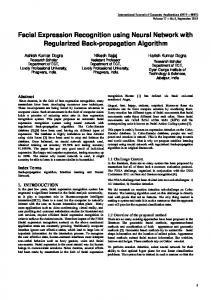
Water maze task Acquisition of the place task, animals were assessed eight weeks after being born. They were trained to find a clear Plexiglas platform (14 cm diameter) submerged 1.5 cm under the surface of water in a water maze modified according to that described by Morris (31). This water maze was a circular pool (146 cm diameter, 45 cm high filled with room temperature water (30 cm depth). The invisible escape platform was placed in the middle of one of the quadrants equidistant from the sidewall and middle of the pool. For the acquisition test, rats were given 4 trials /day with a 30 min interval each for five days. Two different starting positions were chosen semi-randomly, where the distance between the start position and the platform was set to be equal (Fig. 1). Every training trial started from one of the two starting points, used in a random sequence similarly for each rat. A trial began by plac-
324 a : Control
R. Zhang, et al. Changes in learning and L 1 after X-irradiation
b : Irradiated
maze task. The level of statistical significance was set at p-value of less than 0.05.
Histological examination
Fig. 1. Representative swimming paths taken on the fifth block (4 trails) in the water maze. (a) Control rat. (b) Prenatally irradiated rat with severe learning deficient. S : Starting position ; P : Platform.
ing the rat into the water facing the wall of the pool at one of the starting points. If the rat failed to escape within 120 sec, it was guided to the platform by the experimenter. Once the rat reached the platform, it was allowed to stay there for 30 sec and, then, placed in a holding cage for an intertribal interval for 30 min. In each trail, the latency to escape onto the platform was recorded with a cut-off time within 120 sec. The swim path was recorded by a CAT-10 image analyzing system (Muromachi Co., LTD, Japan) and a video camera mounted in the ceiling above the center of the pool, which recorded the behaviors of the animals : the latency of individual rat to reach the platform (swimming time), the swimming distance and swim speed. The swim speed which came from distance /latency provided an index of motor function. In order to analysis the relationship between behavioral changes (learning and spatial cognition in the present study) and brain damage, the rats received prenatal irradiation were divided into the following tree groups based on the mean of swimming time for the 5th block. They were grade 1, slightly damaged group, which reached the platform with 20 sec ; grade 2, moderately damaged group, which reached the platform between 21 sec and 45 sec ; grade 3, severely damaged group, which reached the platform between 46 sec and 120 sec.
Statistical analysis Escape latencies collected during acquisition and cue training were analyzed as trial blocks per day using an ANOVA (analysis of variance between groups) with repeated measures. All results were expressed as mean ! SEM (standard error of the mean). The significance of the differences between the prenatal irradiated group and the control group was analyzed by an unpaired t-test for each block in the water
We wanted to evaluate whether behavioral changes (learning and spatial cognition) of the animals were related to their hippocampal lesions, all of the rats in the grade 1-3 groups described in above were selected for a histological study after the water maze test was completed. These rats and their controls were deeply anesthetized with ether, and perfused with Zamboni’s fixitive solution (4% formaldehyde and 0.2% picric acid in 0.1 M phosphate buffer, pH 7.3) with a rotary pump via the left cardiac ventricle. Following perfusion, the brain was removed and immersed in freshly made Zamboni’s fixitive solution for 1 week. Tissue samples were embedded in paraffin and sectioned coronally at 5 μm with a microtome. Serial coronal sections were prepared from each brain. Every fourth section was kept from the serial sections, stained with hematoxylin and eosin and examined under a light microscope.
SDS-PAGE and Western blot analysis L1 expression in the hippocampus was detected by SDS-PAGE (Sodium dodecyl sulfate-polyacrylamide gel electrophoresis) and Western Blot Analysis. The tissue blocks were prepared from the hippocampus of the rats when their behavioral tests were finished. Particular attention was given to the dissection and orientation of the tissue blocks in this study. The tissue blocks through the full thickness of the hippocampus were dissected as nearly as possible perpendicular to the longitudinal axis (LA) of the cerebral hemisphere, which was 1.5 mm away from both sides of 1/3 point of LA’s total length. This point was local in 1/3 of LA from back side. This tissue block contained a biggest region of hippocampus and could be easy to make. Tissue homogenate (100 μg) was separated by SDS-PAGE under reducing conditions and transferred to nitrocellulose membranes (32). SDS-PAGE analysis was carried out on 7% polyacrylamide gels according to Laemmli (33). Membranes were blocked with Superblock (Pierce Chemical Company) overnight at room temperature. Membranes were then incubated with ant-L1 monoclonal antibody (1 : 250) (Chemicon) for 120 min, and next incubated with peroxidase-labeled goat anti-mouse IgG (1 : 4000) for 90 min. All membranes were visualized using the enhanced chemiluminescence (ECL) and exposure to ECL Hyperfilm (Amersham Life Science, Inc). The immunoblots of the homogenates
The Journal of Medical Investigation
that were incubated with ant-L1 gave blots with multiple bands.
Vol. 54 August 2007
325
radiated group than in the sham-treated group.
Brain histological characteristics
RESULTS Water maze performance All the animals were capable of swimming around the pool. Total thirty irradiated and twenty-six shamtreated rats were trained in 20 sessions to find an underwater invisible platform in the water maze. The control groups swam straight to the reach the hidden platform after training of 2-3 days, while the irradiated rats took a proportion time across the central area to escape onto the platform or went to near the platform of the pool. Representative swimming paths taken on the fifth block (4 trails) were shown in Fig. 1. The acquisition learning curve of the rats in water maze indicates a gradual decrease in escape latency with progressive training both in the controls and the irradiated rats. In the control groups, the rats even reached the platform within 10 sec at the last day of trial. However, the irradiated rats spent a longer time and distance to find the platform than the sham-treated groups (Fig. 2a). The latency to the platform and swimming distance in the irradiated rats significantly increased as compared with the control groups (Fig. 2a, 2b). Significant difference in the swimming speed was not found between the controls and the irradiated rats (Fig. 2c). Posthoc analysis revealed that the latency to the platform and swimming distance during the first to fifth blocks were significantly higher in the prenatal ir-
Compared with the brain in control animals, the rats received prenatal irradiation on E15 showed an atrophic cerebral cortex with a partially preserved lamination and dilation of the cerebral ventricle. The brain size of the exposed rats was smaller, and thickness of the cerebral cortex was reduced. The weight of the whole brain significantly decreased by about 36% in the rats exposed to irradiation compared with control rats, but significant difference in the brain weigh was not found among the grade 1-3 groups (Data not shown). Hippocampus with a well-defined cytoarchitecture and laminar-specific organization were observed in the control rats. It could be subdivided into three fields : CA1, CA2 and CA3 (Cornu Ammonis, CA) according to Lorente de NÓ’s definitions (Fig. 3a). However, the principal cellular layer called the pyramidal cell layer became thin, short and broken in the brain of irradiated rats. CA1, CA2, CA3 fields and the dentate gyrus (DG) were also deranged, and the abnormal cell mass which is known as the ectopic neuronal mass formed partially so as to replace the CA1 region (Fig. 3b, 3c). Based on the position of the abnormal neuronal mass in the irradiated brain, we have defined this kind of the mass as “hippocampal ectopia” in our previous study (5). Such hippocampal ectopias consisted of displaced pyramid neurons and were confined to the dorsal part of the CA1 field of Ammonis horn. These neurons extended in to the adjacent stratum oriens. In order to explore the relationship between his-
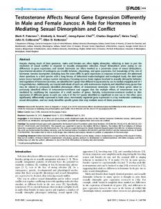
Fig. 2. Acquisition learning curve of rats on the water maze task. Blocks of 4 trials/day were represented in the horizontal axis in each graph.(a). Repeated measures of two-way ANOVA indicated that the mean escape latency (sec) was significant difference between the sham-treated group and the prenatally irradiated group, (*p!0.001). (b). Repeated measures of two-way ANOVA reveled that the mean swimming distance (cm) was significant difference between the sham-treated group and the prenatally irradated group, (*p!0.001). (c). Difference in the swimming speed (cm/sec) on the cued version of the water maze task was not found between the sham-treated group and the prenatally irradiated group. Post-hoc analysis showed that the latency to the platform and swimming distance during the first to fifth blocks were significantly higher in the prenatallly irradiated group than in the sham-treated group (*p!0.001). Mean ! SEM.
326
R. Zhang, et al. Changes in learning and L 1 after X-irradiation
Fig. 3. Representive micrograghs of the hippocampus of 8-week old rats. (a). A normal laminar organization, three fields (CA1, CA2 and CA3) and dentate gyrus (DG) in the control rats. (b) and (c). Typical hippocampal ectopias (arrow) appeared in the CA1 of the hippocampus in the irradiated rats. Their relative sizes were judged by the experimenters and marked as. + : smaller (blue) ; ++ : bigger (black).
tological changes of the hippocampus and spatial learning impairment, we compared number, size and positions of the hippocampal ectopias in slightly damaged group, moderately damaged group and severely damaged group, the results were shown in Table 1. The degree of the spatial learning impairment depended on abnormal structures : number, size and positions of the hippocampal ectopias in the brain.
Expression of neural cell adhesion molecule L1 The expression of neural cell molecule L1 in the hippocampus was examined with SDS-PAGE and Western blot analysis. The controls with non-L1 antibody or with the secondary antibody were negative. The results of L1 expression were shown in the Fig. 4. The immunoblots revealed specific bands of L1 at 200, 180, 140 and 80 kDa both in the shamoperated rat and the irradiated group. However, the bands in the irradiated group were very weak when compared with the control, even at the place of 80 kDa protein marker, the band had disappeared (Fig. 4). The result suggested that prenatal irradiation exposure resulted in weak and rare L1 expression.
Fig. 4. SDS-PAGE and Western blot analysis showing expression of neuronal cell adhesion molecule L1. The immunoblots revealed specific bands of L1 at 200, 180, 140 and 80 kDa. However, the bands in the irradiated rat were very weak when compared with the control. C : Control ; R : Irradiated.
Table 1. Relationship between learning impairment and hippocampal ectopias in the rats prenatally exposed to X-irradiation Spatial learning impairment
Hippocampal ectopia
Slight (5) *4
Moderate (9)
Severe (16)
Number*1
- (3), !(1), + (1)
- (2), + (7)
+ (3), ++ (13)
Size*2
+ (2)
+ (5), ++ (2)
++ (13)
Position*3 (in CA1)
Dorsal + (2) Rostral - (2)
Dorsal + (7) Rostral + (2)
Dorsal + (13) Rostral + (10)
*1 Total numbers of the ectopias (EP) in the bilateral hippocampus (HP). - : no EP ; !: one EP in the unilateral HP ; + : one EP in the bilateral HP ; ++ : two EPs in the bilateral HP. *2 Relative size of EP, judged by the experimenters. Please refer to Fig 3b and 3c to get an image. + : smaller (blue) ; ++ : bigger (black). *3 EP located in the dorsal and/or rostral region(s) of HP. - : no EP in any region ; + : EP in either one region or two regions. *4 ( ) indicated the number of the animals. Total 30 irradiated rats were examined.
The Journal of Medical Investigation
DISCUSSION Water maze is a behavioral procedure designed to test learning, spatial memory. Our study showed that all of the rats prenatally irradiated to X-irradiation on E15 failed to learn the water maze task in the whole training procedure. They swam a long distance and spent much time to find the hidden platform under water, while their swim speed which was calculated from distance/latency was no so difference when it is compared to the sham-control animals. The results clearly indicated that prenatal irradiation exposure did not cause an obvious motor dysfunction, but it selected to affect the function in spatial cognition. It is thought that water maze task depend on the proper cytoarchitecture and function of the hippocampus (34), The rats exposed on E15 in the present study showed an abnormal laminar formation in the cerebral cortex and a characteristic malformation in the hippocampus, where hippocampal ectopias formed and displaced pyramidal neurons. The reasons is that E15 for rats is a critical stage for histogenesis of the cerebral cortex and the hippocampus, which corresponds to a time when an apparent dramatic surge normally results in neurogenesis along with the establishment of architectonic stratification of the cerebral wall and the hippocampal structure (6, 35-38). Therefore, X-irradiation exposure on E15 could cause extensive cell death in the rat brain and an increase in the incidence of malformation and neurological dysfunction. The histological alterations (hippocampal ectopias) induced by X-rays in the hippocampus were involved in the learning disability. We compared the histological alterations in the hippocampus and found that almost of hippocampal ectopias located in the dorsal parts of the CA1 (Table. 1). The current knowledge from neuroanatomical studies have shown that, the dorsal hippocampus is a crucial structure for spatial learning, in which there are a greater number of complex spike cells (pyramidal cells) with finely tuned spatial receptive fields (39), supporting the essential nature of the dorsal hippocampus in successful navigation (40). These conclusions could help us to explain our results, why irradiated animals with dorsal hippocampal lesions lost abilities for learning the localization of a hidden escape platform in the pool during both a reference memory task and a working memory task. One outstanding issue also raised as to whether the degrees of spatial learning impairment were cor-
Vol. 54 August 2007
327
related to the histological changes of the hippocampus. Further comparison of the latency in the each group (grade 1, 2 or 3 group divided by swimming time) and their data of histological examination (Table 1), we interestingly found that the smaller ectopic neuron masses appeared at CA1 of the hippocampus in the some rats of grade 1 (2/5) ; extensive and larger ectopic neuron masses located and occupied much more CA1 regions in the most cases of the grade 2 (7/9) ; while in the grade 3 group, two ectopic neuron masses associated with severely deranged laminar structures were commonly observed to exit in the bilateral dorsal hippocampus (13/16). The results revealed a clear structure-function relation : extensive damage of the hippocampcal regions was consistent with severe deficient in the memory processes. It also strongly supported the view that the dorsal hippocampus was related to cognitive function. These ectopic neuron masses in the hippocampus hampered normal processes in learning development and transmission of information in the hippocampus, finally resulted in learning and cognitive deficient. The degrees of spatial learning impairment could be attributed number, size and positions of the hippocampal ectopias. Cell adhesion molecules of the immunoglobulin superfamily, such as the neural cell adhesion molecule and L1, are cell-surface macromolecules that, through their recognition and adhesion properties, regulate cell-surface interaction and have been reported to play an important role in cognitive functioning. L1 is expressed primarily on the surface of axonal shafts and growth cones of developing neurons (41-43), and axonal tracts such as thalamocortical and hippocampal commissural projections (29, 44, 45). L1 appears to be important in establishing fasciculated axonal pathway. L1-deficient mice display septal and hippocampal abnormalities characterized by small septal nuclei at the medial line and a reduced number of hippocampal cells (46). L1 is the only neuronal cell adhesion molecule of its class known to be associated with a human disease. Mutations in the human L1 gene have been linked to the MASA syndrome (consisting of mental retardation, aphasia, shuffling gait, and adducted thumbs) (47-49). It is known to that L1 is present on hippocampal tracts at a time at which the septohippocampal pathway develops (30, 50) and spatial learning requires the appropriate septohippocampal pathways which involve in regulating functions of the hippocampus such as cognition and hippocampal arousal. Such the pathway that is well known to
328
R. Zhang, et al. Changes in learning and L 1 after X-irradiation
atrophy in Alzheimer’s disease patients who have primary symptoms including memory loss, disorientation, confusion, and problems with reasoning and thinking. Interestingly, the rats prenatally irradiated to X-rays in our experiments showed a lower content of L1 in the hippocampus when it was compared with their control rats. We presumed that septohippocampal pathways were disorganized due to L1 reduction following irradiation exposure. In addition, leaning and spatial cognition also involved in many factors (neurotophic factor, nerve growth factor and some transmission, etc). In the animal experiments, formation of the hippocampal ectopias had changed expression and transmission of some factors concerning learning and cognitive function. Rats prenatally treated with chemical drugs (methylazoxymethanol, MAM) presented hippocampal ectopias associated with significantly increasing in the brain nerve growth factor (NGF) and in the brain-derived neurotrophic factor (BDNF), and reducing in choline acetyltransgerase (ChAT) immunoreactivity in cholinergic neurons. Both NGF and BDNF are neurotrophic factors important in the development and maintenance of neurons and synaptic connectivity, and ChAT is the enzyme that catalyses acetylcholine formation. All of these proteins have been shown to be important in cognition and maintenance of the septohippocampal pathway (51-53). Changes in NGF, BDNF and ChAT produced cognitive deficients. Moreover, primarily acetylcholinergic septohippocampal pathway, including choline acethyltransferase and muscarinicacethylcholine (mACh) receptor, has been shown to play important roles in learning and spatial cognition. In our previous experiments, we have confirmed that mACh receptor binding was selectively decreases about 10% in the hippocampus, but not in the cerebral cortex after prenatal irradiated to Xirradiation in the rat model which was the same as using in this study. Irradiation might be producing significant neurotrophic factor and enzymatic changes along this pathway that might result in cognitive impairment on the water maze. Leaning and spatial cognition is a complex developing process and also highly sensitive to various physical including to irradiation. Any disturbance of the normal process during the embryonic stage may result in disorder of leaning and spatial cognition. The finding reported here have shown some mechanisms of leaning and spatial cognitive impairment, which may be attributed to the formation of hippocampal ectopias and redaction of L1 follow-
ing prenatal exposure to X-irradiation, but other damages in the septohippocampal pathways and hippocampus need further studies.
REFERENCES 1.
Sun XZ, Inouye M, Takagishi Y, Hayasaka S, Yamamura H : Body and brain development following exposure to 60Co gama-irradiation during pregnancy in mice. Environ Med 38 : 111114, 1994 2. Kameyama Y, Inouye M : Irradiation injury to the developing nervous system : Mechanisms of neuronal injury. Neuro Toxicology 15 : 75-80, 1994 3. Sun XZ, Inouye M, Fukui Y, Hisano S, Sawada K, Muramatsu H, Muramatsu T : An immunohistochemical study of radial glial cells in the mouse brain prenatally exposed to gammairradiation. J Neuropathol Exp Neurol 56 : 13391348, 1997 4. Sun XZ, Takahashi S, Fukui Y, Hisano S, Kubota Y, Sato H, Inouye M : Neurogenesis of heterotopic gray matter in the brain of the microcephalic mouse. J Neurosci Res 66 : 10831093, 2001 5. Sun XZ, Takahashi S, Kubota Y, Sato H, Chui C, Fukui Y, Inouye M : Types and Threedimensional distribution of neuronal ectopias in the brain of mice prenatally subjected to Xirrasiation. J Radiat Res 43 : 89-98, 2002 6. Sun XZ, Inouye M, Takagishi Y, Hayasaka S, Yamamura H : Follow-up study on the histogenesis of microcephaly associated with ectopic gray matter induced by prenetal gammairradiation in the mouse. J Neuropathol Exp Neurol 55 : 357-365, 1996 7. Schull WJ, Nishitani H, Hasuo K, Kobayashi T, Goto I, Otake M : Brain abnormalities among the mentally retarded prenatally exposed atomic bomb survivors. RERF TR : 13-91, 1991 8. Schull WJ, Norton S, Jensh RP : Ionizing radiation and the developing brain. Neurotoxicol and Teratol 12 : 249-260, 1990 9. Venero C, Tilling T, Hermans-Borgmeyer I, Herrero AI, Schachner M, Sandi C : Water maze learning and forebrain mRNA expression of the neural cell adhesion molecule L1. J Neurosci Res 75(2) : 172-181, 2004 10. Sienkiewicz ZJ, Haylock RG, Saunders RD : Prenatal irradiation and spatial memory in mice :
The Journal of Medical Investigation
11.
12.
13.
14.
15.
16.
17. 18.
19.
20.
21. 22.
23.
investigation of dose-response relationship. In I Radiat Biol 65 : 611-618, 1994 Bayer SA, Brunner RL, Hine R, Altman J : Behaviors effects of interference with the postnatal acquisition of hippocampal gramule cells. Na New Biol 242 : 222-224, 1973 Nishima K, Iwasaki K, Tsukikawa H, Matsumoto Y, Egashira N, Abe K, Egawa T, Fujiwara M : The scopolamine-induced impairment of spatial cognition parallels the acetylcholine release in the ventral hippocampus in rats. Jpn J Phamacol 84 : 163-173, 2000 O’Carroll CM, Martin SJ, Sandin J, Frenguelli B, Morris GR : Dopaminergic modulation of the persistence of one-trial hippocampus-dependent memory. Learn Mem 13 : 760-769, 2006 Squire LT : Memory and the hippocampus : a synthesis from findings with rats, monkeys, and humans. Psychol Rev 99 : 195-231, 1992 Morris PGM, Frey U : Hippocampal synaptic plasticity : role in spatial learning or the automatic recording of attended experience? Philos Trans R Soc Lond B Biol Sci 352 : 14891503, 1997 Eichenbaum H, Dudchenko P, Wood E, Shapiro M, Tanila H : The hippocampus, memory, and place cells : is it spatial memory of a memory space? Neuron 23 : 209-226, 1999 O’Keefe J, Nadel L : The hippocampus as a cognitive map. Oxford : Clarendon, 1978 Chen G, Chen KS, Knox J, Inglis J, Bernard A, Martin SJ, Justice A, McConlogue L, Games D, Freedmen SB, Morris RG : A learning deficit related to age and beta-amyloid plaques in a mouse model of Alzheimer’s disease. Nature 408 : 975-979, 2000 D’Hooge R, De Deyn PP : Applications of the Morris water maze in the study of learning and memory. Brain Res Rev 36 : 60-90, 2001 Foster TC, Sharrow KM, Masse JR, Norris CM, Kumar A : Calcineurin links Ca2+ dysregulation with brain aging. J Neurosci 21 : 40664073, 2001 Jeffery KJ : LTP and spatial learning-where to next? Hippocampus 7 : 95-110, 1997 Poe GR, Teed RG, Insel N, White R, McNaughton BL, Barnes CA. Partial hippocampal inactivation : effects on spatial memory performance in aged and young rats. Behav Neurosci 114 : 940949, 2000 Zeng H, Chattarji S, Barbarosie M, Rondi-Reig L, Philpot BD, Miyakawa T, Bear MF, Tonegawa
24.
25.
26.
27.
28.
29.
30.
31.
32.
Vol. 54 August 2007
329
S : Forebrain-specific calcineurin knockout selectively impairs bidirectional synaptic plasticity and working/episodic-like memory. Cell 107 : 617-629, 2001 Klementiev B, Novikova T, Novitskaya V, Walmod PS, Dmytriyeva O, Pakkenberg B, Berezin V, Bock E : A neural cell adhesion molecule-derived peptide reduces neuropathological signs and cognitive impairment induced by Abeta25-35. Neuroscience 45(1) : 209224, 2007 Moos M, Tack R, Scherer H, Teplow D, Fruh K, Schachner M : Neural adhesion moleculeL 1 as a member of the immunoglobulin superfamily with binding domains to fibronectin. Nature 334 : 701-703, 1988 Lagenaue GA, Lemmon V : An L1-like molecule, the 8D9 antigen, is apotent substrate for neurite extension. Proc Nat Acad Sci USA 84 : 77537757, 1987 Rathjen FG, Schachner M : Immunocytological and biochemical characterization of a new neuronal cell surface component (L1 antigen) which is involved in cell adhesion. EMBO J 3 : 1-10, 1984 Barallobre MJ, Del Río JA, Alcántara S, Borrell V, Aguado F, Ruiz M, Carmona MA, Martín M, Fabre M, Yuste R, Tessier-Lavigne M, Soriano E : Aberrant development of hippocampal circuits and altered neural activity in netrin 1-deficient mice. Development 127(22) : 4797-4810, 2000 Fujimori KE, Takeuchi K, Yazaki T, Uyemura K, Nojyo Y, Tamamki N : Expression of L1 and TAG-1 in the corticospinal, callosal, and hippocampal commissural neurons in the developing rat telencephalon as revealed by retrograde and in site hybridization double labeling. J Comp Neurol 417 : 275-288, 2000 Wilson MT, Snow DM : Chondroitin sulfate proteoglycan expression pattern in hippocampal development : potential regulation of axon tract formation. J Comp Neurol 424(3) : 532546, 2000 Morris R : Developments of a water-maze procedure for studying spatial learning in the rat. J Neurosci Methods 11 : 47-60, 1984 Mathews AM, Roberts DW, Hinson JA, Pumford NR : Acetaminophen-induced hepatotoxicity : Analysis of total covalent binding versus specific binding to cysteine. Drug Metab Dispos 24 : 1192-1196, 1996
330
R. Zhang, et al. Changes in learning and L 1 after X-irradiation
33. Laemmli UK : Cleavage of structural protein during the assembly of the head bacteriophage T4. Nature 227 : 680-685, 1970 34. Morris RG, Garrud P, Rawlins JN, O’sKeefe J : Place navigation impaired in rats with hippocampus lesions. Nature 297 : 681-683, 1982 35. Rodier PM : Developing brain as a target of toxicity. Environ Health Perspect 103 (6) : 7376, 1995 36. Rodier PM : Correlations between prenatallyinduced alterations in CNS cell populations and postnatal function. Teratology 16(2) : 235-246, 1977 37. Rakic P : Specification of cerebral cortical areas : the radial unit hypothesis. Science 241 : 170-176, 1988 38. Rakic P : Genesis of the dorsal lateral geniculate nucleus in the rhesus monkey : Site and time of origin, kinetics of proliferation, routes of migration. J Comp Neurol 176 : 23-52, 1977 39. Jung MW, Wiener SI, McNaughton BL : Comparison of spatial firing characteristics of units in dorsal and ventral hippocampus of the rat. J Neurosci 14 : 7347-7356, 1994 40. Norman MW, Stephane G : Dorsal hippocampus function in learning and expressing a spatial discrimination. Learning & Memory 13 : 119122, 2007 41. Persohn E, Schachner M : Immunohistological localization of the neural adhesion molecules L1 and N-CAM in the developing hippocampus of the mouse. J Neurocytol 19 : 807819, 1990 42. van den Pol AN, Kim WT : NILE/L1 and NCAM-polysialic acid expression on growing axons of isolated neurons. J Comp Neurol 332 : 237-257, 1993 43. Frappe I, Wang C, Gaines G, Rideout-Gros S : Cell adhesion molecule L1 promotes neurite outgrowth of septal neurons. J Neurosci Res 75 : 667-677, 2004 44. Shults CW, Alberti L, Kimber TA, Stallcup WB : Mesostriatal dopaminergic axons transiently express high levels of NLE during development. Exp Neurol 116 : 275-285, 1992 45. Fukuda T, Kawano H, Ohyama K, Li HP, Yakeda Y, Oohira A, Kawamura K : Immunohistochemical localization of neurocan and L1
46.
47.
48.
49.
50.
51.
52.
53.
in the formation of thalamocortical pathway of developing rats. J Como Neurol 382 : 141-152, 1997 Demyanenko GP, Tsai AY, Maness PF : Abnormalities in neuronal process extension, hippocampal development, and the ventricular system of L1 knockout mice. J Neurosci 19 (12) : 4907-4920, 1999 Kenwrick S, Doherty P : Neural cell adhesion molecule L1 : Relating disease to function. Bioessays 20(8) : 668-675, 1998 Jouet M, Rosenthal A, Armstrong G, MacFarlane J, Stevenson R, Paterson J, Metzenberg A, Ionasescu V, Temple K, Kenwrick S : X-linked spastic paraplegia (SPG1), MASA syndrome and X-linked hydrocephalus result from mutations in the L1 gene. Nat Genet 7(3) : 402-407, 1994 Vits L, Van Camp G, Coucke P, Fransen E, De Boulle K, Reyniers E, Korn B, Poustka A, Wilson G, Schrander-Stumpel C, et al : MASA syndrome is due to mutations in the neural cell adhesion gene L1CAM. Nat Genet 7(3) : 408-413, 1994 Kocsis B, Kaminski M : Dynamic changes in the direction of the theta rhythmic drive between supramammillary nucleus and the septohippocampal system. Hippocampus 16(6) : 531-540, 2006 Van der Zee EA, Compaan JC, Bohus B, Luiten PG : Alterations in the immunoreactivity for muscarinic acetylcholine receptors and colocalized PKC gamma in mouse hippocampus induced by spatial discrimination learning. Hippocampus 5(4) : 349-362, 1995 Dekker AJ, Gage FH, Thal LJ : Delayed treatment with nerve growth factor improves acquisition of a spatial task in rats with lesions of the nucleus basalis magnocellularis : evaluation of the involvement of different neurotransmitter systems. Neuroscience 48(1) : 111119, 1992 Schaaf MJ, Workel JO, Lesscher HM, Vreugdenhil E, Oitzl MS, de Kloet ER : Correlation between hippocampal BDNF mRNA expression and memory performance in senescent rats. Brain Res 915(2) : 227-233, 2001