We thank Steve Robinow for advice during the early phase of this work. We also thank Drs. Sam ...... disease and Down syndrome. Proc Nat1 Acad Sci USA ...
The Journal
of Neuroscience,
December
1990,
lO(12):
38493881
Identification, Secretion, and Neural Expression of APPL, a Drosophila Protein Similar to Human Amyloid Protein Precursor Liqun Luo, Linda E. Martin-Morris, Department
and Kalpana
of Biology, Brandeis University,
White
Waltham, Massachusetts
A Drosophila gene [ amyloid protein precursor-like (Appl)] has recently been identified whose predicted amino acid sequence (APPL) shares extensive homology with the &amyloid protein precursor (APP) associated with Alzheimer’s disease. Characterization of proteins encoded by the Appl gene was initiated with the expectation that this simple model system might help elucidate the basic function provided by APPL and APP proteins. In this report, we identify 2 forms of the APPL protein in embryonic extracts, primary cultures, and transfected cells. APPL is synthesized as a 145kDa membrane-associated precursor that is converted to a 130-kDa secreted form that lacks the cytoplasmic domain. Both forms are N-glycosylated. Pulse-chase and subcellular localization studies suggest that the conversion is very rapid. The similarities of biogenesis between APP and APPL provide further evidence that APPL and APP might be functionally homologous, and that the secretion event is of physiological significance. lmmunocytochemical studies show that the APPL proteins are first detected in developing neurons concomitant with axonogenesis and remain associated with differentiated neurons. APPL immunoreactivity was observed in neuronal cell bodies, axonal tracts, and neuropil regions. In the embryo, APPL proteins are expressed exclusively in the CNS and PNS neurons, consistent with the Appl transcript localization. The expression pattern of APPL proteins suggests an ancestral function for this protein in the nervous system.
Our understandingof the molecular events in the pathogenesis of Alzheimer’s diseasehas been greatly facilitated by the isolation of /3-amyloid protein, a major proteinaceouscomponent of senileplaques(Glenner and Wong, 1984; Masterset al., 1985) and the subsequentcloning of amyloid protein precursor (APP, Goldgaber et al., 1987; Rang et al., 1987; Robakis et al., 1987; Tanzi et al., 1987; for review, seeMtiller-Hill and Beyreuther,
Received May 14, 1990; accepted Aug. 2, 1990. We thank Steve Robinow for advice during the early phase of this work. We also thank Drs. Sam Gandy and Joseph Buxbaum and all members of the White lab for helpful discussions. We gratefully acknowledge Michael Rosbash’s generosity for allowing us to use the tissue culture facilities and the help from Hildur Colot and Marie Laure Samson during the cell culture experiments. We appreciate the comments on this manuscript from Isaac F&y, Pat Parmenter, Michael Rosbash, and Larry Zwiebel. This research was supported by NIH GM-33205 and by an Alzheimer’s Disease and Related Disorders grant to K.W. L.M.-M. was supported by NIH Predoctoml Training Grant NS-07292 and NIMH Predoctoral Fellowship MH-09824. Correspondence should bc addressed to Dr. Kalpana White, Department of Biology, Brandeis University, Waltham, MA 02254. Copyright 0 1990 Society for Neuroscience 0270-6474/90/123849-13$03.00/O
02254
1989; Selkoe, 1989). A Drosophila gene, amyloid protein pre(Appl), hasrecently been identified whosepredicted amino acid sequence(APPL) sharesextensive homology with APP throughout the molecule(Rosenet al., 1989; Martin-Morris and White, 1990). Both APP and APPL have single-membrane-spanningdomainsnear their respective carboxyl termini. Notable features of the homology between APP and APPL include 2 highly conserved regions in the extracellular domain, El and E2. There are 12 cysteine residuesconservedin the El region and a conservedN-glycosylation site in the E2 region. A highly conserved cytoplasmic domain was also observed between APP and APPL, with a stretch of 15 amino acids containing 13 identities in the carboxyl terminus. However, there is no significant primary sequencehomology in the transmembrane domain or in the extracellular domain near the membrane-spanningregion; therefore, the P-amyloid sequence,which spansthe border of extracellular and transmembranedomains of APP, is not found in APPL. The mammalian APP is expressedin many tissuesand encodesa family of proteins consistingof several isoforms, some of which contain an additional proteaseinhibitor domain (Kitaguchi et al., 1988; Ponte et al., 1988; Tanzi et al., 1988). They appear to be generatedby alternative splicing (Kitaguchi et al., 1988; Ponte et al., 1988; Tanzi et al., 1988; de Sauvage and Octave, 1989) aswell asby further processingof primary polypeptides (Weidemann et al., 1989). In contrast, only a singlesize transcript hasbeen found in Drosophila, which is expressed in a neural-specificmanner as determined by Northern and in situ hybridization analysis (Rosen et al., 1989; Martin-Morris and White, 1990). Characterization of APPL protein was initiated with the expectation that this simplemodel systemmight help elucidate the basicfunction provided by this classof molecules. By generatingpolyclonal antibodies againstdifferent parts of APPL, we have identified 2 forms of APPL in Drosophila embryos, embryonic primary cultures, and transfected Schneider cells: a 145-kDa membrane-associatedform and a 130-kDa secretedform. We have shown in cell culture studiesthat the 145-kDa form is a precursor form that is rapidly transformed to the 130-kDa secretedform, and that both forms are N-glycosylated. Furthermore, immunocytochemical studiesdemonstratethat APPL is expressedin differentiated neurons,and that the subcellularlocalization of APPL is consistentwith biochemical data. Our results provide further evidence that APP and APPL might be functionally homologous in their respective organismsand suggestan ancestralnervous systemfunction for this classof molecules. Some of the data presentedhere have been reported previously in brief form (Luo et al., 1989).
cursor-like
3850
Luo et al.
Materials
l
Drosophila APPL
Protein
Secretion
and
Neural
Expression
and Methods
Plasmid constructions. The vector for constructing the T7-fusion protein pGDD7,E was a gift from Dr. M. Palazzolo (California Institute of Technology). To make the T7-APPL’ fusion, cDNA c4 (Rosen et al., 1989) in Bluescript SK+ vector (Stratagene) was digested with Hind III and Xba I and ligated to Hind III and Xba I-diaested nGDD7-E. A single Not I site acthe polylinker was cut, filled in, and religated taalign the T7 coat protein and APPL in frame. To make T7-APPL*, oligodirected mutagenesis was used to change nucleotide 2477 ofAppl(Rosen et al., 1989) from T to C, to create a Not I site on the c4-pGDD7,E hybrid plasmid described above. The plasmid was cut with Not I and religated to delete the extracellular domain. Both junctions were verified by sequencing. To make the o-gal-APPLr fusion protein, cDNA c4 was cloned into the plasmic pUR 288 (provided by Dr. B. Mtlller-Hill) EcoRI site, which would generate an in-frame fusion of fl-galactosidase-APPL’ fusion protein. To make the hsp-Appl construct, a 5.5-kb Appl cDNA (S2) was inserted into the EcoRI site of a plasmid containing the hsp70 promoter and @-tubulin polyadenylation site (provided by Dr. J. Lis). This construct contains the entire open reading frame (ORF) of Appl, and its orientation was confirmed by restriction analysis. Antibody generation and afinity puriJication. The T7-APPLr and T7APPL2 constructs were used to transform E. coli strain Bal 2 1-pLysS (provided by Dr. F. W. Studier). IPTG (final concentration, 1 mM) was added to 500 ml of bacterial culture with an OD,,, of 0.5. After 2 hr of induction, cells were collected by 1000 x g spin, and the pellet was resusvended in 40 ml 50 mM Tris (vH. 8.OV2 mM EDTA. After freezethawing, the cells were sonicated 4~ 15 set and centrifuged at 13,000 x g for 15 min. The insoluble T7-fusion proteins in the pellet were resuspended in 2 ml 50 mM Tris (pH, 8.0)/2 mM EDTA plus 3 ml of 2 x sample buffer (Harlow and Lane, 1988) and loaded on 7.5% preparative SDS-PAGE gels. Induced fusion proteins were excised from the gels and electroeluted into the protein running buffer (Harlow and Lane, 1988) at 400 V for 1 hr. One hundred twentv-five or 250 na of vrotein was used to immunize each rat or rabbit, respectively, and the same amount of protein was used for subsequent boosting. For affinity purification, rat LU-APPL serum was first passed through an Affi-gel 10 (Bio-Rad) column covalently linked with bacteria total proteins with pUR288 expressing B-galactosidase. The flowthrough was loaded on the affinity column of purified P-gal-APPL’ fusion protein (Rio et al., 1986) covalently linked with Al&gel 10. The affinity-purification procedure was as described (Harlow and Lane, 1988). Glycine (0.1 M, pH 2.5) was used to elute the antibodies from the column. Preparation of embryonic extracts and embryonic primary cultures. Unless otherwise mentioned, embryonic extracts were prepared according to the following procedure. After dechorionation in 50% Clorox, staged embryos (at 25°C) were homogenized in a Dounce homogenizer on ice in PBS (Harlow and Lane, 1988) supplemented with 0.5% NP-40, 1 mM EGTA, 0.5 pg/ml leupeptin, 1 &ml pepstatin, 100 KIUI ml avrotinin. and 0.2 mM PMSF. The homogenate was centrifuaed for 5 mm at lOdO x g, and the supematant was-collected. For fractionation of embryonic extracts, dechorionated embryos were homogenized in l/10 x PBS supplemented with 1 mM EGTA and protease inhibitors as above (no detergent). The homogenate was centrifuged for 5 min at 1000 x g, and the supematant was further centrifuged at 40,000 x g for 1 hr. The supematant was regarded as the soluble fraction. The pellet was resuspended in PBS supplemented with 2% NP-40, the resuspended part was centrifuged again; and the supernatant was regarded as the membrane fraction. Fractionation of S2-Appl cells into soluble and membrane fractions were modified after Simon et al. (1989). SZ-Appl cells were heat-shocked for 20 min and rested at room temperature for 40 min. Cells were pelleted and washed with cold PBS, and the cell pellet was frozen in -70°C. After thawing the cells in l/10 x PBS (hypotonic) supplemented with 1 mM EGTA and protease inhibitors (0.5 &ml leupeptin, 1 pg/ ml pepstatin, 100 KIU/ml aprotinin, 0.2 mM PMSF) and extraction in Dounce homogenizer, samples were spun at 1000 x g for 5 min, and the supematants were centrifuged at 40,000 x g for 30 min. The supematant of this spin was regarded as the soluble fraction. The pellet was resuspended in PBS with protease inhibitors as above, supplemented with 2% NP-40. The extraction was subsequently centrifuged at 12,000 x g for 15 min, and the solubilized supematant was regarded as the membrane fraction.
Preparation of primary cultures from embryos was modified from the procedures of Wilcox (1986) and Kidd et al. (1989). Staaed embrvos were dechorionated, washed; and homogenized in 10 x vi1 M3 me*dia (Harvard Biolab) in Dounce homogenizer with 4 strokes. The homogenate was passed through 3 layers of Nitex (35-pm mesh) and spun for 5 min at 500 x g at 4°C. The cell pellet was washed twice in M3 media and, finally, resuspended at a density of lo8 cells/ml in Coming cell wells. For metabolic labeling, (met, cys)-free M3 media (Harvard Biolab) was used instead of complete M3 media during extraction. After plating the cells in the culture wells (10’ cells/ml), cells were given a 45min starvation before adding Tra@S label (ICN) at 500 j&i/ml for 2 hr before immunoprecipitation analysis (see below). Protein gel electrophoresis and immunoblot. Unless otherwise mentioned, all protein gels were 7.5% SDS-PAGE gels. For immunoblotting, gels were electrotransferred onto nitrocellulose with standard nrocedures (Harlow and Lane, 1988) overnight at a current of 150 mA. Immunoreaction was visualized using alkaline phosphatase reaction with Promega’s Western-blotting system. All the antibody reaction steps were according to manufacturer’s specifications (Promega). Transfection, metabolic labeling, and glycosylation. Transfection of Schneider cells with hsp-Appf was according to Rio and Rubin (1985). Two d after transfection, the transfected cultures were heat-shocked for 20 min at 37°C and rested at room temperature (RT) for 40 min. Cells were collected by centrifugation. After washing with PBS, the cell pellet was resuspended in an equal vol PBS and 2 x SDS sample buffer and boiled for 5 min before SDS-PAGE/immunoblot analysis. For metabolic labeling, cells were heat-shocked at 37°C for 20 min, collected from culture dishes, and centrifuged at 500 x g for 5 min. The cell pellet was washed twice with (met, cys)-free M3 media and, finally, resuspended in (met, cys)-free media at a density of 2 x lo6 cells/ml. After 30 min starvation at RT, TranYj label (ICN) was added at a specific activity of 500 &i/ml. Cells were labeled for 1 hr, then collected into microfuge tubes in which media and cells were fractionated by centrifugation. After washing with M3 media, cells were lysed in PBS with 1% SDS and boiled for 3 min, and NP-40 was added to 3% for immunoprecipitation. Media from the labeling was treated in the same wav (SDS added to 1%. boiled. and NP-40 added to 3%). For the pulse-chase labeling experiment, the procedures were basically as above, except that, after 10 min of labeling with TraQ5S label at a specific activity of 1 mCi/ml in 50-ml Falcon tubes, complete M3 media was added, and cells were pelleted and resuspended in complete M3 media. Cell suspension aliquots of 300 ~1 were incubated in Eppendorf tubes for various times at RT (23°C). Further treatment was identical to the metabolic labeling procedure described above. Immunoprecipitation was performed as described below. For the glycosylation experiment, tunicamycin was added at a concentration of 10 pg/ml from the start of heat-shock (37”C, 20 min) and through the starvation (RT, 30 min) and labeling (RT, 2 hr) period. Immunoprecipitation. For immunoprecipitation of labeled proteins, samples were precleared by incubating with 20 pl GammaBind G-Agarose (Genex Corporation) in PBS for 30 min at 4°C and spun at 12,000 x g for 2 min. The recovered supematant was incubated-with 10 & rat a-APPL serum for 1 hr at 4”C, and 20 ~1 of GammaBind G-Agarose was added for a further incubation of 30 min at 4°C. The immunoprecipitated complex was first washed with PBS supplemented with 0.5% Triton, then twice with PBS. The final pellet was resusnended in 2 x sample buffer and subjected to SDS-PAGE. Subsequent fixing and amplification were according to manufacturer’s specification (Amersham). Amplified gels were dried and exposed to Kodak x-ray film. For immunoprecipitation of embryonic extracts (see Fig. 3A), the procedure was essentially the same as above. One mg of total proteins was used in each immunoprecipitation reaction. The SDS-PAGE gel was transferred to nitrocellulose, and the immunoblot reaction was carried out as described above. Immunocytochemistry. Whole-mount embryo staining was performed according to Thomas et al. (1988). Devitellinized embryos were rehydrated through a methanol series and incubated in PBT (0.1 M sodium phosphate buffer, pH 7.2, with 0.3% Triton) with 5% normal goat serum for 2 hr at RT. The primary antibody reaction was carried out at 4°C overnight, and Vector ABC kit was followed for subsequent steps. For larval-brain and eye-disk staining, hand-dissected third-instar larval brains and disks were fixed in 4% parafomraldehyde overnight, washed in PBT twice for 5 min, washed once in methanol for 15 min, washed twice for 5 min in PBT, and incubated for 2 hr in PBT with
The Journal of Neuroscience,
December
1996, fO(l2)
3951
A Extracellular 1
198
APPL’
417
:4-886 El
576
886
303
APPL*
7-886
1
2
3
180116845848-S-
36-S26.6-
5% normal goat serum before primary antibody incubation at 4°C overnight. Vector ABC kit was followed for subsequent steps. Some whole-mount immunostained preparations were further embedded in parathn and sectioned as described (White et al., 1986). For salivary-gland staining of the transformant lines carrying the hspAppl construct, larvae were collected into Eppendorf tubes and heatshocked at 37°C for 20 min, then allowed to rest at RT for 40 min. Salivary glands were dissected, fixed in 4% paraformaldehyde for 1 hr at RT, washed twice in PBT for 5 min each, washed once in methanol for 15 min, washed twice for 5 min in PBT again, and incubated for 30 min .in PBT with 5Oh normal goat serum. After primary antibody incubation at 37°C for 1 hr, samples were washed in PBT 4 x 5 min, incubated with fluorescein isothiocyanate- (FITC) conjugated goat antirat secondary antibody (Cappel) for 1 hr at 37°C washed in PBT 4 x
4
5
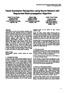
. Figure 1. Generation of htsion proteins. A, Schematic drawings of the APPL structure predicted from the primary sequence and portions of the molecule that were used to make fusion proteins for generating antibodies are shown. Hatched bars indicate the 3 regions most homologous to APP. APPLI and APPL* represent 2 regions of APPL used to generate T7-fusion proteins. B, Coomassie-stained SDSPAGE gels of proteins from bacterial cells expressing different fusion constructs. Lane I, molecular-weight marker (Sigma); lane 2, induced cells with T7-APPLI fusion construct; the doublet around 116 kDa corresponds to T7-APPL’ fusion; lane 3, induced cells with vector (pGDD7,E) alone; lane 4, uninduced cells; lane 5, induced cells with T7-APPL* construct; the novel 37kDa band corresponds to T7-APPL* fusion.
5 min, and washed once in 4 mM sodium bicarbonate (pH, 9.5) for 10 min before mountina. Fly stocks. All the embryo extracts and primary cell cultures were made from wild-type Canton-S flies that were reared on cornmeal-agarmolasses food at 25°C. The deficiency Ilies used as a negative control for immunostaining in Figure 7C, Dffl)srv~Dp24, were made as an interstitial deletion that uncovers 4 known lethal complementation groups in the genomic interval and Appl (K. White, unpublished observations). Df(l)srv~ Dp24/ + females were crossed to wild-type males to obtain embryos deleted for the Appf genomic region. To generate the transformants used in the salivary gland staining experiment (see Fig. 8C,D), the hsp-Appl construct described above was inserted at the Xba I site into Casper transformation vector (Pirotta,
3552
Luo et al.
l
Drosophih
APPL
Protein
Secretion
and
Neural
Expression
B
A
-180-116-1 -84-' -558~" -48-
I P
Figure 2. APPLexpression duringembryonicstages. A, Developmen-
tal immunoblotof APPL in embryonicstagesprobedwith affinitypurifieda-APPL.Proteinextractsweremadefrom stagedembryos(at 25°C)of 3-hr intervalsspanningDrosophilaembryogenesis. B, Immunoblotof 15-l 8-hr embryonicextract probedwith or-APPLserumat a dilution of 1:200.
1988).This DNA wasinjectedinto Dfl)white embryos,andindependently transformedflieswereobtained(Rubin and Spradling,1982). Two of theselines(oneon the secondchromosome, the other on the third chromosome) wereusedto createthe doublehomozygous strain usedin the experiment. Results Generation of anti-APPL antiserum The Appl ORF predicts an 886-amino acid polypeptide with a putative transmembrane domain near the carboxyl terminus (Fig. 1A). cDNA c4 of Appl, which codes from amino acid residue303 to the C terminus, wasusedin a bacterial expression vector systemto generatea T7 coat-protein APPL’ (T7-APPL’) fusion (Studier and Moffat, 1986; Fig. 1A). A fragment of c4, which codes for the cytoplasmic domain along with 7 amino acid residuesin the transmembranedomain, was also used in the sameexpressionsystem to make a T7-APPL* fusion (Fig. 1A). Both fusion constructs were usedto transform E. coli cells allowing for induced expression(seeMaterials and Methods). Compared to the uninduced cells (Fig. lB, lane 4), induced bacterial cells bearing the vector alone expressedthe T7 coat protein that migratesaround 30 kDa (Fig. lB, lane 3). Induction of cells bearing the T7-APPL’ fusion construct resulted in the expressiona doublet of approximately 116 kDa (Fig. lB, lane 2), while cellsbearingthe T7-APPLZ construct expresseda novel 37-kDa protein (Fig. lB, lane 5). These induced proteins were excised from preparative SDS-PAGE gelsand used as immu-
nogensto inject rats and rabbits. The seraobtained are termed (u-APPL and a-APPLC (for cytoplasmic domain), respectively. In both cases,the generation of anti-APPL antibodies was confirmed by immunoblot recognition of a P-gal-APPLI fusion protein (seeMaterials and Methods) and immunoprecipitation of the in vitro translatedproduct from anAppl cDNA containing the entire ORF (data not shown).Rat a-APPL serumwasaffinity purified using a /3-gal-APPL’ fusion protein column (seeMaterials and Methods). APPL occursas a 145-kDa and a 130-kDa form in late embryogenesis To identify APPL protein(s), extracts from stagedembryos were analyzed on developmental immunoblots with affinity-purified a-APPL (Fig. 2A). Two bands with approximate molecular weights of 145 kDa and 130 kDa correspond to major APPL proteins because(1) thesebands showenrichment when probed with affinity-purified antibody compared to serum (cf. Fig. 2A with 2B); and (2) they are first detected in 9-l 2-hr embryos and persist thereafter, as would be predicted from the expression pattern OfAppZtranscript (Rosenet al., 1989).The 90-kDa band, which has a similar developmental profile and is weakly recognized by affinity-purified antibodies, might representa degradation product formed during the extraction procedure, asthe relative intensity varies between extractions, and it is not seen in extracts from embryonic primary culture and transfectedcells (seebelow). The APPL proteins migrate with apparent molecular weights higher than that predicted from the primary sequence (98 kDa). This is indicative of posttranslational modifications, aswill be describedbelow. The 130-kDa form of APPL lacks the cytoplasmic domain and is secretedinto the culture media of primary embryonic cells Several lines of evidence in mammalian systemsindicate the existenceof APP isoformswithout the cytoplasmic domain (de Sauvageand Octave, 1989; Palmer?et al., 1989; Weidemann et al., 1989). To test if a similar situation exists in Drosophila, immunoprecipitations with CY-APPLand a-APPLC were performed in parallel. Extracts from lO-19-hr embryos were immunoprecipitated with either a-APPL or c+APPLCserum. The precipitated proteins were detected on immunoblots using a-APPL serum.While c~-APPLcould precipitate both the 130kDa and the 145kDa proteins with approximately equal efficiency (Fig. 3A, lane l), (u-APPL~ could only precipitate the 145kDa protein (Fig. 3A, lanes 2, 3). As a negative control, antiserum generatedin the sameexpressionsystemagainstanother Drosophila protein, ELAV (Robinow et al., 1988; Robinow, 1989),failed to precipitate proteins recognizedby (u-APPL (Fig. 3A, lane 4). Basedon these findings, we suggestthat the 130-kDa band representsa form of APPL that lacks the cytoplasmic domain. If the difference betweenthe 2 forms of APPL is solely at the carboxyl terminus, the molecular weight difference betweenthe 2 forms suggeststhat the 130-kDa form would also lack the membrane-spanningdomain and therefore may representa secreted form. In order to test this possibility, a primary culture systemfrom embryos was developed (seeMaterials and Methods). Cells dissociatedfrom 1l-14-hr embryos and cultured in M3 media continue to synthesize APPL for at least 5 hr as detected by immunoblot analysis or metabolic labeling (data not shown). As shown by a typical time course in Figure 3B,
The Journal
of Neuroscience,
December
1990,
IO(12)
3853
A 1
2
3
4 -180
* "xur
I -116 -
84 58 48 Figure 3. The 130-kDa form lacks cy-
B ,-Time
(min)
Cell
030601202100
I
I-Media-, 3060
120
210
EE -180 -1 16 -84
after culturing the cells for various times, cell lysates contain the 145kDa form and, to a lesser extent, the 130-kDa form. Between 30 and 60 min after plating, the 130-kDa form is observed in the culture media, and it accumulates thereafter throughout the culture period. This experiment suggests that the 130-kDa cytoplasmic-domain-free form is a secreted form. A single cDNA transfected into Schneider cells produces both forms of APPL There are several likely explanations for the different forms of APPL observed (Fig. U). They could result from posttranslational modification of a single polypeptide, they could be generated from alternatively spliced Appl mRNA, or they could be encoded by separate but homologous genes in Drosophila. To distinguish between these possibilities, we transfected Schneider S2 cells with a construct of Appl cDNA that has the entire ORF of 886 amino acids under the control of heat-shock promoter (see Materials and Methods). By immunoblot analysis, the APPL protein is not detected in nontransfected S2 cells (Fig. 4A, lanes 1, 3). S2 cells transfected with the hsp-Appl construct (S2-Appl cells), on the other hand, express both the 145~kDa and the 130kDa forms that corn&rate with the forms observed in extracts prepared from embryos (Fig. 4A, lanes 2, 4; cf. lane 5). The expression of APPL protein is enhanced by heat-shock treatment (Fig. 4A, cf. lane 4 with lane 2). Because only a single cDNA from the Appl locus is used for transfection, the 2 forms
toplasmic domain and is secreted into culture media of embryonic cells. A, 1O19-hr embryo extracts were immunoprecipitated with rat a-APPL serum (lane I), (u-APPLc sera from 2 different rats (lanes 2,3), and rat (u-ELAV serum (lane 4). The precipitated proteins were run on a 7.5% SDS-PAGE gel. transferred to nitrocellulose, and prabed with rabbit (u-APPL serum (1:250). B. Time course of APPL secretion in primary cell cultures made from 1l-14-hr embryos. At different times(min) after the plating of cells in M3 media, equal aliquots were withdrawn. Cells were pelleted. Cell lysates and media supernatants were collected, run on a 7.5% SDS-PAGE gel, transferred to nitrocellulose, and stained with a-APPL serum (1:250). EE, lO-19-hr embryonic extracts.
must be derived from the samegene. The result is consistent with 2 forms of APPL being different posttranslational modification products, though at this stage,the alternative splicing explanation is not ruled out. Immunoprecipitation studies of S2-Appl cells labeled with 35S-(met,cys) confirmed the above observations.S2 cellsdo not expressAPPL endogenously(Fig. 4B, lanes 3, 4, 7, 8). Conversely, in S2-Appl cultures, the 145-kDa APPL could be immunoprecipitated from the lysed cellsby (Y-APPL and (u-APPL~ (Fig. 4B, lanes 1, 5), while the 130-kDa form could be immunoprecipitated only by (u-APPL from the media (Fig. 4B, lanes 2,6) and, to a lesserdegree,from the cells(Fig. 4B, lane 1). This experiment substantiatesthe observation in primary cultures that the 130-kDa form is secreted,and that it lacks the cytoplasmic domain. S2-Applcells were separatedinto membraneand solublefractions by lysing the cellsthrough freeze-thawing and subsequent extraction in hypotonic solution in the absenceof detergent (Simon et al., 1989). In this preparation, the 145-kDa form is exclusively presentin the membrane fraction (Fig. 4A, lane 6), while the 130-kDa form is predominantly in the solublefraction (Fig. 4A, lane 7). The underrepresentationof the amount of the 145-kDa band in Figure 4, lane 6 (cf. the ratio betweenthe 145kDa and the 130-kDa forms in lanes6, 7 vs. that in lane 4), is due to the incomplete extraction of the membrane fraction in 2% NP-40 (seeFig. 4, legend). Fractionation of embryonic ex-
3554
Luo et al. - Dmsophi/a
APPL Protein Secretion
and Neural Expression
A Transfection Heat-shock
-
* -
EE
+
M
S
EE .,;
-180-1 1 6-
I
“’ “I
7
8
-84\ Lane Figure 4. APPL expression and secretion in transfected Schneider cells. A, Immunoblot of Schneider cell oroteins probed with CX-APPL serum. L&e I, S2 cells without heat-shock; lane 2, S2-Appl cells without heat-shock: lane 3. S2 cells with heat-shock; lane i, SZ-AipZ cells with heat-shock, lanes 5 and 8, 1O-l 9hr embryo extract; lane 6, membrane fraction of heat-shocked SZ-AppZ cells; lane 7, soluble fraction of heat-shocked S2-AppZ cells. The heat-shock regime for the above experiment is 37°C for 20 min, RT for 40 min. Samples loaded in lanes 14 were from equal number of cells (5 x 1O4cells/lane). Lanes 6 and 7 also represent samples from equal number of cells. The amount of the 145kDa protein in lane 6 is underrepresented due to the incomplete solubilization of the membrane because the pellet of 2Oh NP-40 extraction contains the 145~kDa form also (data not shown). EE, lO-19-hr embryonic extracts. B, Immunoprecipitations of 35S-(met, cys)labeled culture. Lanes I and 5, S2-AppZ cells; lanes 2 and 6, SZ-AppZ culture media; lanes 3 and 7, S2 cells; lanes 4 and 8, S2 culture media. Lanes 1-4 were immunoprecipitated by CY-APPL, and lanes 5-8 were immunoprecipitated with a-APPLC.
1
2
3
4
5
6
B Anti body Transfection Cell Media
+
a-APPL + -
+
+
-
+
w-APPLC + -
+
+
+
+
-
+
+
-205:b:6- -66-45Lane
tracts by similar proceduresgeneratedsimilar results (data not shown). Theseexperiments suggestthat the 145-kDa form representsthe 886-amino acid membrane-spanningform predicted from the cDNA ORF. The 145-kDa membrane-associated form is a precursor that is rapidly converted to the I30-kDa secretedform in S2-Appl cells In order to establish the relationship between the 2 forms of APPL, pulse-chaselabeling experiments were conducted in S2AppZ cells.Heat-shockedS2-Appl cellswere given a lo-min 35S(met, cys) pulse,briefly washed,and further incubated in complete M3 media. Samplesfrom the cell lysatesand culture media were collected at 0, 10, 20, 30, 60, and 90 min for immunoprecipitation analysis (Fig. 5A). During the first 20-min chase
1
2
3
4
5
6
7
8
period, the 145~kDa form is observed in the cell lysates, and the amount of radioactivity reachespeak values at 30 min (Fig. 5B). After 30 min chase,the labeled 145~kDa form starts to decline in cell lysatesconcurrent with the accumulation of the labeled 130~kDaform in the media. By 90 min chase,the majority of the labeled 145~kDaform has beenconverted into the 130~kDasecretedform. In addition, the rate of appearanceof the 130~kDaform is similar to the rate of disappearanceof the 145~kDaform, indicating a product-precursor relationship. This experiment strongly suggeststhat the 145~kDacell-associated form is the precursor that is converted to the cytoplasmic-domain-free secreted form, presumably by proteolytic cleavage within its extracellular domain. From the size difference, the cleavagesite might be closeto the membrane-spanningdomain. Moreover, the kinetics suggestthat the conversion is a rapid event.
A’
Cell
I Time
(min)
,I
I----
10 20 30 60 90
0
Media
1
01020306090
205-
-205
11697-
-1 16 -97
66-
-66
45 Lane
8
1234567
9
10
11
12
13
600
cell
” . Y
0
20
40
Time
60
80
media
100
(min)
Figure’ 5. Pulse-chase labeling study of APPL in SZ-Appl cells. A, S2-Appl cells were labeled with 35S-(met, cys) for 10 min and chased for O-90 min in complete M3 media. Cell lysates (lanes I-6) and media (lanes 8-13) were immunoprecipitated with a-APPL. Lane 7 is blank. B, Quantitative analysis of pulse-chase labeling of the same gel as in A with Ambis radioanalytic imaging system. Circles represent the cpm of cell lysate points, and triangles represent the cpm of media points.
3858
Luo et al.
l
Drosophi/a
APPL Protein Secretion
Tunicamycin Cell Media
+
+ +
+
and Neural Expression
+ + 4205
-1 16 97
66
Lane
1
2
3
4
Figure 6. Both 145~kDa and 130~kDa forms are N-glycosylated. S2Appl cells were labeled with YS-(met, cys) for 2 hr in the absence (lanes 1 and 3) and presence (lanes 2 and 4) of 10 &ml tunicamycin. The cell lysates (lanes I and 3) and culture media (lanes 2 and 4) were immunoprecipitated with (Y-APPL serum. Compared with lanes1 and 3, in lanes2 and 4, there are apparent molecular-weight decreases for APPL proteins, indicating the inhibition of N-glycosylation.
Both the precursor and the secretedforms of APPL are N-glycosylated Studies of mammalian systemshave shown that APP is both N- and 0-glycosylated (Weidemannet al., 1989).Sequencecomparison has revealed a conserved N-glycosylation site between APP and APPL (Rosen et al., 1989). In order to test whether APPL is also N-glycosylated, S2-Appl cells were metabolically labeled after heat-shock in the presenceof 10 pg/ml tunicamycin, which is an N-glycosylation inhibitor. Both the precursor and the secretedforms have apparent-molecular-weight shifts in the presenceof tunicamycin (Fig. 6, cf. lanes 1, 3 with lanes 2, 4), suggestingthat APPL is N-glycosylated, and that the inhibition of N-glycosylation doesnot prevent the formation of the secreted form. Similar molecular-weight shifts were observed in metabolically labeled primary embryonic cellsin the presenceof tunicamycin (data not shown). APPL is expressedin dlyerentiated neurons Next, we studied the distribution of APPL during Drosophila development. The Appl transcript is restricted to the nervous system(Martin-Morris and White, 1990); however, the protein distribution may differ in light of its secretion.Affinity-purified (u-APPL was used for immunocytochemical studiesin embryonic stages.Staining above background is first detected at stage 13 (Campos-Ortegaand Hartenstein, 1985), when germ-band shortening is complete and axonogenesisstarts (Jacobs and Goodman, 1989; data not shown). The staining increaseswith ageand persiststhroughout embryonic development (data not
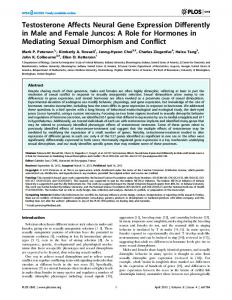
shown), which is consistent with the developmental immunoblot (Fig. 2A) and RNA analysis (Rosen et al., 1989; MartinMorris and White, 1990). APPL immunoreactivity is restricted to, and presentthroughout, the CNS and PNS during embryonic stages(Fig. 7A), both in cortical and neuropil regions(Fig. 7A,C). In particular, the axon tracts are heavily stained, ascan be seen in the central commissuresof the brain lobes (Fig. 7B) and in the longitudinal and transversecommissuresof the ventral cord (Fig. 7A,C). Through all sectionsof the embryo, no immunoreactivity was detected outside the CNS and PNS (Fig. 7B,C), though this doesnot exclude the possibility that there might be very low levels of APPL elsewhere. By staining whole-mount third-in&r larval nervous systems and imaginal disks,APPL protein is detectedin the third-instar larval brain and ventral ganglion (data not shown). APPL immunoreactivity is also present in eye disks. Figure 70 shows APPL expressionpattern in developing photoreceptor cells in eye disks. APPL immunoreactivity is present in cells posterior to the morphogenetic furrow, which indicates the border between differentiated and undifferentiated neurons (Ready et al., 1976), and in the bundle of photoreceptor axons in the optic nerve. Furthermore, there is a clear gap of several columns between the morphogenetic furrow and cells with APPL immunoreactivity. This gapmight correspondto preclusteredcells between the morphogenetic furrow and mature photoreceptor clusters(Ready et al., 1976). These observations are consistent with the in situ analysis of the AppZ transcript (Martin-Morris and White, 1990) and clearly demonstrate that, in this region where neuronal differentiation events are well documented, APPL is expressedonly in differentiated neurons. To addressthe specificity of the affinity-purified (w-APPLserum, immunostaining was carried out on a population of embryos from a genetic crossin which 1/4of the total would consist of embryos hemizygous for a deletion that includes the Appl gene(seeMaterials and Methods). In this population, ‘/4of the embryos should lack APPL immunoreactivity. Indeed, 27.6% (78 of 283) embryos showedno staining. Figure 7C.depicts an APPL- embryo along with its APPL+ sibling, strongly suggesting that APPL immunoreactivity is associatedwith the Appl gene product. Subcellular localization of APPL BecauseAPPL is synthesized as a membrane precursor and converted into a secretedform, it will be of interest to determine the subcellular localization of APPL protein. Relative to the small size of Drosophilaneurons,salivary glandsconsistof large cells ideal for subcellular localization studies (Vincent et al., 1989). As APPL expressionis restricted to the nervous system, we made use of a transformant line that is homozygous for 2 P-elements bearing the hsp70-Appl cDNA construct (seeMaterials and Methods). After heat-shock induction, ubiquitous expression of APPL should be achieved (Lis et al., 1983), including expressionin salivary glands. Salivary glands from third-instar larvae of Drosophila melanogasterare composedof large cells; somecan be as large as 50 pm (Fig. 8A). While salivary glandsfrom heat-shockedwildtype third-instar larvae did not show APPL immunoreactivity (Fig. 8B; cf. phase-contrastphotomicrograph in Fig. 8A), salivary glandsfrom hsp-Appl transformants showedstrongstaining (Fig. 8C). APPL immunoreactivity is strongestsurrounding the nuclear region and extending outwards, reminiscent of endoplasmic reticulum (ER) and Golgi apparatus localization (Fig.
,. ,
Figure 8. Subcellular localization ofAPPL in flies bearing transformed hsp-AppI. Salivary glands from heat-shocked larvae were dissected, stained with affinity-purified (u-APPL, and visualized with PITC-conjugated secondary antibodies under fluorescence optics. A and B, These photomicrographs are of a salivary gland from a wild-type larva viewed with phase-contrast (A) and fluorescence (B). C, Immunofluorescence of a salivary gland from a larva homozygous for 2 P-element inserts containing hsp-Appl. D, A high magnification of part of C. Anterior is to the left. Scale bar, 261 pm, A, B; 200 pm, C; 50 pm, D.
The Journal
80; Lippincott-Schwartz et al., 1990). No strong signal is visible at the cellular border (cf. Vincent et al., 1989). These findings, admittedly in non-neuronal cells, are nevertheless compatible with the above biochemical analyses of APPL, indicating that APPL is synthesized as a membrane protein, but is rapidly converted to a secreted form. Discussion Identification of APPL proteins Several criteria suggest that the 145-kDa and 130-kDa forms are the major products ofAppl. First, both forms start to appear on developmental immunoblots from 9-12-hr embryos and continue throughout embryogenesis (Fig. 2A), consistent with the expression pattern of the Appl transcript (Rosen et al., 1989). Second, both forms show enrichment using an affinity-purified ol-APPL antibody compared to serum (cf. Fig. 2A with B). Third, the antibody was demonstrated to specifically recognize an antigen derived from the genomic region where Appl is located (Fig. 7C). Finally, in embryonic culture cells and in Schneider cells transfected with hsp-Appf, 2 bands corn&rating with the embryonic bands on SDS-PAGE gels are observed (Figs. 3B, 4A). Especially in the case of S2-Appl cells, the 2 bands are specific for transfected cells (Fig. 4A, cf. lanes 1, 3 with lanes 2, 4) and are heat inducible (cf. lane 2 with lane 4), as would be predicted from the construct used for transfection. This strongly suggests that the 2 forms are derived from the Appl transcript. Taken together, the evidence validates the identification of the 145-kDa and 130-kDa forms as the major Appl products. Biogenesis of APPL By using the cytoplasmic domain-specific antibody, we have subsequently shown that the 130-kDa form in embryonic extracts lacks the cytoplasmic domain (Fig. 3A). Embryonic culture studies further demonstrate that the 130-kDa form is secreted into the media (Fig. 3B). Studies of S2-Appl cells confirm these findings and suggest that the 2 forms are derived from the same cDNA (Fig. 4A,B). Further, fractionation of S2-Appl cells shows that the 145-kDa protein is exclusively membrane associated, while the 130-kDa protein is mainly in the soluble fraction (Fig. 4A, lanes 6-8). Fractionation of embryo extracts made in hypotonic solution in the absence of detergent shows a similar distribution of the 2 forms as in S2-Appl cells (data not shown). These results are consistent with the view that APPL is synthesized as a membrane-associated 145kDa protein, which correlates with the 886-amino acid polypeptide deduced from the cDNA sequence. It is then converted into the cytoplasmicdomain-free 130-kDa secreted form. Our pulse-chase experiment confirms this view and indicates that such a conversion is very rapid (Fig. 5). Most cell-surface or secretory proteins require more than 30 min from their synthesis on the surface of rough ER through ER, Golgi, and transporting vesicles to reach their final destiny (see, e.g., Wieland et al., 1987). In the case of APPL, while the labeled cellular protein level peaks between the 20- and 30-min chase periods (Fig. 5B), the secreted form already starts to be detected during the same period (Fig. 5B). This indicates that APPL exists as the 145-kDa precursor only for a very short period of time before its conversion to the secreted form. The subcellular immunostaining study in large salivary glands supports this conclusion. The strongest staining is observed intracellularly, along with the pathways of membrane and secreted protein synthesis (Fig. 80). On the other hand, cell-surface staining could not be observed,
of Neuroscience,
December
1990,
1O(12)
3859
suggesting that either the 145~kDa protein has a very transient appearance on the surface, or it has been converted to the 130kDa form before it reaches the cell surface. From the molecularweight difference between the 2 forms, together with the fact that the secreted form lacks the cytoplasmic domain, we propose that the 130~kDa form could be generated by proteolytic cleavage at its extracellular domain near the membrane-spanning region. The precise cellular compartment in which the cleavage might occur could not be clearly determined from our data. The pulsechase experiments imply that the 145 kDa to 130 kDa conversion happens at or very close to the cell surface, because only the 145-kDa protein band is seen associated with the cells (Fig. 5A). However, several other experiments suggest that the conversion may happen intracellularly as the secreted form is seen associated with the cells in (1) immunoblot analysis of S2-Appl cells (Fig. 4A), (2) the immunoprecipitation experiments of cells labeled for a longer duration (Fig. 4B, lane I), and (3) in embryonic cell cultures (Fig. 3B). The association of the 130-kDa form with the cells in these later situations could also be explained by specific or nonspecific binding of the secreted 130kDa form to the cells when the concentration of the 130-kDa form increases to a certain degree in the media. In light of the close relationship between the abnormal cleavage and the pathogenesis of Alzheimer’s disease, the precise cleavage site and the nature of the cleavage is certainly worthy of further investigation. Metabolic labeling of S2-Appl cells in the presence of tunicamycin, an N-glycosylation inhibitor, results in a molecular weight decrease for both forms of the APPL protein. This suggests that APPL is N-glycosylated, like its mammalian counterpart (Weidemann et al., 1989). In addition, in the presence of 10 &ml tunicamycin, which should completely inhibit the N-glycosylation, APPL still migrates as broad bands. This result was confirmed by 3 independent experiments (data not shown). This might suggest that, in addition to N-glycosylation, there could be other posttranslational modifications, such as O-glycosylation and tyrosine sulfation. Both of these posttranslational modifications have been demonstrated in mammalian APP studies (Weidemann et al., 1989). A point of difference in the pulse-chase and glycosylation experiments between APPL and APP is noteworthy. In the case of APP, Weidemann et al. (1989) first observed the N-glycosylated form early in the chase period, and N- and 0-glycosylated form was observed later. In the case of APPL, there is no apparent molecular-weight change of the 145-kDa form during the early chasing period. This difference may be accounted for by more pronounced temporal uncoupling of the N-glycosylation and other modifications (O-glycosylation) in the PC-12 cells as compared to S2 cells. Comparison between APP and APPL The similarities between mammalian APP and Drosophila APPL have been extended from sequence homology (Rosen et al., 1989) to the properties of protein biosynthesis. In both cases, the proteins are synthesized as glycosylated transmembrane precursor proteins (Figs. 3-6; Weidemann et al., 1989). Secreted forms that lack the cytoplasmic domain are cleaved from the precursor and released into the media (Figs. 3-5; Palmert et al., 1989; Schubert et al., 1989a; Weidemann et al., 1989). Even the half-life of the membrane-associated precursor in pulsechase experiments appears to be similar (Fig. 5; Weidemann et al., 1989). These observations provide further evidence that
3860
Luo et al.
l
Drosophila
APPL
Protein
Secretion
and
Neural
Expression
APP and APPL might be functionally homologous in their respective organisms. In the case of secretion, the comparison is especially intriguing. There is no apparent primary sequence homology between APP and APPL in either the membranespanning region or the extracellular region bordering the membrane where the putative cleavage sites are most likely located, nor did we observe any conserved protease cleavage site. Yet, the secretion event itself is conserved. Moreover, in developing Drosophila embryos, the secreted form always coexists with the membrane-associated form (Fig. 2A). These observations strongly suggest that the secretion event is of physiological significance. In fact, secreted forms of APP in mammalian systems have been shown to produce a variety of effects (see below). Do APP and APPL function as receptors, as was initially predicted from the APP sequence structure (Kang et al., 1987), or alternatively, could the secreted form be the active form, which plays some function in the extracellular matrix or serves as a ligand? We have no direct evidence to distinguish between these 2 possibilities, but they are certainly worth pursuing. If the latter case is true, then the strong homology between the cytoplasmic domain of APP and APPL could mean either that the extracellular secretion is regulated cytoplasmically, or that the remaining part, and/or the precursor, may play yet other functions. Despite the striking similarities described above, there are apparent differences between APP and APPL. Multiple differentially spliced products have been found in APP, including 2 classes of APP that have, in the extracellular portion, an additional domain encoding a Kunitz-type protease inhibitor (Kitaguchi et al., 1988; Ponte et al., 1988; Tanzi et al., 1988). In Drosophila, a single transcript of 6.5 kb has been identified for Appl (Rosenet al., 1989) and all the protein products identified could be accounted for by a singlecDNA that encodesthe 886amino acid transmembraneprotein (Fig. 4). Although we cannot completely rule out the possibility of the existence of multiple alternatively splicedtranscripts that have similar sizesand generate different proteins of similar size, a proteaseinhibitor domain was not found in cDNA analyses(L. Luo and K. White, unpublished observations). Therefore, the restricted nervous system expression of the Appl transcript (Martin-Morris and White, 1990) and the APPL protein (Fig. 7) resemblesan isoform of APP, APP,,,, which is expressedin a neural-specific fashion (Ponte et al., 1988; Neve et al., 1989; Weidemann et al., 1989) and lacks the proteaseinhibitor domain. Taken together, these comparisons suggestthat APPL and APP,,, are more similar and may represent an ancestral nervous-system function for this classof molecules.The proteaseinhibitor function, associatedwith the mammalian gene that has the ubiquitous tissue expression, may have evolved during vertebrate development. Alternatively, protease inhibitor function may have been lost during invertebrate evolution. Towards the biologicalfunction of APPL Immunocytochemical analysis of the APPL protein in developing embryos demonstratesthat APPL expressionis restricted to the nervous system,and that its onset coincideswith axonogenesis.The expression pattern of APPL in developing eyes further substantiatesthe correlation between APPL expression and neuronal differentiation. This suggestsa possiblerole for APPL in neural development. Various studies of mammalian APP have shed some light on its possible function. Secreted forms of APP have been shown to have growth-promoting activities on cultured fibroblasts (Saitoh et al., 1989). Fragments
of APP have been implicated in either the enhancement of neuronal survival (Whitson et al., 1989)or neurotoxicity (Yankner et al., 1989). Recently, Schubert et al. (1989b) showedthat, in PC-12 cells, the secretion of APP without the proteaseinhibitor domain (APP,,,) increasesupon treatment with NGF or FGF, which inducesmorphologicaldifferentiation of PC- 12cells. Furthermore, the secretedform of APP could enhancethe adhesion of PC-12 cells (Schubert et al., 1989b). On the basisof the similarities between APPL and APP (especially APP& as discussedabove, we can start to speculateon the possibleroles of APPL in neuronaldifferentiation. The secretedform of APPL may function in developmental processessuch as stimulating neurite outgrowth or helping neurite adhesion, or it may help in the maintenanceof neurons. Further insightsinto the in vivo function of APPL shallawait the mutational analysisof the Appl gene. References Campos-Ortega JA, HartensteinV (1985) The embryonicdevelopmentof Drosophila melanogaster. Berlin:Springer. de SauvageF, OctaveJN (1989) A novel mRNA of the A4 amyloid precursorgenecodingfor a possiblysecretedprotein.Science245: 651-653. GlennerGG, WongCW (1984) Alzheimer’sdisease: initial reportof the purificationandcharacterizationof a novel cerebrovascular amyloid protein.BiochemBiophysResCommun120:885-890. GoldgaberD, LermanMI, McBride OW, Saffiotti U, GajdusekDC (1987) Characterizationandchromosomal localizationof a cDNA encodingbrainamyloidofAlzheimer’sdisease. Science 235:877-880. HarlowE.LaneD (1988) Antibodies.alaboratorvmanual.ColdSnring _ Harbor,NY: Cold SpringHarborLaboratory.. JacobsJR, GoodmanCS (1989) Embryonicdevelopmentof axon pathwaysin the Drosophila CNS. II. Behavior of pioneergrowth cones.J Neurosci9:2412-2422. KangJ, LemaireHG, UnterbeckA, SalbaumJM, MastersCL, Gneschik KH, Multhaup G, BeyreutherK, Miiller-Hill B (1987) The precursorof Alzheimer’sdisease amyloidA4 proteinresembles acellsurfacereceptor.Nature325:733-736. Kidd S,BayliesMK, GasicGP, Young M (1989) Structureand distribution of the Notch nroteinin develoningDrosophila. GenesDev 3:1113-1129. KitaguchiN, TakahashiY, TokushimaY, Shiojiri S, Ito H (1988) Novel precursorof Alzheimer’sdisease amyloidproteinshowsproteaseinhibitory activity. Nature 331:530-532. Lippincott-SchwartzJ, DonaldsonJD, SchweizerA, BergerEG, Hauri H-P, Yuan LC, KlausnerRD (1990) Microtubule-dependent retrogradetransportof proteinsinto theER in thepresence of Brefeldin A suggests an ER recyclingpathway.Cell60:821-836. Lis JT, Simon JA, Sutton CA (1983) New heat shockpuffs and fl-galactosidase activity resultingfrom transformationof Drosophila with an hsp70-1acZ hybrid gene.Cell 35:403-410. Luo L, Martin-Morris L, White K (1989) Temporaland spatialdistribution of a Drosophila proteinwhichresembles p-amyloidprecursor.Sot NeurosciAbstr 15:1375. Martin-Morris LE, White K (1990) TheDrosophila transcriptencoded by the p-amyloid protein precursor-like geneis restrictedto the nervoussystem.Development,in press. MastersCL, SimmsG, WeinmanNA, Multhaup G, McDonald BL, BeyreutherK (1985) Amyloid plaquecore protein in AIzheimer disease andDownsyndrome.ProcNat1AcadSciUSA 82:4245-4249. MtiIler-Hill B, BeyreutherK (1989) Molecularbiologyof Alzheimer’s disease. Annu Rev Biochem58:287-307. Neve RL, Finch EA, DawesLR (1988) Expression of the Alzheimer amyloid precursorgenetranscriptsin the humanbrain. Neuron 1: 669-677.
PalmertMR, PodlisnyMB, Witker DS, OltersdorfT, You&in LH, SelkoeDJ, You&in SG (1989) The @-amyloid proteinprecursorof Alzheimerdisease hassolublederivativesfoundin humanbrainand cerebrospmal fluid. ProcNat1AcadSci USA 86:6338-6342. PirottaV (1988) Vectorsfor P-mediatedtransformations in Drosophila. In: Vectors:a surveyof molecularcloningvectorsandtheir uses
The Journal
(Rodriguez RL, Reinhardt DT, eds), pp 437456. Boston: Butterworths. Ponte P, Gonzalez-DeWhitt P, Schilling J, Miller J, Hsu D, Greenberg B, Davis K, Wallace W, Lieberburg I, Fuller F, Cordell B (1988) A new A4 amyloid mRNA contains a domain homologous to serine proteinase inhibitors. Nature 331525-527. Ready DF, Hanson TE, Benzer S (1976) Development of the Drosophilu retina, a neurocrystalline lattice. Dev Biol 53:217-240. Rio DC, Rubin GM (1985) Transformation of cultured Drosophila melunoguster cells with a selectable marker. Mol Cell Biol 5: 18331838. Rio DC, La&i RA, Rubin GM (1986) Identification and immunochemical analysis of biologically active Drosophila P element transposase. Cell 44:21-32. Robakis NK. Ramakrishna N. Wolfe G. Wisniewski HM f 1987) Molecular cloning and characterization of a cDNA encoding the cerebrovascular and the neuritic plaque amyloid peptide. Proc Nat1 Acad Sci USA 84:41904 194. Robinow S (1989) The eluv gene of Drosophila melunogaster encodes a neuron-specific RNA binding protein which is required for the development and maintenance of the nervous system. PhD thesis, Brandeis University. Robinow S, Campos AR, Yao K-M, White K (1988) The elav gene product of Drosophila, required in neurons, has three RNP consensus motifs. Science 242: 1570-l 572. Rosen DR, Martin-Morris L, Luo L, White K (1989) A Drosophila gene encoding a protein resembling the human fl-amyloid protein precursor. Proc Nat1 Acad Sci USA 86:2478-2482. Rubin GM, Spradling AC (1982) Genetic transformation of Drosophila with transposable element vectors. Science 218:348-353. Saitoh T, Sundsmo M, Roth J-M, Kimura N, Cole G, Schubert D, Olterdorf T, Schenk DB (1989) Secreted form of amyloid p protein precursor is involved in the growth regulation of fibroblasts. Cell 58: 6 15-622. Schubert D, LaCorbiere M, Saitoh T, Cole G (1989a) Characterization of an amyloid @ precursor protein that binds heparin and contains tyrosine sulfate. Proc Nat1 Acad Sci USA 86~2066-2069. Schubert D, Jin L-W, Saitoh T, Cole G (1989b) The regulation of amyloid /3 protein precursor secretion and its modulatory role in cell adhesion. Neuron 3:689-694. Selkoe DJ (1989) Biochemistry ofaltered brain proteins in Alzheimer’s disease. Annu Rev Neurosci 12:463-490.
of Neuroscience,
December
1990,
fO(12)
3861
Simon MA, Botwell DD, Rubin GM (1989) Structure and activity of the sevenless protein, a protein tyrosine kinase receptor required for photoreceptor development in Drosophila Proc Nat1 Acad Sci USA 86~8333-8337. Studier FW, Moffat BA (1986) Use of bacteriophage T7 RNA polymerase to direct selective high-level expression of cloned genes. J Mol Biol 189:113-130. Tanzi RE, Gusella JF, Watkins PC, Bruns GAP, St George-Hyslop P, Van Keuren ML, Patterson D, Pagan S, Kumit DM, Neve RL (1987) Amyloid p protein gene: cDNA, mRNA distribution, and genetic linkage near the Alzheimer locus. Science 235:88&884. Tanzi RE, McClatchey AI, Lamperti ED, Villa-Komaroff L, Gusella JF, Neve RL (1988) Protease inhibitor domain encoded by an amyloid protein precursor mRNA associated with Alzheimer’s disease. Nature 331:528-530. Thomas JB, Crews ST, Goodman CS (1988) Molecular genetics of the single-minded locus: a gene involved in the development of the Drosophila nervous system. Cell 52: 133-l 4 1. Vincent WS III, Gregory RJ, Wadsworth SC (1989) Embryonic expression of a Drosophila src gene: alternate forms of the protein are expressed in segmental stripes and in the nervous system. Genes Dev 3:334-347. Weidemann A, Konig G, Bunke D, Fischer P, Salbaum JM, Masters CL, Beyreuther K (1989) Identification, biogenesis, and localization of precursors of Alzheimer’s disease A4 amyloid protein. Cell 57: 115-126. White K, Hurteau T, Punsal P (1986) Neuropeptide-FMRFamidelike immunoreactivity in Drosophila: development and distribution. J Comp Neurol 247:43w38. Whitson JS, Selkoe DJ, Cotman CW (1989) Amyloid p protein enhances the survival of hippocampal neurons in vitro. Science 243: 1488-1490. Wieland F, Gleason ML, Serafini TA, Rothman JE (1987) The rate of bulk flow from the endoplasmic reticulum to the cell surface. Cell 50:289-300. Wilcox M (1986) Cell surface antigens. In: Drosophila, a practical approach (Roberts DB, ed), pp 2431277. Oxford: IRL Press. Yankner BA. Dawes LR. Fisher S. Villa-Komaroff L. Oster-Granite ML, Neve ‘RL (1989) ’ Neurotoxicity of a fragment ‘of the amyloid precursor associated with Alzheimer’s disease. Science 245:417-420.