Please verify that (1) all pages are present, (2) all figures are acceptable, (3) all fonts and special characters are correct, and (4) all text and figures fit within the margin lines shown on this review document. Return to your MySPIE ToDo list and approve or disapprove this submission.
Statistically Based, Spatially Adaptive Noise Reduction of Planar Nuclear Studies A. Hans Vijaa*, Timothy R. Gosnellb, Amos Yahilb†, Eric G. Hawmana, John C. Engdahlc a Siemens Medical Solutions USA, Inc., Nuclear Medicine Group, 2501 N. Barrington Rd., Hoffman Estates, IL 60195; b Pixon LLC, P.O. Box 312, East Setauket, NY 11733; c Bradley University, College of Engineering and Technology, 1501 W. Bradley Ave., Peoria, IL 61625 ABSTRACT The data-driven Pixon noise-reduction method is applied to nuclear studies. By using the local information content, it preserves all statistically justifiable image features without generating artifacts. Statistical measures provide the user a feedback to judge if the processing parameters are optimal. The present work focuses on planar nuclear images with known Poisson noise characteristics. Its ultimate goals are to: (a) increase sensitivity for detection of lesions of small size and/or of small activity-to-background ratio, (b) reduce data acquisition time, and (c) reduce patient dose. Data are acquired using Data Spectrum’s cylinder phantom in two configurations: (a) with hot and cold rod inserts at varying total counts and (b) with hot sphere inserts at varying activity-to-background ratios. We show that the method adapts automatically to both hot and cold lesions, concentration ratios, and different noise levels and structure dimensions. In clinical applications, slight adjustment of the parameters may be needed to adapt to the specific clinical protocols and physician preference. Visually, the processed images are comparable to raw images with ~16 times as many counts, and quantitatively the reduced noise equals that obtained with ~50 times as many counts. We also show that the Pixon method allows for identification of spheres at low concentration ratios, where raw planar imaging fails and matched filtering underperforms. Conclusion: The Pixon method significantly improves the image quality of data at either reduced count levels, or low target-to-background ratios. An analysis of clinical studies is now warranted to assess the clinical impact of the method. Keywords: data-driven, image processing, noise reduction, nuclear medicine, planar imaging, Poisson statistics, scintigraphic images, spatially adaptive, statistical methods
1. INTRODUCTION Noise reduction in nuclear medicine can be achieved by increasing the counts or by filtering the image. The former choice requires an increase in acquisition time and/or administered dose, which in many clinical cases is suboptimal. In clinical practice, the acquisition time for a particular nuclear medical application is a compromise between image quality, workflow economics, patient safety and patient comfort. The same considerations apply to the dose, which should be minimized as far as possible, particularly in pediatric nuclear medicine. Image filtering is often employed to improve image quality, simply because constraints on acquisition time and/or dose are such that the diagnostic value of the image is too low, and some filtering may help to increase confidence in the diagnosis of features observed in the image. At the same time, filtering should not create false negatives by suppressing real features or false positives by generating spurious artifacts. Pixon LLC has developed a unique method for the reduction of random noise in planar images subject to Poisson counting statistics. Based on techniques initially developed for astronomical applications,1-3 the Pixon® method has been applied to nuclear medicine4 and is now being implemented in collaboration with the Nuclear Medicine Group of * †
[email protected]; phone: phone 1 847 304-7104; fax 1 847 304-7706; medical.siemens.com
[email protected]; phone: phone 1 631 223-4665; fax 1 509 271-9275; pixon.com
5747-65 V. 2 (p.1 of 12) / Color: No / Format: Letter / Date: 2005-01-20 19:15:53 SPIE USE: ____ DB Check, ____ Prod Check, Notes:
Please verify that (1) all pages are present, (2) all figures are acceptable, (3) all fonts and special characters are correct, and (4) all text and figures fit within the margin lines shown on this review document. Return to your MySPIE ToDo list and approve or disapprove this submission.
Siemens Medical Solutions USA, Inc. The method is a statistically rigorous procedure for noise reduction that avoids generation of spurious artifacts yet preserves all statistically justifiable image features resident in the raw counts. The method optimizes noise reduction by taking full advantage of the local information content of the image and avoiding global noise-reduction criteria,5-8 which may not be locally optimal.3 Pixon processing is especially well suited to imaging at low signal-to-noise ratio and is therefore particularly attractive for applications in nuclear medicine. Investigations are ongoing to assess the best uses of the Pixon method in this field. The present work focuses on Pixon processing of planar nuclear images with the ultimate goals of (a) increasing sensitivity for detection of lesions of small size and/or of small activity-to-background ratio, (b) reducing data acquisition time, and (c) reducing patient dose. We present results based on phantom studies, which demonstrate the potential of the Pixon method and lay the foundation for more comprehensive clinical evaluations. This study sets out to investigate noise reduction by the Pixon method in planar nuclear imaging and to compare it with other known methods. It investigates the following aspects of Pixon noise reduction: (a) accuracy, (b) precision, (c) adaptability, (d) robustness, and (e) dependence of processed image quality on acquisition time and contrast ratio. Accuracy and precision are measured by comparing the processed data to reference standards. Adaptability is assessed by observing whether the method can automatically adjust the tradeoff between noise reduction and preservation of resolution across the image: The method should utilize the local image information content to achieve at each image location the maximum noise reduction possible and it should do so without either losing resolution by oversmoothing the image or creating spurious artifacts. Robustness is demonstrated by the ability of the method to provide optimal noise reduction for a variety of images differing in acquisition times and contrast ratios without having to adjust the processing parameters. Finally, we evaluate how well the Pixon method fares as acquisition time and/or contrast ratios are varied, particularly when they are reduced relative to normal clinical practice. A large number of filtering and nonparametric fitting techniques have been developed to reduce noise in images3. These techniques make no assumptions about the structures that are observed and can therefore be applied in clinical nuclear medicine when the structure and biodistribution of the radiopharmaceutical are not known. Alternatively, when the structure is known or a particular structure is sought, one of the best filters is considered to be the matched filter, which is designed with the knowledge of both the imaging process and the imaged structure. For the phantom images in this study, the imaged structures are, of course, known. A particularly strong test of the Pixon method is therefore to compare it with matched filtering. The test is particularly powerful because of the different assumptions made by the methods. The matched filter assumes a given signal structure, but does not care about the statistics of the counts, while the Pixon method knows nothing a priori about the signal structures, discovering them from the data utilizing its knowledge of the (Poisson) statistics of the counts. Other general, spatially adaptive filters, such as the Wiener and wavelet filters, are also investigated but are found to be inferior to Pixon processing, as shown in other investigations.3 For the sake of brevity, we concentrate on the comparison between Pixon processing and matched filters, and do not report on other filters.
2. METHODS 2.1. Statistically driven, spatially adaptive Pixon noise reduction The key objective of the Pixon method is to generate an output image that is as smooth as possible yet statistically consistent with the raw counts. The method is a nonparametric, spatially adaptive smoothing technique, in which the width of the smoothing kernel at each image location is adjusted to the local image conditions2-3. Where the underlying image is uniform, or the signal-to-noise ratio is not high, it is possible to smooth over a wider span of pixels without loss of information; where the underlying image varies significantly relative to the noise, the smoothing kernel is restricted to fewer pixels to avoid loss of resolution. The local information content of the image, and only the local information content, determines the best smoothing scale. The two-dimensional array comprising the kernel widths chosen at each pixel, called the Pixon map, offers a visual representation of the image information content and may have clinical value of its own, beyond that of the image itself. Details of the Pixon method2-3 and its application to nuclear medicine4 are reported elsewhere.
5747-65 V. 2 (p.2 of 12) / Color: No / Format: Letter / Date: 2005-01-20 19:15:53 SPIE USE: ____ DB Check, ____ Prod Check, Notes:
Please verify that (1) all pages are present, (2) all figures are acceptable, (3) all fonts and special characters are correct, and (4) all text and figures fit within the margin lines shown on this review document. Return to your MySPIE ToDo list and approve or disapprove this submission.
2.2. Data acquisition and setup Data are acquired using Data Spectrum’s cylinder phantom in two separate configurations: (a) with the hot and cold rod (HCR) inserts and (b) with the hot sphere (HS) inserts. The rod diameters are 12.7 mm, 11.1 mm, 9.5 mm, 7.9 mm, 6.4 mm, 4.8 mm, and the inner diameters of the spheres are 31.3 mm, 24.8 mm, 19.7 mm, 15.6 mm, 12.4 mm, and 9.9 mm, with corresponding volumes of 16.0 ml, 8.0 ml, 4.0 ml, 2.0 ml, 1.0 ml, and 0.5 ml, respectively. The thickness of the walls of the spheres is 2 mm. The acquisition times are varied to yield a range of total counts and thus a range of signalto-noise ratios, whereby the tradeoff between image accuracy and acquisition time or dose can be assessed. The cylinder’s flat surface is placed on the scan bed of an e.cam Signature series with the detector heads in anterior and posterior position. The HCR images are acquired simultaneously with a pixel size of 0.6 mm and a 1024x1024 matrix with both LEHR and LEAP collimators. The acquisitions are stopped on counts measured in the detector facing the hotrod sector, and doubled for each acquisition period beginning at 0.1 Mcts and continuing through 6.4 Mcts, as shown in Table 1. In the case of the HS measurements, spheres are filled with activity and the activity-to-background ratio is varied, thus yielding a measure of the detection sensitivity. The spheres are screwed into the cylindrical vessel, where the equatorial planes of all spheres are coplanar and 7 cm off the bottom. The cylinder is filled with 5.5 l of water resulting in a water depth of 15.2 cm, where the displacement of the spheres and supporting rods is taken into account. The distance from the spheres’ reference plane to detector 1 is 15 cm, and to detector 2 is 24 cm. The images are acquired with a pixel size of 0.6 mm and a 1024x1024 matrix with the LEHR, which has an angular acceptance angle of 0.0495 radians. The spheres are filled with 6.3 µCi/ cm3 (0.2 Mbq/cm3), and the background is subsequently filled with 1222 µCi (45.2 MBq), 5530 µCi (204.6 MBq), 6420 µCi (237.5 MBq), and 5400 µCi (199.8 MBq), resulting in total background activities of 1222.4 µCi, 6752 µCi, 13172 µCi, 18572 µCi, respectively. The activity contrast ratio between the concentration in the spheres and in the background are then 45:1, 8:1, 4:1, and 3:1, respectively. The lid of the phantom is left off during the acquisition so as not to bias the data with a superimposed structure, as seen from detector 1. LEHR LEHR LEAP LEAP
Hot Rods (Mcts) Cold Rods (Mcts) Hot Rods (Mcts) Cold Rods (Mcts)
0.10 0.16 0.10 0.16
0.20 0.32 0.20 0.32
0.40 0.64 0.40 0.64
0.80 1.29 0.80 1.26
1.6 2.56 1.6 2.51
3.18 5.11 3.19 5.02
6.35 10.23 6.37 10.05
Table 1: Stop counts from LEHR and LEAP acquisitions of the hot-cold rods.
Parameters controlling Pixon processing in nuclear medicine,4 are kept fixed for all acquisitions to strain the test of how adaptive and data driven the method is. They are smoothing parameters of 2.0 for the HCR data—for both the hot and cold rods—and 2.5 for the HS data. The widths of the Pixon smoothing kernels were spaced in equal logarithmic intervals (geometric series) with three kernels per octave (factor of two in width) and 3 octaves for the HCR data and 5 octaves for the HS data. These parameters need not be finely tuned. The difference between the smoothing parameters used for the HCR and HS data sets is minor, and any parameter in the range of 2.0–2.5 would work well for either data set. Similarly, the larger number of octaves used for the HS data has a minor effect in smoothing more strongly larger background areas in those images. In both cases the ranges of the parameters correspond to the expected variation due to physician preference. It is also possible that adjustment of these parameters in clinical applications to acquisition time, radiopharmaceutical, or other variables might provide additional improvement. 2.3. Analysis of data from the hot-cold-rods phantom Accuracy and precision are demonstrated by statistical consistency between the processed images and the raw counts. Since the variance of the Poisson counts depends on the expected signal, which is unknown and varies across the image, the images differenced are ones in which the pixel counts N are replaced with X=(N+0.25)1/2. For a Poisson variable N with mean λ, the expectation value and standard deviation of X are asymptotically λ1/2 and 0.5, respectively, in the limit of large λ. (The X variable is specifically designed to converge rapidly to the asymptotic limit by setting the first-order correction to the asymptotic values to zero. For λ>2 the relative errors of the expectation value and standard deviation
5747-65 V. 2 (p.3 of 12) / Color: No / Format: Letter / Date: 2005-01-20 19:15:53 SPIE USE: ____ DB Check, ____ Prod Check, Notes:
Please verify that (1) all pages are present, (2) all figures are acceptable, (3) all fonts and special characters are correct, and (4) all text and figures fit within the margin lines shown on this review document. Return to your MySPIE ToDo list and approve or disapprove this submission.
are, in fact, smaller than 0.2% and 2.5%, respectively.) A second test analyzes the image formed from the cumulative probability distribution functions of the raw counts, N, under the null hypothesis that the processed image, P, is the underlying truth image, CDF=Prob(Count ≥ N | P). Statistical consistency is established if the pixel values of the CDF are uncorrelated and uniformly distributed between 0 and 1. Significant CDF correlations or a nonuniform CDF distribution indicate a discrepancy. Precision is tested by quantifying the noise reduction brought about by Pixon processing. To that end, consider the difference images ∆=µ1/2A–B, where A is the X image of a single acquisition, which can be raw or processed, B is the raw X image of the sum of all the other acquisitions, and µ is the ratio between the total counts of B and A. From the above asymptotic approximation it follows that for a raw A image the expectation value of ∆ is 0 and the variance is V(∆)=0.25(µ+1). The part of the variance attributable to the sum image is 0.25 and may not be neglected, so a good noise parameter for the single acquisition is the part of the variance attributable to it, ν=V(∆)–0.25=0.25µ. For the Pixon image, ν is smaller than 0.25µ because of the noise reduction. The ratio (νraw/νPixon)1/2 is the noise reduction factor between the raw and Pixon X images and also approximately equals the noise reduction factor for the images themselves. Another, more direct way to assess noise reduction is to ask how much larger the total count needs to be for ν in the raw image to equal ν in the Pixon image. 2.4. Analysis of data from the hot-spheres phantom The HS phantom is analyzed using both matched filters and Pixon processing and both are compared to the raw data. The matched filters are designed to take the imaging physics and the structure of the objects into account. The filters in this case are the projections of spheres with diameters equal in turn to each of the diameters of the six spheres in the phantom. These projections are then convolved with the point-spread function of the LEHR collimator at the distances of the spheres from the collimators of each camera. The filters are normalized to unit sum to conserve counts in the filtering process. The spheres reside in 60° sectors, and the entire image is filtered with the 6 corresponding matched filters. Sectors with matching filter and sphere diameters are extracted and a composite image is constructed. The images are shown in two ways: (a) with a global windowing range with the minimum and maximum obtained from the entire image, and (b) with a local windowing range, where each sector has been windowed to its minimum and maximum value. In this way, the results can be viewed over a larger dynamic range. Inverted grayscale (peak intensity black) is chosen for easier translation of the result to clinical bone scans, which are also usually viewed in inverted grayscale. Data from both detectors is acquired and processed, but only the data from detector 1 is shown. Noise-reduction is measured with the same ν parameter used in the HCR case.
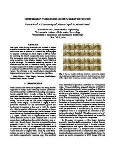
3. RESULTS 3.1. Hot-cold-rods phantom Figure 1 (hot rods) and Figure 2 (cold rods) show raw and processed images for the HCR phantom. In both figures, the top row shows the raw data with increasing counts (left to right), each image containing double the counts of the previous image. The bottom row shows the Pixon images. The Pixon images are seen to improve image quality in a way that can be achieved by the raw images only by increasing the count by a factor ~16 (four frames to the right). It is particularly telling that images of cold rods can be improved just as well as those of hot rods. Figure 3 shows additional information about Pixon processing for both the hot (top row) and cold (bottom row) rods. The first two columns reproduce the raw counts and the Pixon processed images from the rightmost columns of Figure 1 and Figure 2. The third column displays the Pixon map used, showing for each pixel the Pixon kernel used to smooth that particular pixel, where dark represents little or no smoothing and white represents maximum smoothing. The fourth and fifth columns show the ∆ and CDF images, respectively. All four panels look like random white noise. As far as we can tell from the data, therefore, the Pixon images are the true underlying images. More precisely, the raw counts are statistically consistent with being Poisson random deviates of the smooth Pixon images.
5747-65 V. 2 (p.4 of 12) / Color: No / Format: Letter / Date: 2005-01-20 19:15:53 SPIE USE: ____ DB Check, ____ Prod Check, Notes:
Please verify that (1) all pages are present, (2) all figures are acceptable, (3) all fonts and special characters are correct, and (4) all text and figures fit within the margin lines shown on this review document. Return to your MySPIE ToDo list and approve or disapprove this submission.
Finally, Figure 4 shows the noise parameter ν as a function of 1/µ, a measure of the total count. The raw counts show the expected dependence. ν=0.25µ, a simple consequence of Poisson statistics. The noise in the Pixon images is lower by an order of magnitude. Put differently, the noise level obtained by the Pixon images at the lowest count level is equivalent to that obtained by the raw counts only with ~50 times as many counts. Furthermore, the same noise reduction is obtained for the hot and cold rods.
Figure 1: LEHR hot-rod images obtained over the acquisition periods specified in Table 1. Top row: raw planar images. Bottom row: Pixon images. Columns contain data with different total counts increasing by approximately a factor of two per column from left to right, ranging from 0.1 Mcts on the left to 6.4 Mcts on the right (Table 1). Visual inspection shows the quality of the Pixon images to be approximately equivalent to the quality of the raw counts four columns to the right, a factor of 16 in total counts.
Figure 2: LEHR cold-rod images obtained over the acquisition periods specified in Table 1. Top row: raw planar images. Bottom row: Pixon images. Columns contain data with different total counts increasing by approximately a factor of two per column from left to right, ranging from 0.1 Mcts on the left to 6.4 Mcts on the right (Table 1). Visual inspection shows the quality of the Pixon images to be approximately equivalent to the quality of the raw counts four columns to the right, a factor of 16 in total counts. It is particularly noteworthy that the cold rods show the same image improvement as the hot rods shown in Figure 1.
3.2. Spheres phantom Figure 5 shows raw, matched and Pixon images for a fixed (volume) concentration ratio of 8:1 and for different acquisition times. The figure is broken up into three top rows and three bottom rows, which differ only in the way in which the images are autoscaled. In the top three rows each image is autoscaled in its entirety. In the bottom three rows, each sector in each image is autoscaled separately. The purpose of the latter autoscaling is to provide maximum use of the dynamic range of the grayscale for each sphere. It corresponds to what a radiologist might do interactively while adapting the contrast of a display to a particular suspected lesion. In each of the groups of three rows, the top row shows the raw data, the middle row the matched images and the bottom row the Pixon images. The columns correspond to different acquisition times, from left to right 1 minute, 2 minutes, 4 minutes, 8 minutes, and 15 minutes (total of the other
5747-65 V. 2 (p.5 of 12) / Color: No / Format: Letter / Date: 2005-01-20 19:15:53 SPIE USE: ____ DB Check, ____ Prod Check, Notes:
Please verify that (1) all pages are present, (2) all figures are acceptable, (3) all fonts and special characters are correct, and (4) all text and figures fit within the margin lines shown on this review document. Return to your MySPIE ToDo list and approve or disapprove this submission.
columns). Inspection of the figure shows that spheres that are barely seen in the raw counts are clearly seen in the Pixon images and somewhat less well in the matched images.
Figure 3: Top row: LEHR hot-rod planar data and Pixon results. Bottom row: LEHR cold-rod planar data and Pixon results. From left to right in both rows: raw data, Pixon image, Pixon map, difference image ∆, and cumulative distribution function (CDF). The Pixon maps are visual representations of the information content of the images. The two rightmost images demonstrate that the Pixon images are consistent with the raw counts because they do not have recognizable features.
Figure 4: Noise parameter ν for raw and Pixon images of a single acquisition. It is computed by comparing with the truth image made up of the sum of the raw images of all the other acquisitions, whose total count exceeds that of the single acquisition by a factor µ. (See §2.3 for details.) Pixon processing is seen to reduce noise by an order of magnitude, so the noise level in a Pixon image is equivalent to that of a raw image whose acquisition time is ~50 times longer.
The concentration ratio of 8:1 in Figure 5 is actually smaller than a typical one in a bone scan, which would be several times larger. Figure 6 shows how Pixon and matched processing fare at different concentrations. The rows in Figure 6 have the same meaning as the rows in Figure 5. The columns correspond, left to right, to concentrations of 3:1, 4:1, 8:1, and 45:1. Note that these are volume concentration ratios, not the projected contrasts, which are much smaller. Again, Pixon images offer much better signal-to-noise ratio and detectability than raw images, with matched images somewhere in between. The precision of the matched and Pixon methods is studied in the same way as for HCR. Figure 7 shows the noise parameter ν for raw, matched and Pixon images as a function of acquisition time for HS. The noise reduction by Pixon processing is similar to that seen in Figure 4 for the hot-cold rods. The adaptive Pixon processing reduces noise more than matched filters for low counts, where statistics allow stronger smoothing, and less for higher counts to avoid oversmoothing. The matched filters, by contrast, are insensitive to statistics and oversmoothed for larger counts, resulting in loss of accuracy.
5747-65 V. 2 (p.6 of 12) / Color: No / Format: Letter / Date: 2005-01-20 19:15:53 SPIE USE: ____ DB Check, ____ Prod Check, Notes:
Please verify that (1) all pages are present, (2) all figures are acceptable, (3) all fonts and special characters are correct, and (4) all text and figures fit within the margin lines shown on this review document. Return to your MySPIE ToDo list and approve or disapprove this submission.
Figure 5: Fixed concentration of 8:1 for hot spheres with different acquisition times. Rows from top to bottom: (1) raw counts, (2) counts filtered by matched filters, (3) Pixon images, (4–6) same as 1–3 but with each sector autoscaled separately. Acquisition times in columns from left to right: (1) 1 minute, (2) 2 minutes, (3) 4 minutes, (4) 8 minutes, (5) 15 minutes (total of 1–4). Spheres that are barely seen in the raw counts are clearly seen in the Pixon images and somewhat less well in the matched images.
5747-65 V. 2 (p.7 of 12) / Color: No / Format: Letter / Date: 2005-01-20 19:15:53 SPIE USE: ____ DB Check, ____ Prod Check, Notes:
Please verify that (1) all pages are present, (2) all figures are acceptable, (3) all fonts and special characters are correct, and (4) all text and figures fit within the margin lines shown on this review document. Return to your MySPIE ToDo list and approve or disapprove this submission.
Figure 6: Fixed acquisition time of 8 minutes for hot spheres with different concentration ratios. Rows from top to bottom: (1) raw counts, (2) counts filtered by matched filters, (3) Pixon images, (4–6) same as 1–3 but with each sector autoscaled separately. Concentration ratios in the columns from left to right: (1) 3:1, (2) 4:1, (3) 8:1, (4) 45:1. Spheres that are barely seen in the raw counts are clearly seen in the Pixon images and somewhat less well in the matched images
5747-65 V. 2 (p.8 of 12) / Color: No / Format: Letter / Date: 2005-01-20 19:15:53 SPIE USE: ____ DB Check, ____ Prod Check, Notes:
Please verify that (1) all pages are present, (2) all figures are acceptable, (3) all fonts and special characters are correct, and (4) all text and figures fit within the margin lines shown on this review document. Return to your MySPIE ToDo list and approve or disapprove this submission.
Figure 7: Noise parameter ν for raw, matched and Pixon images as a function of acquisition time for the hot spheres. Dashed curve: raw counts, dot-dashed curves: matched images for different spheres from smallest (highest curve) to largest (lowest curve), solid curve with measured points: Pixon images. Noise reduction by Pixon processing is similar to that seen in Figure 4 for the hot-cold rods. Being adaptive, it reduces noise more than matched filters for low counts, where statistics allow stronger smoothing, and less for higher counts to avoid oversmoothing. The matched filters, by contrast, are insensitive to statistics and oversmooth for larger counts, resulting in loss of accuracy.
3.3. Adaptability The poorer performance of the matched images might seem surprising, since the structures of the spheres are known. The reason is that the same matched filter is applied everywhere, whereas Pixon processing is careful to adapt the filter even within the projected area of a single sphere. In fact, the matched filter must, by necessity, oversmooth the spheres, because a convolution of a structure with itself results in a broader and shallower structure. This is borne out in Figures 8 and 9, which show the CDF distributions of the matched (top row) and Pixon (bottom row) images for the parallel images in Figures 5 and 6, respectively. The Pixon CDF images are perfectly random, while the matched CDF images show the effect of oversmoothing by the matched filters. 3.4. Robustness The robustness of the Pixon method is demonstrated by the fact that the processing parameters of the method have not been changed when all the images where processed, regardless if the hot or cold section, the high-or low total counts data or the small or large sector in the data is processed. The resulting CFD images are essentially white-noise images.
5747-65 V. 2 (p.9 of 12) / Color: No / Format: Letter / Date: 2005-01-20 19:15:53 SPIE USE: ____ DB Check, ____ Prod Check, Notes:
Please verify that (1) all pages are present, (2) all figures are acceptable, (3) all fonts and special characters are correct, and (4) all text and figures fit within the margin lines shown on this review document. Return to your MySPIE ToDo list and approve or disapprove this submission.
Figure 8: Cumulative distribution function (CDF) for the matched and Pixon images shown in Figure 5. Top row: matched CDF, bottom row: Pixon CDF. The columns have the same meaning as in Figure 5. The Pixon CDF are completely random, while the matched CDF show loss of accuracy due to oversmoothing. (They also show some edge effects, but these are not important medically.
Figure 9: Cumulative distribution function (CDF) for the matched and Pixon images shown in Figure 6. Top row: matched CDF, bottom row: Pixon CDF. The columns have the same meaning as in Figure 6. The Pixon CDF are completely random, while the matched CDF show loss of accuracy due to oversmoothing. (They also show some edge effects, but these are not important medically.)
4. DISCUSSION Figures 1, 2, 5 and 6 show visually that Pixon processing significantly improves the quality of scintigraphic images. The range of acquisition times for HCR show that the Pixon images reach quality only available in raw images with roughly 16 times as many counts. Figures 3, 8 and 9 further show that the Pixon images are statistically consistent with the raw
5747-65 V. 2 (p.10 of 12) / Color: No / Format: Letter / Date: 2005-01-20 19:15:53 SPIE USE: ____ DB Check, ____ Prod Check, Notes:
Please verify that (1) all pages are present, (2) all figures are acceptable, (3) all fonts and special characters are correct, and (4) all text and figures fit within the margin lines shown on this review document. Return to your MySPIE ToDo list and approve or disapprove this submission.
images and do not introduce artifacts. The medical benefit of the resultant increased sensitivity for detection of lesions of small size and/or of small activity-to-background ratio now needs to be established in clinical trials. Figures 4 and 7 show an order-of-magnitude of measured noise reduction in the Pixon images. Figure 4 shows that the noise level in the Pixon images is equivalent to that in raw images with ~50 times as many counts. Note, however, that noise measure is not the only measure of image quality. Pixon processing deliberately seeks the smoothest image consistent with the raw counts, bypassing less smooth images, which may be equally consistent with the raw counts. Therefore, careful analysis of other quantitative and clinical factors is needed to establish by how much counts can safely be reduced without compromising medical diagnosis. Besides reducing acquisition time, physicians may opt to maintain their current acquisition times and to use Pixon processing to facilitate the identification of lesions with very small target-to-background ratios or small sizes, which would not be identifiable with the desired degree of confidence with the current techniques. Figure 6 shows that, at a given acquisition time, the spheres at low target-to-background ratio are identified by virtue of better noise reduction. The matched images seem to perform well too yet are not adaptive, so they fail to take into account either the sizes of the lesions or the noise level. A filter matched to large structures oversmoothes smaller lesions, while a filter seeking small structures undersmoothes the background. In either case, the lesion may be lost in the background, as can be seen in Figures 8 and 9. This is also illustrated in Figure 7, in which the noise-reduction parameter ν scales with µ for both the raw and matched data, decreasing as counts increase. The intercepts, however, depend on the widths of the filters, the larger matched filter the stronger noise reduction. The Pixon method, by contrast, is adaptive, adjusting noise reduction to the sizes of the features it encounters and to the noise level. Its noise parameter is lower than those of any of the matched filters at the lowest counts, effectively corresponding to a wider filter, yet at higher counts its noise parameter is higher than those of any of the matched filters. It is important to emphasize that, with decreased noise, it may be necessary to record the Pixon images to higher precision than integers. The DICOM format requires integer storage of images, and at low counts rounding off to the nearest integer may undo some of the improvement obtained by Pixon processing. The solution is to increase the dynamic range of the Pixon images by rescaling them by a sufficiently large correction factor before they are rounded off. When the Pixon image is written to a DICOM file, the scaling factor can be incorporated in the header, to be used by applications that require quantitative flux measurements. There is no problem with saturation at the high end, since most DICOM files use 16 bits to record counts—well above any counts obtained in nuclear medicine—allowing a considerable rescaling range.
5. CONCLUSIONS The present work focuses on planar nuclear images with known Poisson noise characteristics. We show that the datadriven, statistically rigorous Pixon method is able to reduce noise by an order of magnitude, thereby achieving the stated goals: (a) increasing sensitivity for detection of lesions of small size and/or of small activity-to-background ratio, (b) reducing data acquisition time, and (c) reducing patient dose, or combinations of these goals. The method is robust, adapting essentially automatically to (a) hot and cold lesions, (b) noise content, and (c) various structure sizes. Statistical tests demonstrate that the raw counts are consistent with being random Poisson deviates of the Pixon images. The method is therefore accurate, providing at each location in the image the maximum degree of noise reduction possible while (a) conserving counts, (b) maintaining resolution, and (c) avoiding the introduction of artifacts. Clinical studies should now assess the clinical impact of the method and translate it into revised procedures.
REFERENCES 1. 2.
3.
R. K. Piña, R. C. Puetter, “Bayesian image reconstruction: the Pixon and optimal image modeling”, Publ. Astron. Soc. Pac., 105, 630-637, 1993. R.C. Puetter, A. Yahil, “The Pixon method of image reconstruction”, in: Astronomical Data Analysis Software and Systems VIII., edited by D. M. Mehringer, R. L. Plante D. A. Roberts, Astronomical Society of the Pacific, San Francisco, ASP Conference Series 172, 307-316, 1999. R. C. Puetter, T. R. Gosnell, A. Yahil, Ann. Rev. Astron. Astrophys., 43, in press, 2005.
5747-65 V. 2 (p.11 of 12) / Color: No / Format: Letter / Date: 2005-01-20 19:15:53 SPIE USE: ____ DB Check, ____ Prod Check, Notes:
Please verify that (1) all pages are present, (2) all figures are acceptable, (3) all fonts and special characters are correct, and (4) all text and figures fit within the margin lines shown on this review document. Return to your MySPIE ToDo list and approve or disapprove this submission.
4.
5. 6. 7. 8.
C. A. Wesolowski, A. Yahil, R. C. Puetter, P. S. Babyn, D. L. Gilday, M. Z. Khan, “Improved lesion detection from spatially adaptive, minimally complex, Pixon® reconstruction of planar scintigraphic images”, Comput. Med. Imaging Graph., 29, in press, 2005. B. A. Gwiazdowska, E. T. Skrzypczak, J. R. Tolwinski, “The evaluation of noise reduction and resolution degradation in scintigraphic images due to smoothing procedures”, Nuklearmedizin, 21, 126-129, 1982. C. C. Kunni, B. H. Hasegawa, W. R. Hendee, “Noise reduction in nuclear medicine images” JNM, 24, 532-534, 1983. C. Riddell, R. E. Carson, J. A. Carrasquillo, et al, “Noise Reduction in oncology FDG PET images by iterative reconstruction: a quantitative assessment” JNM, 42, 1316-1323, 2001. P. Hannequin, J. Mas, "Statistical and heuristic noise extraction (SHINE): a new method for processing Poisson noise in scintigraphic images” PMB, 47, 4329-4344, 2002.
5747-65 V. 2 (p.12 of 12) / Color: No / Format: Letter / Date: 2005-01-20 19:15:53 SPIE USE: ____ DB Check, ____ Prod Check, Notes:


![[cea-00470594, v1] Spatially Adaptive Mixture ... - Semantic Scholar](https://m.moam.info/img/260x300/cea-00470594-v1-spatially-adaptive-mixture-semanti_59eabfd81723dded32744e64.jpg)