May 30, 2018 - ethanol before placing in the biosafety cabinet. 4. Resuspend MEFs in 5ml FM10 per dish (ie: 20ml for 4 dishes), using medium to rinse.
Stem Cell Research Core Facility
HUMAN PLURIPOTENT STEM CELLS
hPSC Maintenance Media
Brigitte Arduini, version 2, 2013-Jun-12 Initially, it was found that pluripotency of human pluripotent stem cells (hPSCs) can be maintained when plated in co-culture with mouse embryonic fibroblasts (MEFs) or in media conditioned by MEFs (MEF-CM). In the latter case, also known as “feeder-free” culture, additional treatment of tissue culture vessels with Matrigel or comparable substrate is necessary (see Matrigel Plate Coating). Subsequently, defined media formulations have been developed and marketed. However, MEF-CM is currently the only medium that allows the maintenance or spontaneous differentiation of hPSCs with the change of a single parameter (ie: conditioned medium versus base medium). At this time, commercially available defined media do not have complementary formulations that lack pluripotency maintenance factors while supporting robust cell survival and differentiation. On the other hand, MEF-CM contains multiple components that are not completely defined, including Knockout Serum Replacement and factors released by the MEFs. The choice of pluripotency maintenance medium, therefore, must be determined by the experimental questions being pursued and the needs of the investigator. Defined media Many attempts have been made to develop defined media for the maintenance of hPSCs, but a 2010 study by the International Stem Cell Initiative indicates that only a handful of these formulations support long-term self-renewal and pluripotency 1. mTeSR1 2 and STEMPRO (both available from Life Technologies) were found to be successful. Another commercially available medium, Nutristem (Stemgent), also appears to maintain pluripotency. However, increasing the bFGF concentration from 4 ng/ml to 20 ng/ml is necessary for sustained growth rates. Finally, a new maintenance medium called Essential8 was developed by the Thompson lab at the University of Wisconsin 3, and is available in prototype from Life Technologies. Early indications suggest that maintenance of hPSCs is supported. 1
International Stem Cell Initiative Consortium, Akopian V, et al. Comparison of defined culture systems for feeder cell free propagation of human embryonic stem cells. In Vitro Cell Dev Biol Anim. 46(3-4):247-58 (2010). doi: 0.1007/s11626-010-9297-z. PMID: 20186512. 2 Chen G, et al. Chemically defined conditions for human iPSC derivation and culture. Nat Methods. 8(5):424-9 (2011). doi: 10.1038/nmeth.1593. PMID: 21478862. 3 Ludwig T, Thomson J. Defined, feeder-independent medium for human embryonic stem cell culture. Curr Protoc Stem Cell Biol. Sep;Chapter 1:Unit 1C.2 (2007). doi: 10.1002/9780470151808.sc01c02s2. PubMed PMID: 18785163.
1
Stem Cell Research Core Facility However, I have found that it is not possible to grow hPSCs to uniform confluence when cultured in Essential8. This observation was confirmed by other stem cell facility directors, and applies to mTeSR1, as well. This is a critical consideration for procedures such as neural induction, as differentiation of central nervous system neurons is optimal with a nearly 100% confluent population of hPSCs to start. Additional considerations: Colony morphology in the various media hPSCs cultured in CM form flat monolayers in adherent culture, whereas colonies in mTeSR1 may be slightly domed. Essential 8 appears to support the flattened colony morphology. Cells are more tightly packed in mTeSR1 and Essential 8 than in MEF-CM. Although pluripotency does not appear to be affected, these features may come into play during imaging. Live cell imaging and FACS Unlike imaging of fixed cells, which may be performed in PBS, live cell imaging requires that cells be kept in the maintenance or differentiation medium of choice. This may have consequences for detection of green and red fluorescent proteins, due to background fluorescence from media components. See Live Cell Imaging and FACS protocols for more information.
2
Stem Cell Research Core Facility
Mouse Embryonic Fibroblast-Conditioned Medium Production
Brigitte Arduini, version 6, 2018-May-30 Citation: Arduini BL, Brivanlou AH. Modulation of FOXD3 activity in human embryonic stem cells directs pluripotency and paraxial mesoderm fates. Stem Cells. 2012 Oct; 30(10):2188-98. Modified from Scott Noggle, 2007-Sep-28 Mouse embryonic fibroblasts (MEFs) may be obtained from multiple commercial sources or isolated from E13 mouse embryos. The quality of this reagent is crucial for the production of reliable hPSC maintenance media, and we have found that GlobalStem (www.globalstem.com) is a consistently dependable source. MEFs must be Mitomycin-C treated or gamma irradiated to inhibit cell division. This will result in more consistent numbers of MEFs during the conditioning process and prevent contaminating MEFs from outgrowing hESC cultures.
Procedure 1: Plating Mouse Embryonic Fibroblasts (MEFs)
Materials: MEFs (GlobalStem, cat# GSC-6001M, 3-4x10⁶ cells/vial or RnD Systems cat# PSC001, 6x10⁶ cells/vial) Filter system (bottle + filter), 0.2µm PES membrane (VWR #73520-990) 15cm tissue culture dishes 0.1% Gelatin (Fisher Scientific #NC9411627) Media components (see below) Procedure:
1. Prepare FM10 culture medium: Component DMEM Fetal Bovine Serum L-glutamine Penicillin-Streptomycin 2-Mercaptoethanol
Catalog No. Final Conc. For 500ml Thermo Scientific #11995-065 88% 440ml Thermo Scientific #16000 10% 50ml Thermo Scientific #25030-081 1X 5ml Thermo Scientific #15140-122 1X 5ml Thermo Scientific #21985-023 0.1mM 900µl Add bFGF as needed (see below) bFGF Thermo Scientific #PHG0023 20ng/ml **FM10 can be stored at 4oC for up to eight days, refreshing bFGF after four days. For longer storage, aliquot FM10 to 50ml conical tubes (45ml maximum volume per tube) and store at -80oC for up to one year. Thaw aliquots at 4oC and refresh bFGF.
2. Prepare dishes by coating with 10ml of 0.1% gelatin at room temperature for 20 – 30 minutes. Aspirate gelatin, add 20ml FM10 to each dish and equilibrate at 37oC. 3. Thaw MEFs rapidly at 37oC in a water bath. Wipe vials with a kimwipe sprayed with 70% ethanol before placing in the biosafety cabinet. 4. Resuspend MEFs in 5ml FM10 per dish (ie: 20ml for 4 dishes), using medium to rinse each vial. Immediately plate MEFs, distributing an equal volume to each prepared plate, taking into account added volume from MEFs (~1ml per vial). 5. Move dish side-to-side to distribute cells, first in the biosafety cabinet and again after transferring to the incubator. Incubate overnight to attach.
3
Stem Cell Research Core Facility
Procedure 2: Conditioning HUESM 1. 2. 3. 4.
Prepare HUESM (see below). Aspirate FM10. Rinse MEFs with HUESM and replace with 30ml HUESM. Incubate overnight. After 24 hours (+/- 1 hour), remove conditioned medium to a conical tube and replace with fresh HUESM. - MEF-CM should be collected in aliquot volumes appropriate for ~1 week of culture. Medium from two dishes may be combined into the same aliquot. Medium conditioned on two consecutive days may be combined to the same aliquot, provided the original tube is kept at 4oC overnight instead of frozen. 5. Observe confluence of MEFs daily. Typically after 4 – 5 days, reduce volume of HUESM being conditioned to 25ml per dish. After an additional 1 – 3 days, further reduce HUESM to 20ml per dish. Dishes may be used to condition HUESM for approximately 7 – 10 days. 6. MEF-CM aliquots should be frozen at -80oC overnight before use. Store MEF-CM aliquots at -80oC for up to one year. hPSC Growth Medium (HUESM)
Medium formulation adapted from original H1 medium by Scott Noggle 1. Modifications: • DMEM base rather than DMEM/F12 • Addition of B27 supplement containing antioxidants (microarray data indicate that hESCs express genes associated with oxidative stress 2) • Increase of bFGF concentration from 4 ng/ml to 20 ng/ml (seems to compensate for variation between lots of MEFs when making conditioned medium) Component Knockout Serum Replacement (KSR) GlutaMAX MEM non-essential amino acids Penicillin-streptomycin (Pen-strep) 2-Mercaptoethanol DMEM
Catalog No. Thermo Scientific 10828 Thermo Scientific 35050 Thermo Scientific 11140-050 Thermo Scientific 15140-122
Final Conc. 20%
For 500ml 100ml
2mM 0.1mM 100U/ml-0.1mg/ml
5ml 5ml 5ml
Thermo Scientific 21985-023 0.1mM 900µl Thermo Scientific 11995-065 75% 375ml Filter prior to adding supplement B27 Supplement w/o Vitamin A Thermo Scientific 12587-010 1X 10ml bFGF Thermo Scientific PHG0021 20ng/ml To complete growth medium, bFGF should be added just prior to use. Additional bFGF should be readded to remaining medium after four days, again to a final concentration of 20 ng/ml. KSR, GlutaMax and Pen-Strep may be thawed at 4oC, aliquoted, and re-frozen once at -80oC (KSR) or -20oC (GlutaMax, Pen-Strep). HUESM may be stored at 4oC for up to two weeks. Pre-warm only what is needed for 20 to 40 minutes at 37oC.
1
James D, Noggle SA, Swigut T, Brivanlou AH. Contribution of human embryonic stem cells to mouse blastocysts. Dev Biol. 2006 Jul 1;295(1):90-102. PMID: 16769046. 2 Sato N, Brivanlou AH. Microarray approach to identify the signaling network responsible for self-renewal of human embryonic stem cells. Methods Mol Biol. 2006;331:267-83. PMID: 16881522.
4
Stem Cell Research Core Facility
Support Protocol 1: Reconstitution of bFGF (100µg/ml)
bFGF/AA10-155 (catalog no. PHG0021/PHG0023) is available in bulk quantities at substantial longterm savings. Aliquots should be made at room temperature, then snap-frozen with liquid nitrogen and stored at -80oC. Once thawed, aliquots should not be re-frozen.
1. Prepare a 10mM solution of Tris-HCl (ph 7.6) + 0.1% bovine serum albumin (BSA): 120µl Tris-HCl pH 7.6 (1M; Sigma #T2444) 1.6ml BSA (7.5%; Sigma #A8412-100ML) 10.2ml Tissue Culture-Grade Water (Life Technologies #15230-162) Filter sterilize with a syringe (VWR #BD301604) & syringe filter (VWR #28143-310) 2. At room temperature, reconstitute bFGF in 1ml of Tris/BSA solution and transfer to a 10ml conical tube. Rinse the bFGF vial with an additional 1ml of Tris/BSA solution and transfer to the same conical tube. 3. Add 8ml of Tris/BSA solution to the reconstituted bFGF and mix thoroughly by pipetting. 4. Aliquot bFGF to microfuge tubes and place tubes directly in a clean plastic freezer boxes (such as Nalgene 9x9; VWR #55709-968). 60 200
100µl aliquots (1 freezer box) 20µl aliquots (3 freezer boxes)
5. Place lid on plastic box and snap freeze aliquots in liquid nitrogen, allowing liquid nitrogen to enter the box through the holes in the bottom. 6. Transfer aliquots directly to -80oC storage.
Support Protocol 2: Medium and Component Storage -
All reagents should be thawed once at 4oC, aliquoted and re-frozen one time. Components may be stored at 4oC for up to one week prior to use in complete medium.
Medium/Component KSR GlutaMax Pen-Strep MEM non-essential amino acids 2-Mercaptoethanol B27 supplement DMEM Fetal Bovine Serum L-Glutamine bFGF HUESM FM10
Aliquot 50ml 5ml
Long-term Storage -80oC 4oC -20oC
Duration Up to 1 year Until expiration Up to 1 year
-
4oC
Until expiration
50ml 5ml Any size -
4oC -20oC 4oC -80oC -20oC o 4 C/-80oC 4oC/-80oC 4oC/-80oC
Until expiration Until expiration Until expiration Up to 1 year Up to 1 year 1 week/up to 1 year 1 week/up to 1 year 1 week/up to 1 year
5
Stem Cell Research Core Facility
Maintenance of hPSC Cultures
hPSC cultures should be observed, curated and fed daily, with the exception of the day after passaging. Differentiating cells (based on morphology) should be scraped from plates using an extruded glass hook or a P10/P2 pipette tip just prior to media changes. To maintain pluripotency, hPSCs should be fed a minimum of 2ml (35mm dish) or 4ml (60mm dish) of pluripotency medium such as MEF-CM+bFGF each day, using larger volumes when cultures are more confluent. 1. Thaw MEF-CM at 4oC. a. It is not necessary to pre-warm MEF-CM before media changes. If pre-warming is desirable, only the volume to be used for that day should be warmed. bFGF is very labile and repeated warm-cool cycles rapidly result in loss of activity. 2. bFGF should be added to MEF-CM just before use, to a final concentration of 20 ng/ml. a. MEF-CM+bFGF may be used for four days. On the fourth day, additional bFGF should be added (20 ng/ml). 3. Manually remove differentiating cells by scraping with a P10/P2 pipette tip just prior to media changes. If clumps are left in plates too long, they may re-attached and differentiate further. 4. Change media. Cultures should be regularly tested for mycoplasma contamination. hPSCs are particularly sensitive to this genus of bacteria, resulting in dramatically reduced growth rates and increased cell death (see Mycoplasma Testing). In addition, extended culture may result in karyotypic abnormalities. Routine karyotype analysis (every 10 – 15 passages) is recommended (see Karyotype Analysis).
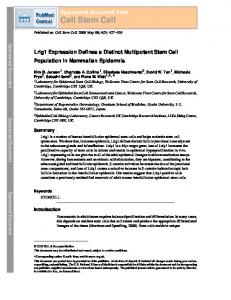
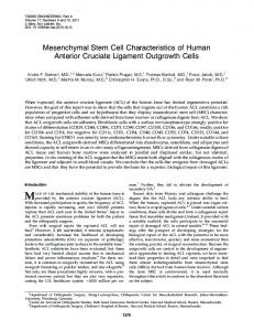
Figure 1.1. Morphological indicators of differentiating human pluripotent stem cells. Colonies exhibiting threedimensional growth (left, arrow) or flattened crater-like morphology (right, arrow) are differentiating and should be removed from cultures. Images taken on an Olympus SZX stereo microscope.
6
Stem Cell Research Core Facility
Matrigel Plate Coating
Brigitte Arduini, version 3, 2016-Jul-25 Citation: Arduini BL, Brivanlou AH. Modulation of FOXD3 activity in human embryonic stem cells directs pluripotency and paraxial mesoderm fates. Stem Cells. 2012 Oct; 30(10):2188-98. Modified from Scott Noggle, 2007-Sep-28 Matrigel (BD Biosciences), Geltrex (Life Technologies) and comparable substrates from other vendors are composed of extracellular matrix proteins derived from Engelbreth-Holm-Swarm (EHS) mouse tumor cells. The predominant component is laminin, but fibronectin, collagen and other proteins are also present. Not all substrates offered are specifically qualified for hESC maintenance. Although we routinely use hESC-qualified Matrigel, others have used alternative formulations such as growth factor-reduced Matrigel with success.
Materials:
Item Matrigel,LDEV-free hESC-qualified matrix (5 ml) DMEM/F12 Cryovials, internal threads Pipette tips, aerosol barrier Serological pipets, 10 ml 15ml or 50ml Conical tube
Vendor Corning
Catalog No. 354277
Thermo Scientific VWR (Nunc)
11330-032 66021-986
All materials that will come in contact with Matrigel during plating must be cold to prevent premature gelling of the matrix. It may be convenient to store pipette tips at -20oC specifically for aliquoting Matrigel.
Procedure 1: Matrigel Aliquots
Each hESC-qualified Matrigel lot is provided with dilution instructions rather than a specific concentration. 1. Thaw 5ml vial of Matrigel overnight at 4oC. 2. Pre-chill cryovials and pipette tips. 3. Aliquot desired volume to cryovials according to Matrigel lot specifications (typically 250400µl for later dilution to 25ml). Half-volume aliquots may also be made for dilution to 12ml. 4. Freeze at -80oC for up to 6 months.
7
Stem Cell Research Core Facility
Procedure 2: Matrigel Coating
1. Thaw one aliquot of Matrigel at 4oC. This may be done over several hours or overnight. 2. Pre-chill conical tube for dilution. 3. Culture vessels to be coated are pre-chilled by placing them on a metal incubator tray seated in a rectangular ice bucket. Alternatively, we use Cool Boxes (Biocision, http://www.biocision.com/products/coolbox-ice-free-cooling/) to avoid placing ice in the biosafety cabinets. 4. Chill a 10ml serological pipet by opening the top of the sleeve slightly and dipping the pipet (still in the sleeve) into liquid nitrogen. 5. Draw up a small amount of DMEM/F12 and use this to rinse the cryovial and collect the Matrigel. Thoroughly mix into the total volume of DMEM/F12. 6. With the same 10ml serological pipet, immediately distribute the diluted Matrigel to waiting tissue culture vessels. 7. Store tissue culture vessels at 4oC until needed (at least one hour).
Suggested Coating Volumes 35mm dishes, such as Falcon or ibidi Standard TC 96well plate, such as Falcon Ibidi 96 well plate
1ml for use within 1 day, otherwise 2ml 0.05 – 0.075 ml per well 0.1ml per well
Procedure 3: Using Matrigel-coated Plates
1. Before plating hESCs, place Matrigel dishes in the incubator for at least one hour. Coated dishes may be kept in the incubator for as long as one week before use, as long as experiments or passaging will be complete within two weeks of plates first being placed at 37oC. Also take care that Matrigel does not evaporate to the point where the centers of the wells/dishes are no longer covered. 2. Check formation of matrix under the microscope prior to plating cells. 3. Aspirate Matrigel, removing as much liquid as possible without scraping the bottom of the dish. It is not necessary to rinse Matrigel-coated dishes. 4. Plate hESCs in desired maintenance medium.
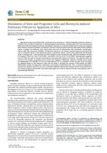
Figure 1.2. Properly polymerized matrigel on polystyrene appears as small dark matter under the microscope (arrows). Imaged with an Olympus IX51 microscope, 10X objective.
8
Stem Cell Research Core Facility
Enzymatic Passaging of hPSCs Human pluripotent stem cells grow as colonies rather than single cells. Most hPSC lines do not tolerate single cell passaging well. Therefore, viability is significantly enhanced by passaging cells in clumps. However, see Protocol 2 for passaging as single cells. Timing of passage is dependent on confluence and appearance of differentiation, but 3 – 5 days is typical.
Protocol 1: Passaging hPSCs as clumps with Dispase or Collagenase, Type V Brigitte Arduini, version 1, 2013-Feb-14 Modified from Scott Noggle, 2007-Sep-28
Materials:
Item Growth Medium Matrigel-coated TC plates DMEM/F12 Dispase Cell Lifters 15ml or 50ml conical tubes P1000 Barrier Pipette Tips
Vendor
Catalog No.
Thermo Fisher Scientific Stem Cell Technologies VWR (Corning #3008)
11330-032 07913 29442-200
Dispase: Thaw overnight at 4oC. Aliquot 2 ml each to 15 ml conical tubes and store at -20oC. Thaw and dilute 1:5 with DMEM/F12 prior to use. Working solutions may be stored at 4oC for up to two weeks, but will lose activity with repeated warming.
Procedure
Before passaging, examine the colonies under the microscope and look for any colonies that are differentiating. Spontaneously differentiating areas of the culture can be removed with a glass tool or a P10 pipette tip. 1. Replace growth medium with dispase. Incubate at 37oC up to 7 minutes. Observe cells under the microscope starting at ~5 minutes. Edges of colonies should begin to lift off the plate while the centers remain attached. **Prolonged dispase treatment will result in entire colonies detaching from the plate. 2. Quickly but gently, wash the dispase off the plate with DMEM/F12. Repeat. **If colonies have completely detached, wash by transferring cells in dispase to a conical tube, diluting in DMEM/F12 and centrifuging to pellet, and then repeating the wash. 3. Add fresh DMEM/F12 and use a cell lifter to gently detach colonies. 4. Transfer cells and medium to a conical tube. 5. Centrifuge at 200 rcf for 4 minutes at room temperature. 6. Aspirate supernatant and resuspend colonies in growth medium with a P1000. 7. Triturate to break up colonies into smaller clumps. 8. Plate a portion of the clumps to a new matrigel-coated plate (or other desired vessel). The proportion of cells passaged onto the new plate depends on the confluency of the previous plate. A 35mm plate at 60% confluence may be passaged approximately 1:6.
9
Stem Cell Research Core Facility
Protocol 2: Passaging hPSCs as Single Cells Brigitte Arduini, version 3.1, 2018-May-30
Materials:
Item Growth Medium Matrigel-coated TC plates ROCK Inhibitor (Y27632) 5mM in TC-grade water, filtered DPBS (no calcium, no magnesium) DMEM/F12 TrypLE or Accutase Cell Strainers (40 µm), optional 15ml or 50ml conical tubes P1000 Barrier Pipette Tips
Vendor
Catalog No.
Millipore or Stemgent Thermo Scientific Thermo Scientific Thermo Scientific Falcon
688000 04-0012 14190-144 11330-032 12605-010 352340
TrypLE (trypsin): Store at 4oC, strictly avoid repeated warming and cooling. Pre-warm only what is needed for the current experiment. Trypsin is inhibited by media components, particularly serum or serum replacement. PBS washes are essential for enzyme activity. Treatment with ROCK inhibitor has been shown to enhance survival efficiency of hPSCs passaged as single cells. However, prolonged and repeated exposure to ROCK inhibitor may increase the frequency of karyotypic abnormalities. Prior to passaging as single cells, an extra plate of hPSCs should be split for maintenance of the cell line by passaging as clumps. One day after single cell passaging, media should be changed to remove ROCK inhibitor. Reconstitute ROCK inhibitor at 5mM in sterile H2O (5mg in 2.956ml). Syringe filter, aliquot and store at -20oC. Thawed aliquots may be stored at 4oC for several days.
Procedure
Before passaging, examine the colonies under the microscope and look for any colonies that are differentiating. Spontaneously differentiating areas of the culture can be removed with a glass tool or a P10 pipette tip. 1. Pre-treat cells for at least one hour with ROCK inhibitor (Y27632) by adding it directly to the media at a final concentration of 10nM. 2. At the time of passaging, wash cells two times quickly in 1X DPBS (-Ca2+, -Mg2+). 3. Add TrypLE to each plate and incubate at 37oC for 10 – 20 minutes. Every few minutes, inspect the plates and slide back and forth to break up cell clumps. Suggested volumes: 0.5ml TrypLE for a 35mm plate, 1ml TrypLE for a 60mm plate 4. Triturate with a P1000 to further dissociate cells. 5. Wash and collect cells with growth medium + 10nM ROCK inhibitor (at least 5 times the volume of the TrypLE used). 6. Optional: Transfer cells and medium to a conical tube using a cell strainer to remove remaining clumps. 7. Mix 10µl of cell suspension with 10µl 0.4% Trypan Blue 8. Transfer 10 µl of cells/trypan blue to a hemocytometer. a. Count the total number of cells in each of the four grids. b. Total number of cells = n/4 * 10 * dilution * volume of cells (µl) 9. Centrifuge at 200 rcf for 4 minutes at room temperature. 10
Stem Cell Research Core Facility 10. Aspirate supernatant and resuspend cells in growth medium plus 10nM ROCK inhibitor with a P1000. 11. Triturate to ensure a single cell suspension, and remove excess cells if necessary. 12. Add an appropriate amount of growth medium plus ROCK inhibitor with a serological pipet and plate the desired number of cells to new matrigel-coated plates (or other desired vessels). 13. Incubate cells overnight at 37oC. Change media on the following day to growth medium without ROCK inhibitor.
11
Stem Cell Research Core Facility
Cryopreservation of hPSCs by Vitrification
Brigitte Arduini, version 3, 2016-Jul-25 Modified from Scott Noggle, 2007-Jan-30
Optimal cryopreservation methods vary from cell type to cell type 1. For human pluripotent stem cells, the Rensselaer Stem Cell Core routinely utilizes a modified vitrification protocol for cryopreservation 2,3. This method utilizes multiple cryoprotectants (DMSO, ethylene glycol, sucrose) and rapid freezing to -150oC. Cells are banked in cryovials rather than straws to facilitate primary container labeling, storage and distribution. With respect to RUES1 and RUES2 hESC lines, vitrification enhances cell viability by 40 - 50% over slow-cooling, DMSO-only procedures, resulting in consistently high recovery (>95%) from frozen vials. This method is also compatible with GMP compliance. Further, we have found that other hPSC lines, including WiCell and iPSC lines, can also be successfully preserved using this method. A seed lot system is used to maintain and conserve cell line resources. Cell line records of frozen stocks include cell line, passage number, date and method of freeze, and location.
Materials:
Item Growth Medium Ethylene glycol Dimethyl Sulfoxide (DMSO) Sucrose 1M HEPES solution Cryovials, internal threads Cryo storage box(es) Liquid Nitrogen in an ice bucket
Vendor
Cat No.
Sigma Sigma Sigma Thermo Fisher Scientific VWR (Nunc) Nalgene 9x9
102466 D2650 S7903 15630 66021-986 5026-0909
Procedure 1: Freezing hPSCs Prepare Media
Prepare all media fresh, sterile filter, and maintain on ice while working. Note: ethylene glycol and DMSO become very viscous when combined with sucrose solution. It is easier to combine these two reagents (1:1) and filter before adding other components. DO NOT filter these reagents individually. HM (Growth Medium + 20mM HEPES) Growth Medium (ex: MEF-CM or mTeSR1) 20mM HEPES
HM+Sucrose 3.42g sucrose in 10ml HM
VS2 30% HM 30% HM+Sucrose 20% ethylene glycol 20% DMSO
VS3 40% HM+Sucrose 30% ethylene glycol 30% DMSO
1
See also https://www.atcc.org/~/media/PDFs/Cryopreservation_Technical_Manual.ashx Richards M,et al. An efficient and safe xeno-free cryopreservation method for the storage of human embryonic stem cells. Stem Cells. 22(5):779-89 (2004). Erratum in: Stem Cells. 23(4):604 (2005). 3 James D, Noggle SA, Swigut T, Brivanlou AH. Contribution of human embryonic stem cells to mouse blastocysts. Dev Biol. 295(1):90-102 (2006). 2
12
Stem Cell Research Core Facility Procedure
Work quickly. hPSCs cannot be exposed to the cryoprotectants for very long or they will die/differentiate upon thawing. Steps 5-7 must be timed accurately. 1. Prepare cryopreservation solutions based on the number of vials to be frozen, adding 10% for loss of solution during filtering, etc. 2. Label cryovials ahead of time, including 1-2 extra just in case. 3. Place a cryo storage box in 1-2 inches of LN2 in a large rectangular ice bucket, within easy reach of the biosafety cabinet. 4. Harvest hPSCs in clumps by manual dissection or collagenase/dispase treatment. 5. Centrifuge at 200 rcf, remove supernatant, and resuspend clumps in HM. total volume = (# vials + 2) x 40µl **Cells can be kept at room temperature in HM for up to 20 minutes. Prolonged incubation will result in clumping and reduced attachment after thawing. 6. Transfer 40µl of cells per sterile cryovial at room temperature. **Steps 5 – 7 must be completed in no more than 1 minute. Handle only as many tubes as can be processed in this amount of time (typically two vials). Remember that handling and capping tubes will require some time. Pro tip: use two P200 pipettors, one set fo 40µl and one set at 160µl, to eliminate time spent on changing volumes. 7. Processing 2 cryovials at a time, add 40µl of VS2 and mix by gentle pipetting. 8. Add 160µl VS3 and mix by gentle pipetting. 9. Submerge the tubes quickly in LN2 and swirl while freezing. The frozen solution should have a pink glass-like appearance, while a thin layer on the top might be opaque. Be sure caps are tightened and place vials in LN2 storage boxes. 10. Transfer entire box to LN2 storage.
13
Stem Cell Research Core Facility
Procedure 2: Thawing hPSCs Prepare Media & Plates
WS3 Growth medium + 1M sucrose Prepare at room temperature and vortex periodically to completely dissolve sucrose. Filter-sterilize and store on ice during the procedure. Growth medium, pre-warmed to room temperature (12ml per cell line to be thawed) 35mm dish per cell line to be thawed (with MEFs, Matrigel or other desired substrate)
Procedure
Steps 1-3 must be performed quickly so that cells are not exposed to high concentrations of cryoprotectants for too long. The incubation times in steps 4-7 remove sucrose slowly and prevent osmotic shock and lysis of the cells. 1. Remove a tube from LN2 and quickly submerge bottom of tube in warm sterile water. 2. Quickly wipe with 70% EtOH-soaked kimwipe. 3. Immediately add 800µl of cold WS3, mix gently by stirring with the pipette tip, and let sit for 30 seconds. 4. Add 1ml of growth medium, mix as above, let sit for 2 minutes. 5. Transfer to 15ml conical tube. 6. Rinse cryotube two times with 1ml each of growth medium and add to 15ml tube, mixing gently. Let sit for 1 minute. 7. Add 6ml growth medium, slowly dropwise to cells over about 2 minutes. Mix thoroughly between additions of medium. 8. Spin at 200 rcf for 4 minutes. 9. Resuspend gently in 1 ml of growth medium with a P1000. 10. Using a P1000, transfer to a 35mm plate containing 1ml of growth medium. Colonies should recover and show signs of growth within one week, and be ready for passage between 7 and 14 days. Change medium daily.
14