ABSTRACT. Numerous procedures have been described for the operative management of acromioclavicular joint inju- ries, but surprisingly little information is ...
0363-5465/100/2828-0103$02.00/0 THE AMERICAN JOURNAL OF SPORTS MEDICINE, Vol. 28, No. 1 © 2000 American Orthopaedic Society for Sports Medicine
Structural Properties of the Intact and the Reconstructed Coracoclavicular Ligament Complex* Richard I. Harris, PhD, Andrew L. Wallace, MBBS, PhD, FRACS, Gareth D. Harper, MBBS, FRCS, Jerome A. Goldberg, MBBS, FRACS, David H. Sonnabend, MBBS, BSc(Med), FRACS, and William R. Walsh,† PhD
From the Department of Orthopaedic Surgery, Prince of Wales Hospital, University of New South Wales, Sydney, New South Wales, Australia and least stiff, and augmentation with another form of coracoclavicular fixation is recommended. These results provide a useful baseline for comparison of the initial performance of reconstructive techniques with the performance of the native coracoclavicular ligament.
ABSTRACT Numerous procedures have been described for the operative management of acromioclavicular joint injuries, but surprisingly little information is available on the ultimate mechanical behavior of the native coracoclavicular ligament complex or on the various methods of reconstruction. We tested 19 fresh-frozen cadaveric bone-ligament-bone preparations of the coracoclavicular ligament in uniaxial tension at 25 mm/min until failure. Seven specimens were left intact, six had the trapezoid ligament sectioned, and six had the conoid ligament sectioned. Reconstruction of the coracoclavicular ligament was achieved using coracoacromial ligament transfers, woven polyester slings, suture anchors, and Bosworth screws; all reconstructions were also tested to failure. The intact coracoclavicular ligament failed by avulsion or midsubstance tear at 500 (⫾134) N, with a stiffness of 103 (⫾30) N/mm and elongation to failure of 7.7 (⫾1.9) mm. There was no significant difference between the contributions of the conoid or trapezoid ligaments in this loading configuration. Coracoclavicular slings and suture anchors provided strength similar to that of the coracoclavicular ligament, but with significantly greater deformations (14 to 26 mm). Screw fixation resulted in comparable stiffness and superior strength to the coracoclavicular ligament, but only if bicortical purchase was obtained. Coracoacromial ligament transfers were the weakest
Higher-grade acromioclavicular joint injuries (Rockwood types III through VI) represent failure of the coracoclavicular ligament complex, which is formed by the conoid and trapezoid ligaments. This complex has been termed the primary suspensory structure of the upper limb.4, 22 In the more severe types of injuries (types IV through VI), surgical stabilization of the acromioclavicular joint has been recommended to prevent disabling pain, weakness, and deformity.16 The management of type III injuries remains controversial. There are numerous reports, including a number of randomized trials, that show satisfactory outcome with either nonoperative or operative treatment.1, 9, 15 However, in younger active patients, particularly high-level athletes and manual laborers who perform overhead tasks, surgical intervention is often preferred to allow more rapid rehabilitation.11 There is probably no other joint for which so many techniques for stabilization have been described as the acromioclavicular joint,22 and it can be inferred from this observation that there is no ideal method of reconstruction. The surgical approaches have been classified into five main types16: 1) fixation of the acromioclavicular joint using pins, wires, or plates; 2) fixation of the coracoid to the clavicle using screws, synthetic slings, or tapes; 3) ligament substitution using the coracoacromial ligament; 4) dynamic muscle-tendon transfers; and 5) excision of the lateral end of the clavicle. In addition, combinations of these techniques have been used frequently, with a high percentage of good and excellent clinical results, although
* Presented at the 24th annual meeting of the AOSSM, Vancouver, British Columbia, Canada, July 1998, at which it won the Aircast Award for Basic Science. † Address correspondence and reprint requests to William R. Walsh, PhD, Department of Orthopaedic Surgery, Prince of Wales Hospital, Randwick Sydney NSW, Australia 2031. No author or related institution has received any financial benefit from research in this study.
103
104
Harris et al.
many reports are retrospective and based on relatively small series of patients.7, 14, 27 Despite these numerous options for reconstruction, there is a scarcity of biomechanical knowledge of the acromioclavicular joint and its stabilizing structures.5, 8, 25 A thorough search revealed no information regarding the ultimate mechanical properties of the intact coracoclavicular ligament complex or its conoid and trapezoid components, nor were there any biomechanical studies in the English literature comparing the ultimate properties of different methods of reconstruction. Consequently, there were no biomechanical guidelines within which to predict the amount of initial postoperative loading the reconstructed coracoclavicular ligament would tolerate without failure. The purpose of this study was to document the ultimate tensile structural properties of the intact coracoclavicular ligament complex, its individual components, and several commonly used coracoclavicular fixation techniques.
MATERIALS AND METHODS
American Journal of Sports Medicine
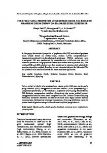
Figure 1. Intact coracoclavicular ligament complex mounted before testing. The scapula, including the coracoid, is fixed to the load cell. A custom-made jig with multiple loading points was used to distract the clavicle in tension along the axis of the conoid ligament until specimen failure.
Specimen Preparation Coracoclavicular bone-ligament-bone specimens were harvested from 19 fresh-frozen human cadavers. Ten right and nine left unpaired shoulders were obtained from donors with a mean age of 70 years (range, 55 to 90). All specimens were free from disease or injury to the acromioclavicular joint and all donors had died from causes unrelated to the musculoskeletal system. Shoulder specimens were thawed at room temperature for 24 hours before use. The skin, subcutaneous tissues, and muscles were removed, and the acromioclavicular ligaments and capsule were divided, leaving the coracoacromial ligament and coracoclavicular ligament complex intact. The specimens were then divided into three groups: 1) entire coracoclavicular ligament complex intact (N ⫽ 7), 2) isolated conoid ligament (trapezoid ligament divided, N ⫽ 6), and 3) isolated trapezoid ligament (conoid ligament divided, N ⫽ 6). Immediately after dissection, the body of the scapula was embedded in an open-top steel box using molten metal (lead-cadmium alloy), allowing the upper part of the spine, glenoid, and coracoid to protrude (Fig. 1). Care was taken to embed each specimen in the correct anatomic position, with the long axis of the clavicle and the scapular plane oriented at approximately 90° to one another. During preparation and testing, specimens were kept appropriately hydrated with physiologic saline at room temperature. Experimental Procedure Unidirectional tensile loading was performed on an MTS 858 materials testing machine (MTS Corporation, Minneapolis, Minnesota). The scapular box was secured to a stationary load sensor, while the clavicle was secured using a custom-made jig with multiple loading points on either side of the coracoclavicular ligament complex. Ten-
sile loading was applied to the clavicle along the axis of the conoid ligament at a rate of 25 mm/min until specimen failure. The mode of failure was characterized as midsubstance, avulsion (including a bone fragment), or insertional (without a bone fragment) from either the coracoid or clavicle. After ligament failure, the coracoclavicular ligament was reconstructed by a number of methods and the reconstruction was tested to failure using the same protocol. Coracoacromial Ligament Transfer (N ⫽ 7): The acromial end of the coracoacromial ligament was released from its insertion under the acromion and transferred to the medullary canal of the clavicle, which had been osteotomized as described by Weaver and Dunn.28 The transferred ligament was secured through two 1.6-mm drill holes in the superior cortex of the clavicle using an Ethibond No. 2 suture (Ethicon, Inc., Somerville, New Jersey) that had been inserted in the free end of the coracoacromial ligament using a Bunnell-type weave before transfer. Coracoclavicular Screw (N ⫽ 7): An original Bosworth coracoclavicular screw (6.4-mm thread diameter, 3.6-mm shank diameter) was inserted at the intersection of a parasagittal plane bisecting the base of the coracoid and the plane located at the midpoint between the anterior and posterior borders of the clavicle, after predrilling with a 3.2-mm drill. In three specimens (unicortical), only the superior cortex of the coracoid was fixed. In another four (bicortical), the inferior cortex was also breached by two threads of the screw, with the appropriate screw lengths determined using a depth gauge. Coracoclavicular Sling (N ⫽ 4): A sling was fashioned using an 8-mm diameter straight-woven polyester vascular prosthesis (CRBard Inc., Billerica, Massachusetts) and passed under the coracoid process, as described by Goldberg et al.10 One end of the loop was passed through a
Vol. 28, No. 1, 2000
Structural Properties of the Coracoclavicular Ligament Complex
105
TABLE 1 Modes of Failure of the Coracoclavicular Ligament (CCL) Complex and Its Components Mode of failure
Midsubstance rupture Coracoid avulsion Clavicular avulsion Coracoid insertion
Intact CCL (N ⫽ 7)
Isolated conoid (N ⫽ 6)
3
3
3
2
Isolated trapezoid (N ⫽ 6)
1 1
1
1
4
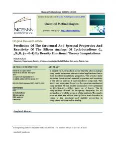
Figure 2. Comparative load-displacement curves for the isolated conoid and isolated trapezoid components. 5-mm drill hole through the clavicle and tied to the other free end using a square knot. The knot was secured using an Ethibond No. 1 suture. Coracoid Suture Anchor (N ⫽ 5): Suture anchor reconstructions were prepared in accordance with the technique of McCann.17 Two Mitek G4 Superanchors (Johnson & Johnson Inc.) were inserted in the superior cortex of the coracoid and secured to the clavicle with Ethibond No. 5 sutures passed through three 2.0-mm drill holes positioned at the points of an equilateral triangle in the clavicle directly above the coracoid. Data Analysis Load-displacement values were analyzed for each test to determine structural properties, that is, peak load (in newtons), stiffness (in newtons per millimeter), elongation at peak load (in millimeters), and energy absorbed at failure (in newton-meters). Energy absorption values were not recorded for the reconstructed coracoclavicular ligament complexes. Data were statistically compared using Statistica software (Statsoft, Inc., Tulsa, Oklahoma). A one-way analysis of variance was applied to find any differences between groups, followed by a post hoc multiple comparison technique (Duncan’s multiple range test) to determine specific differences. Statistical significance was attained when P was less than 0.05.
tional failure occurred in one case. Clavicular insertional failure was not seen. Similar patterns were found with the isolated conoid ligament. In the isolated trapezoid ligament group, there were no midsubstance ruptures. The trapezoid ligament failed mainly by coracoid avulsion or at the coracoid insertion, with one specimen failing at the clavicular insertion. Ultimate tensile strength, tensile stiffness, elongation at failure, and energy absorbed at failure are presented in Table 2. The load to failure values (mean ⫾ SD) for the intact coracoclavicular, isolated conoid, and isolated trapezoid ligaments were 500 (⫾134) N, 394 (⫾170) N, and 440 (⫾118) N, respectively. Elongation at failure ranged from 4.64 to 13.30 mm across the groups. Isolated conoid ligaments had the greatest mean stiffness (105 ⫾ 45 N/mm) but, as with the peak load and elongation at failure, no statistically significant differences between groups were found. Values for energy absorbed at failure were significantly higher for the isolated trapezoid ligaments (1.9 ⫾ 0.53 N䡠m) than for the isolated conoid ligaments (1.1 ⫾ 0.6 N䡠m) (P ⬍ 0.05), but not in comparison with the intact coracoclavicular ligament (1.6 ⫾ 0.62 N䡠m). An example of the load-versus-displacement for the isolated conoid and trapezoid ligaments is presented in Figure 2.
RESULTS
Reconstructions
Coracoclavicular Ligament Complex
Failure mechanisms for the reconstructed specimens were limited to damage to the implant material or to a bone component of the shoulder girdle. Coracoacromial ligament transfers failed either by suture rupture at the point of exit from the clavicular medullary canal (2 of 7) or, more commonly, suture pullout from the coracoacromial liga-
Distinct failure patterns were observed across the three groups and are summarized in Table 1. Intact coracoclavicular ligament complexes failed mainly by midsubstance rupture or coracoid avulsion, although coracoid inser-
TABLE 2 Structural Properties of the Coracoclavicular Ligament (CCL) Complex and Its Components (Means ⫾ SD)
a
Structure
Strength (N)
Stiffness (N/mm)
Elongation at failure (mm)
Energy absorbed at failure (N䡠m)a
Intact CCL (N ⫽ 7) Isolated conoid (N ⫽ 6) Isolated trapezoid (N ⫽ 6)
500 (⫾134) 394 (⫾170) 440 (⫾118)
103 (⫾30) 105 (⫾45) 84 (⫾19)
7.7 (⫾1.9) 7.1 (⫾2.1) 9.2 (⫾2.6)
1.6 (⫾0.62) 1.1 (⫾0.6) 1.9 (⫾0.53)
Isolated trapezoid and isolated conoid group were significantly different (P ⬍ 0.05).
106
Harris et al.
American Journal of Sports Medicine TABLE 3 Structural Properties of the Coracoclavicular Reconstructions (Means ⫾ SD) Methoda
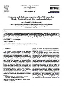
Figure 3. Comparative load-displacement curves for the native coracoclavicular ligament (CCL) and the Dacron sling, Bosworth screw, Weaver-Dunn coracoacromial ligament transfer (WD), and the Mitek suture anchor. ment (5 of 7). All coracoclavicular screws pulled out of the coracoid, irrespective of uni- or bicortical placement. All coracoclavicular slings failed by fracture of the coracoid at its base. Coracoid suture anchors failed either in the suture material (3 of 5) or by anchor pullout from the coracoid (2 of 5). In terms of ultimate tensile strength, coracoclavicular slings and coracoid suture anchor reconstructions provided strength comparable with the native coracoclavicular ligament complex, while superior strength was afforded when the Bosworth screw engaged both cortices of the coracoid (927 ⫾ 64 N). The coracoacromial ligament transfers and unicortical coracoclavicular screw failed at significantly lower loads (145 ⫾ 107 N and 229 ⫾ 99 N, respectively) (P ⬍ 0.001). Only the unicortical Bosworth screw had stiffness similar to that of the native coracoclavicular ligament complex (103 ⫾ 63 N/mm). Coracoacromial ligament transfers, coracoclavicular slings, and coracoid suture anchors were less than half as stiff as the intact coracoclavicular ligament (P ⬍ 0.005). The bicortical Bosworth screw provided superior stiffness (176 ⫾ 26 N/mm) (P ⬍ 0.001). These results were also reflected in the elongations at failure (Table 3), in which the coracoclavicular slings, in particular, performed poorly, failing at more than three times the displacement of the intact coracoclavicular ligament complex. Figure 3 shows a comparison of the load-versusdisplacement for the different reconstructions.
DISCUSSION This study presents, to our knowledge, the first report of the ultimate structural properties of the native coracoclavicular ligament complex and its constituent ligaments in the English literature. In the Polish literature, Milka18 reported a similar experiment in which the high-load mechanical behavior of the intact coracoclavicular ligament as an isolated clamped ligament was compared with a single technique of reconstruction using a carbon fiber implant. He found values for strength and stiffness very similar to the results in our study. The mechanical testing protocol developed for our experiments also provided functional insights into the conoid and trapezoid ligaments
CA ligament transfer (N ⫽ 7) CC sling (N ⫽ 4) Suture anchor (N ⫽ 4) Unicortical CC screw (N ⫽ 3) Bicortical CC screw (N ⫽ 3)
Tensile strength (N)
Tensile stiffness (N/mm)
Elongation at failure (mm)
145 (⫾107)b
8 (⫾4)b
11.65 (⫾3.6)
423 (⫾169)
23 (⫾7)b
26.12 (⫾6.8)b
366 (⫾96)
28 (⫾4)b
14.52 (⫾2.6)b
229 (⫾99)b
103 (⫾63)
3.37 (⫾1.1)
927 (⫾64)b
176 (⫾26)b
6.63 (⫾0.9)
a
CA, coracoacromial; CC, coracoclavicular. Significantly different from intact coracoclavicular ligament complex at P ⬍ 0.05. b
and comparison with a variety of coracoclavicular fixation techniques. Division of either the conoid or trapezoid ligaments made little difference to the ultimate tensile strength of the coracoclavicular ligament complex, raising the question of the true function of these individual components. Until recently, knowledge of the respective functions of the conoid and trapezoid ligaments has been limited. Cadenat5 performed the first ligament sectioning experiments, and he claimed that the coracoclavicular ligament acted as a pivot for anteroposterior rotation of the clavicle, with the conoid ligament acting as a limit to posterior translation and the trapezoid ligament the limit to anterior translation of the scapula. More detailed investigations by Urist25 confirmed that division of both the acromioclavicular and coracoclavicular ligaments would produce acromioclavicular joint dislocation, but that division of either the conoid or trapezoid ligaments in isolation had no specific effect. He concluded that in their contribution to joint stability, the coracoclavicular ligaments were, therefore, not analogous to the cruciate ligaments of the knee. Quantitative analysis of the contribution of the acromioclavicular and coracoclavicular ligaments to acromioclavicular joint stability at prefailure loads was undertaken by Fukuda et al.8 In their experiment, cadaveric specimens were loaded in orthogonal planes and rotated after sequential ligament sectioning. These authors concluded that the conoid ligament was the primary restraint to superior elevation of the clavicle at large displacements, whereas the trapezoid ligament was found to act as a restraint to compression along the axis of the clavicle. In the present study, the observed loading response of the isolated conoid ligament recapitulates these findings. When tested along its long axis, the conoid ligament had proportionately higher stiffness and lower energy absorption and elongation at failure than the trapezoid ligament,
Vol. 28, No. 1, 2000
Structural Properties of the Coracoclavicular Ligament Complex
suggesting a primary loadbearing role at “physiologic” loads (Fig. 2). However, equivalent peak load values for the isolated components suggest that the trapezoid ligament may also contribute significantly to stability of the acromioclavicular joint in this orientation. Energy absorption and elongation at failure were greater for the trapezoid ligament, indicating that it may act as an important secondary restraint during “pathologic” loading. Consequently, inadvertent damage to the more laterally placed trapezoid ligament, for example, during lateral clavicular excision for degenerative acromioclavicular joint disease, should have minimal effect on joint mechanics during normal activities of daily living, but it would clearly increase the risk of dislocation in athletic patients. None of the reconstruction techniques analyzed in the present study were able to restore the normal mechanical function of the intact coracoclavicular ligament complex. Earlier biomechanical studies using fairly simple methods of analysis have shown that reconstruction using wires, coracoclavicular slings, or transfixion pins can reduce the extent of vertical displacement or “step deformity” of the acromioclavicular joint.2 Using a more sophisticated loading environment, Kiefer et al.13 showed that fixation of the acromioclavicular joint with cerclage wires or plates reduced strain in the conoid ligament. Ligament substitution using the coracoacromial ligament provides an attractive biologic solution for the coracoclavicular ligament-deficient shoulder, but loss of reduction has been reported in up to 29% of cases (5 of 17), especially in those treated late.29 The structural properties of the coracoacromial ligament have recently been documented, and comparison with our results indicates that the coracoacromial ligament has only about half the strength and stiffness of the native coracoclavicular ligament.23 Together with the very low strength and stiffness of the Weaver-Dunn procedure, these findings suggest that augmentation with some form of coracoclavicular fixation should be considered for a coracoacromial ligament transfer.7, 21 However, at present, the potential effects of stress shielding of the transferred ligament by an augmentation device remain unknown. Coracoclavicular fixation using a polyester sling or suture anchor was comparable with the intact coracoclavicular ligament in terms of strength. Neither technique, however, provided sufficient stiffness, demonstrating less than 30% the stiffness of the native coracoclavicular ligament. These materials may not be appropriate for reconstruction of the more severe types of acromioclavicular joint dislocation where large displacements have occurred, as there is a significant risk of recurrent subluxation.26 Despite satisfactory clinical reports,10 the use of nonabsorbable slings has been associated with coracoid and clavicular erosion and infection in rare cases.20, 24 Unfortunately, newer absorbable sling materials have also been associated with high rates of recurrent deformity3, 12 and sterile discharge.6 To date, there have been no reports of clinical outcome with suture anchors, but an experimental study has demonstrated marginal improvements in reduc-
107
tion of laxity compared with coracoacromial ligament transfer.19 The mechanical performance of coracoclavicular screw fixation was closest to that of the native coracoclavicular ligament. If bicortical purchase was obtained, ultimate strength was 80% higher than that in the intact ligament. However, if only one cortex was breached, strength was reduced by 50% compared with the intact ligament, indicating the critical importance of correct screw placement. Despite the biomechanical advantages, complications of the Bosworth technique include screw pullout, infection, and irritation over the screwhead.9 Screw breakage has also been reported.11 However, the risk of early implant removal to prevent implant failure should be balanced against the risk of recurrent deformity, which may be as high as 35% if the implant is removed at 6 weeks after surgery.1 In considering the clinical applications of the present study, several significant limitations should be recognized. First, because of limited availability, mechanical testing was performed on older specimens, in which agerelated bone density changes are common. Failure mode and magnitude may not be the same in a younger athletic population in which these injuries typically occur. Second, tensile loading was performed in one axis, for a single loading cycle, at a strain rate much lower than that likely to occur during injury, and in the absence of other dynamic factors such as muscle loads. Third, we did not assess the cyclic and static viscoelastic properties of the coracoclavicular ligament at physiologic loads, or the fatigue properties of the various reconstructive materials, and these factors are also clinically relevant. Finally, testing of the reconstructions was performed on specimens in which the intact coracoclavicular ligament or its components had been failed immediately beforehand, and therefore there may have been a predisposition to bone failure in some specimens. Nonetheless, the results in the present study provide useful quantitative information about the immediate mechanical behavior of a number of reconstructive techniques in comparison with the intact coracoclavicular ligament complex. Understanding the mechanical limits of each reconstruction may also indicate the loads and range of shoulder motion tolerable during rehabilitation. From the data in this study we concluded that 1) division of either the conoid or trapezoid ligament has little effect on the overall strength of the coracoclavicular ligament, 2) the trapezoid ligament is an important secondary loadbearing structure in the vertical axis, 3) coracoacromial ligament transfers are initially weak and should be augmented with some form of coracoclavicular fixation during the healing process, 4) coracoclavicular slings are strong but very elastic, undergoing marked deformation at low load, and may not be suitable for severe injuries with large scapuloclavicular dissociation (that is, Rockwood type V injuries), and 5) coracoclavicular screw fixation provides immediate strength and stiffness comparable with the intact coracoclavicular ligament but these properties are dependent on accurate placement.
108
Harris et al.
ACKNOWLEDGMENTS The authors express their gratitude to Mr. Tim Letters for his technical assistance and Dr. Ralph Stanford for his assistance with the surgical reconstructions. REFERENCES 1. Bannister GC, Wallace WA, Stableforth PG, et al: The management of acute acromioclavicular dislocation: A randomised prospective controlled trial. J Bone Joint Surg 71B: 848 – 850, 1989 2. Bargren JH, Erlanger S, Dick HM: Biomechanics and comparison of two operative methods of treatment of complete acromioclavicular separation. Clin Orthop 130: 267–272, 1978 3. Blatter G, Meier G: Augmentation of the coraco-clavicular ligament suture. Comparison between wire cerclage, Vicryl tape, and PDS cord [in German]. Unfallchirurg 93: 578 –583, 1990 4. Bosworth BM: Acromioclavicular separation: New method of repair. Surg Gynecol Obstet 73: 866 – 871, 1941 5. Cadenat FM: The treatment of dislocations and fractures of the outer end of the clavicle. Int Clin 1: 145–169, 1917 6. Clayer M, Slavotinek J, Krishnan J: The results of coraco-clavicular slings for acromio-clavicular dislocation. Aust N Z J Surg 67: 343–346, 1997 7. Copeland S, Kessel L: Disruption of the acromioclavicular joint: Surgical anatomy and biological reconstruction. Injury 11: 208 –214, 1980 8. Fukuda K, Craig EV, An K-N, et al: Biomechanical study of the ligamentous system of the acromioclavicular joint. J Bone Joint Surg 68A: 434 – 440, 1986 9. Galpin RD, Hawkins RJ, Grainger RW: A comparative analysis of operative versus nonoperative treatment of grade III acromioclavicular separations. Clin Orthop 193: 150 –155, 1985 10. Goldberg JA, Viglione W, Cumming WJ, et al: Review of coracoclavicular ligament reconstruction using Dacron graft material. Aust N Z J Surg 57: 441– 445, 1987 11. Guy DK, Wirth MA, Griffin JL, et al: Reconstruction of chronic and complete dislocations of the acromioclavicular joint. Clin Orthop 347: 138 –149, 1998 12. Hessmann M, Gotzen L, Gehling H: Acromioclavicular reconstruction augmented with polydioxanonsulphate bands: Surgical technique and results. Am J Sports Med 23: 552–556, 1995 13. Kiefer H, Claes L, Burri C, et al: The stabilizing effect of various implants on the torn acromioclavicular joint: A biomechanical study. Arch Orthop Trauma Surg 106: 42– 46, 1986
American Journal of Sports Medicine 14. Lancaster S, Horowitz M, Alonso J: Complete acromioclavicular separations: A comparison of operative methods. Clin Orthop 216: 80 – 88, 1987 15. Larsen E, Bjerg-Nielsen A, Christensen P: Conservative or surgical treatment of acromioclavicular dislocation: A prospective, controlled, randomized study. J Bone Joint Surg 68A: 552–555, 1986 16. Lemos MJ: The evaluation and treatment of the injured acromioclavicular joint in athletes. Am J Sports Med 26: 137–144, 1998 17. McCann PD: Acromioclavicular dislocation and fixation (educational videotape). American Academy of Orthopaedic Surgeons, Rosemont, IL, 1996 18. Milka S: A comparative analysis of tension properties of the coracoclavicular ligament versus carbon fiber implant [in Polish]. Chir Narzadow Ruchu Ortop Pol 58: 453– 458, 1993 19. Moses JM, Wilson DR, Zilberfarb JL, et al: A biomechanical comparison of acromioclavicular joint reconstruction techniques. Trans Orthop Res Soc 23: 740, 1998 20. Neault MA, Nuber GW, Marymont JV: Infections after surgical repair of acromioclavicular separations with nonabsorbable tape or suture. J Shoulder Elbow Surg 5: 477– 478, 1996 21. Neviaser RJ: Injuries to the clavicle and acromioclavicular joint. Orthop Clin North Am 18: 433– 438, 1987 22. Rockwood CA Jr, Williams GR, Young DC: Injuries to the acromioclavicular joint, in Rockwood CA, Green DP, Bucholz RW, et al (eds): Fractures in Adults. Volume 2. Fourth edition. Philadelphia, Lippincott-Raven Publishers, 1996, pp 1341–1413 23. Soslowsky LJ, An CH, Johnston SP, et al: Geometric and mechanical properties of the coracoacromial ligament and their relationship to rotator cuff disease. Trans Orthop Res Soc 18: 139, 1993 24. Takagishi K, Yonemoto K, Tsukamoto Y, et al: A cautionary note to treatment of complete acromioclavicular separation using artificial materials. Orthop Int 4: 343–347, 1996 25. Urist MR: Complete dislocations of the acromioclavicular joint: The nature of the traumatic lesion and effective methods of treatment with an analysis of forty-one cases. J Bone Joint Surg 28: 813– 837, 1946 26. Verhaven E, DeBoeck H, Haentjens P, et al: Surgical treatment of acute type-V acromioclavicular injuries in athletes. Arch Orthop Trauma Surg 112: 189 –192, 1993 27. Walsh WM, Peterson DA, Shelton G, et al: Shoulder strength following acromioclavicular injury. Am J Sports Med 13: 153–158, 1985 28. Weaver JK, Dunn HK: Treatment of acromioclavicular injuries, especially complete acromioclavicular separation. J Bone Joint Surg 54A: 1187– 1194, 1972 29. Weinstein DM, McCann PD, McIlveen SJ, et al: Surgical treatment of complete acromioclavicular dislocations. Am J Sports Med 23: 324 –331, 1995