Journal of General Virology (2005), 86, 1223–1228
Short Communication
DOI 10.1099/vir.0.80351-0
Subcellular distribution of mutant movement proteins of Cucumber mosaic virus fused to green fluorescent proteins Tomas Canto and Peter Palukaitis Scottish Crop Research Institute, Invergowrie, Dundee DD2 5DA, UK
Correspondence Peter Palukaitis
[email protected]
Received 9 June 2004 Accepted 14 December 2004
The subcellular distribution of the movement proteins (MPs) of nine alanine-scanning mutants of Cucumber mosaic virus (CMV), fused to the green fluorescent protein (GFP) and expressed from CMV, was determined by confocal microscopy of infected epidermal cells of Nicotiana tabacum and Nicotiana benthamiana, as well as infected N. benthamiana protoplasts. Only those mutant MPs that were functional for movement in all host species tested localized to plasmodesmata of infected epidermal cells and to tubules extending from the surface of infected protoplasts, as for wild-type CMV 3a MP. Various mutant MPs that were either conditionally functional for movement or dysfunctional for movement did not localize to plasmodesmata and did not form tubules on the surface of infected protoplasts. Rather, they showed distribution to different extents throughout the infected cells, including the cytoplasm, nucleus or the plasma membrane. The CMV 3a MP also did not associate with microtubles.
All five of the proteins encoded by the three genomic RNAs of Cucumber mosaic virus (CMV) affect the movement of CMV (reviewed by Palukaitis & Garcı´a-Arenal, 2003). However, the 3a protein encoded by CMV RNA 3 is considered to be the primary movement protein (MP) (Canto et al., 1997; Kaplan et al., 1995; Nagano et al., 1997). Various observations demonstrated a role for the CMV 3a protein in movement, e.g. the 3a protein bound single-stranded RNA cooperatively in vitro (Kim et al., 2004; Li & Palukaitis, 1996), potentiated the movement of itself, RNA and fluorescent dextran through plasmodesmata (Ding et al., 1995) and localized to plasmodesmata between various cell types and to parietal sieve elements in the phloem (Blackman et al., 1998). The 3a protein fused to green fluorescent protein (GFP) was also able to form tubules extending from the surface of protoplasts (Canto & Palukaitis, 1999a). Natural and artificial mutants of the 3a protein have been shown to affect CMV movement in specific hosts or tissues (Canto & Palukaitis, 1999a, b; Kaplan et al., 1997; Li et al., 2001; Takeshita et al., 2001) and mutants of the 3a protein also have been identified that affect cell-to-cell versus long-distance movement (Li et al., 2001). Various 3a protein mutants generated by alanine-scanning mutagenesis (Table 1; Fig. 1a) were shown to be deficient in various functions. Mutants designated M4–M7 were unable to promote cell-to-cell movement, although CMV containing mutants M4, M5 and M6 could be complemented for cellto-cell and long-distance movement in transgenic tobacco A supplementary figure showing the localization of MP–red fluorescent protein and tubulin–GFP in epidermal cells is available in JGV Online.
0008-0351 G 2005 SGM
Printed in Great Britain
plants expressing the 3a protein (Li et al., 2001). The movement-defective mutant designated M5 was also shown to be deficient for the gating of plasmodesmata, with no cell-to-cell movement of itself, RNA or fluorescent dextran (Ding et al., 1995). The mutant designated M8 was unable to induce tubule formation in infected Nicotiana benthamiana protoplasts or promote the cell-to-cell movement of CMV in the epidermal cells of either Nicotiana tabacum (Canto & Palukaitis, 1999a) or Chenopodium amaranticolor (Canto & Palukaitis, 1999b). However, whether the various other mutations affected the ability of the CMV 3a protein to target plasmodesmata in epidermal cells and to assemble in protruding tubular structures in protoplasts has not been reported. Such might be expected, as mutants of the MPs of Alfalfa mosaic virus (AMV), Cowpea mosaic virus (CPMV) and Tobacco mosaic virus (TMV), generated by alanine scanning, have shown differences in their localization patterns (Huang et al., 2001; Kahn et al., 1998; Pouwels et al., 2003). To examine the importance of subcellular localization vis-a`-vis the proper function of the CMV MP, we have examined the subcellular distribution of the various mutant 3a proteins fused to GFP in epidermal cells of two host species (N. benthamiana and N. tabacum). We also have examined the localization of the various GFP-tagged MP mutants in infected N. benthamiana protoplasts derived from mesophyll cells. The wild-type 3a protein per se, as well as this protein fused to GFP at its C terminus, both localized to plasmodesmata, as determined previously by both electron microscopic immunolocalization and confocal laser-scanning microscopy (Blackman et al., 1998). Therefore, confocal microscopy was 1223
T. Canto and P. Palukaitis
Table 1. Infectivity and subcellular distribution of the wild-type and nine alanine-scanning mutants of the MP of CMV fused to GFP Virus (mutated position)*
Wild-type M1 (R8, T9) M2 (E38, D40) M3 (Y75, D76) M4 (R97, T98) M5 (Y144, D145) M6 (R156, F158) M7 (N191, Y192) M8 (D20, D21) M9 (P60)
InfectivityD
+ + + + 2 2 2 2 ± ±
Distributiond Epidermal cells
Mesophyll protoplasts
Tubule quantification§
Pd Pd Pd Pd, Pm C, N, (c Ag) C, N, (c Ag) C, N, (c Ag) C, (Pm?) C, N, (c Ag) C, N, Pm, (c Ag)
Tubules, ps Ag Tubules, ps Ag Tubules, ps Ag Tubules, ps Ag C, N, (Ag) C, N, (Ag) C, N (Ag) C, Pm C, Pm C, Pm, N
23/100 48/119 56/100 20/100 0/100 0/100 0/100 0/100 0/100 0/100
*The mutants contain alterations to alanine in the 3a protein sequence at the positions indicated. DInfectivity of the wild-type and mutant MPs as determined on six systemic hosts of CMV and two local lesion hosts (Li et al., 2001). +, Infectious on all hosts tested; 2, no detectable movement on any hosts tested; ±, infectious on all systemic hosts at room temperature, but either not infectious on local lesion hosts and not able to move between epidermal cells (M8), or temperature-sensitive for long-distance movement in tobacco (M9). dAbbreviations: C, cytoplasm; c Ag, cytoplasmic aggregates; N, nucleus; Pd, plasmodesmata; Pm, plasma membrane; ps Ag, punctate surface aggregates. §No. protoplasts with filaments/total infected cells counted.
used here to examine the distribution of the mutant 3a proteins fused to GFP (Fig. 1), as was done previously for the wild-type 3a protein and the mutant M8 (Canto & Palukaitis, 1999a; Canto et al., 1997). PCR mutagenesis was used to fuse sequences encoding the GFP to those encoding the various mutant 3a proteins. The fusion proteins were expressed from the corresponding mutated CMV RNAs 3, following inoculation of plants with RNA transcripts of CMV RNAs 1, 2 and the mutated RNAs 3, all as described previously (Canto & Palukaitis, 1999a; Canto et al., 1997; Li et al., 2001). The various mutant 3a proteins were shown previously to accumulate to similar levels in infected N. tabacum protoplasts (Li et al., 2001). In inoculated N. tabacum and N. benthamiana plants, considerable variation was found between mutants in the number of fluorescent foci detected by confocal microscopy between 2 and 5 days post-inoculation (p.i.). In most cases, fluorescence levels were very weak and many infected foci may have escaped detection (data not shown). Nevertheless, the patterns of distribution observed in inoculated epidermal cells were consistent from focus to focus and from plant to plant for a given mutant and, in general, were similar in N. tabacum (Fig. 1b) and N. benthamiana (Fig. 1c). In epidermal cells, those mutants that were functional for movement (M1, M2, M3, M8 and M9) showed two general types of distribution of the MP–GFP fusions (Table 1): 1224
either localizing to plasmodesmata (M1, M2 and in part M3), often spreading to plasmodesmata in cells neighbouring the initially infected cell (for mutants M1 and M2, as for the wild-type 3a–GFP fusions) (Figs 1b and c, arrows) or partitioning between the cell periphery and elsewhere in the cells (M8 and M9). In the case of the M8 and M9 MP–GFP fusions, fluorescence was confined to the initially infected cells, in the form of small aggregates, with M8 distributed uniformly throughout the cytoplasm as well as the nucleus, and M9 distributed in the cytoplasm in a more granular fashion as well as in the nucleus (Figs 1b and c). It was not possible to ascertain whether the fluorescence distributed along the periphery of the cell was also associated with plasmodesmata, although mutant M9 did show several punctate bodies that suggested as such (Figs 1b and c). The absence of an association with plasmodesmata for mutant M8 was not surprising, as this mutant was shown previously to be unable to promote the movement of CMV between epidermal cells (Canto & Palukaitis, 1999a, b). For those mutants that were dysfunctional for movement (M4, M5, M6 and M7), fluorescence was confined to the initially infected cells, both in N. tabacum and in N. benthamiana. The fluorescence pattern of mutants M4, M5 and M6 showed some similarities in their distribution, in that the MP–GFP fusions were not localized to plasmodesmata, but showed partitioning between the nucleus and cytoplasm, including transvacuolar strands (Fig. 1b). Mutant M4 seemed to produce mostly smaller, uniformly sized Journal of General Virology 86
CMV mutant movement protein distribution
(a) M1
M8
M8
M2
M9
M9
M3
M4
M5
M6
M7
(b)
WT
M1
M2
M3
M4
M5
M6
M7
M8
M9
WT
M1
M2
M3
M4
M5
M6
M7
M8
M9
(c)
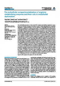
Fig. 1. Subcellular distribution of CMV wild-type (WT) and mutant (M1–M9) 3a–GFP fusions. (a) Schematic map of mutations M1–M9 in the 3a protein of CMV. Mutants listed above the rectangle are competent for movement, whereas those listed below the rectangle are dysfunctional for movement; those mutants both above and below the rectangle are conditionally functional for movement. (b, c) Confocal-microscopy images of WT and mutant 3a–GFP fusions in infected epidermal cells of (b) N. tabacum and (c) N. benthamiana 3 days p.i. Arrows in (b) and (c) indicate plasmodesmatal targeting occurring in cells adjacent to the initially inoculated one. Bars, 100 mm.
fluorescent aggregates than either mutants M5 or M6, both of which produced quite large fluorescent inclusion bodies, although there was some variability between samples. The M7 MP–GFP fusion showed high levels of fluorescence distributed throughout the oppressed cytoplasm (Fig. 1b). The dysfunctional mutant MPs showed a distribution of fluorescence in N. benthamiana (Fig. 1c) similar to that in N. tabacum (Fig. 1b). http://vir.sgmjournals.org
Unlike the situation with TMV (Boyko et al., 2000; Heinlein et al., 1995, 1998; McLean et al., 1995), neither the wild-type CMV MP nor the dysfunctional mutants appeared to be associated with the microtubules. To verify this, the CMV 3a MP was fused to the monomer red fluorescent protein and expressed after agroinfiltration into either transgenic N. benthamiana expressing an a-tubulin–GFP fusion (Gillespie et al., 2002) or N. benthamiana plants co-agroinfiltrated 1225
T. Canto and P. Palukaitis
with a plasmid expressing the a-tubulin–GFP fusion (Ueda et al., 1999). Both approaches gave similar results. Neither the wild-type nor two mutant MPs (M4 or M8) showed co-localization with a-tubulin–GFP (see Supplementary Figure in JGV Online). Given that the MPs of several other viruses also have been shown not to require microtubules for tubule formation (Huang et al., 2000, 2001; Laporte et al., 2003; Pouwels et al., 2002), this is not a unique situation. Moreover, the interaction of the TMV MP with microtubules has been shown to be associated with a degradation pathway (Gillespie et al., 2002; Kragler et al., 2003) and, unlike the TMV MP, the CMV 3a protein did
27
not interact with a microtubule-associated protein designated MPB2C (Kragler et al., 2003). The distribution of the MP mutants within epidermal cells was compared with that in mesophyll protoplasts (Table 1) by preparing N. benthamiana protoplasts and infecting them with CMV RNA transcripts expressing the various mutant MP–GFP fusions, as described previously for the wild-type 3a MP and mutant M8 (Canto & Palukaitis, 1999a). The protoplasts were analysed by fluorescence microscopy at two time points: 27 and 51 h p.i. (Fig. 2). In different experiments, the percentage of infected protoplasts
51
27
WT
M5
M1
M6
M2
M7
M3
M8
M4
M9
51
Fig. 2. Subcellular distribution of CMV wild-type (WT) and mutant (M1–M9) 3a–GFP fusions in infected mesophyll protoplasts of N. benthamiana 27 and 51 h after electroporation. Arrows point to distinct protruding filament structures. Bar, 100 mm. 1226
Journal of General Virology 86
CMV mutant movement protein distribution
varied between 12?5 and 66 %, as measured by fluorescence (data not shown). The distribution of the MP–GFP fusions in the infected protoplasts varied with both the sample time and the mutant. As described previously (Canto & Palukaitis, 1999a), the wild-type 3a–GFP showed the presence of aggregates of MP on the surface of the protoplasts, forming tubules. The number of aggregates and tubules was greater at 51 h p.i. than at 27 h p.i. (Fig. 2 and data not shown). The tubules were very fragile and could break off during preparation of the samples for viewing. Only the MP mutants M1, M2 and M3, all of which were functional for virus movement in all hosts tested, showed the presence of fluorescent tubules on the surface of the infected protoplasts (Fig. 2; Table 1). Interestingly, the tubules were more abundant in the case of mutants M1 and M2 than in the wild-type 3a–GFP fusions (Table 1). The MP mutants that were dysfunctional for movement (M4, M5, M6 and M7) all showed some similar features, including the absence of distinct tubules (Table 1). M4, M5 and M6 all showed the presence of small and/or large aggregates in the cytoplasm and association of fluorescence with transvacuolar strands (Fig. 2). M7 showed some features similar to these, but not large inclusions, and M7 also showed higher fluorescence along the periphery of the protoplasts. None of the mutants M4, M5, M6 or M7 was able to form the discrete, punctate spots that were observed on the surface of protoplasts infected with either the wildtype 3a–GFP fusion or fusions involving the other mutants that could form tubules (M1, M2 and M3). This suggests that the punctate spots are precursors of the tubules, as has been concluded for tubules forming on the surface of protoplasts infected with several other viruses [AMV, Cauliflower mosaic virus (CaMV) and CPMV] expressing MP–GFP fusions (Heinlein et al., 1998; Huang et al., 2000; Pouwels et al., 2002, 2003). The MP mutants M8 and M9, both of which are conditionally functional for movement, also did not show the formation of tubules on the surface of infected protoplasts (Fig. 2; Table 1; Canto & Palukaitis, 1999a). Rather, M8 showed MP distributed throughout the cell, although somewhat more concentrated along the periphery, whilst M9 showed MP distributed mostly along the periphery, i.e. presumably associated with the plasma membrane and with some isolated spots on the surface (Fig. 2). The MPs of both M8 and M9 also showed some association of fluorescence with the nucleus, as was observed for the dysfunctional mutants (Fig. 2; Table 1). The role of the CMV 3a protein in tubule formation remains unclear, as this property was shown not to be essential for CMV movement (Canto & Palukaitis, 1999a), in contrast to the situation with AMV (Huang et al., 2001). Moreover, no tubule-like structures could be detected between cells in CMV-infected plants (Blackman et al., 1998). By contrast, the MP of the ilarvirus Olive latent virus 2, which, like CMV and AMV, is also in the family Bromoviridae, did form such tubules containing virus particles (Grieco et al., 1999). The http://vir.sgmjournals.org
MPs of both CPMV and CaMV form tubules between infected cells containing virus particles, through which it is believed that cell-to-cell movement occurs (Kasteel et al., 1996; Linstead et al., 1988; van Lent et al., 1990). However, the MPs of these viruses also were able to bind RNA in vitro (Carvalho et al., 2004; Citovsky et al., 1991), as were the MPs of CMV (Li & Palukaitis, 1996) and AMV (Schoumacher et al., 1992). Thus, it is conceivable that many viruses retain elements of both major movement pathways, whilst only one pathway is used for a given virus. That these bifunctional elements are still conserved may be a consequence of other host–virus interactions associated with aspects of virus movement that are common to the two pathways, which have yet to be determined.
Acknowledgements This work was supported by a Grant-in-Aid from the Scottish Executive Environment and Rural Affairs Department. The authors wish to thank Dr Kath Wright for helpful technical assistance with the confocal microscope and Dr Petra Boevink for providing us with the tua–GFP binary vector and tubulin–GFP-expressing transgenic plants.
References Blackman, L. M., Boevink, P., Santa Cruz, S., Palukaitis, P. & Oparka, K. J. (1998). The movement protein of Cucumber mosaic
virus traffics into sieve elements in minor veins of Nicotiana clevelandii. Plant Cell 10, 525–537. Boyko, V., Ferralli, J. & Heinlein, M. (2000). Cell-to-cell movement of
TMV RNA is temperature-dependent and corresponds to the association of movement protein with microtubules. Plant J 22, 315–325. Canto, T. & Palukaitis, P. (1999a). Are tubules generated by the 3a
protein necessary for cucumber mosaic virus movement? Mol Plant Microbe Interact 12, 985–993. Canto, T. & Palukaitis, P. (1999b). The hypersensitive response to cucumber mosaic virus in Chenopodium amaranticolor requires virus movement outside the initially infected cell. Virology 265, 74–82. Canto, T., Prior, D. A. M., Hellwald, K.-H., Oparka, K. J. & Palukaitis, P. (1997). Characterization of cucumber mosaic virus.
IV. Movement protein and coat protein are both essential for cell-to-cell movement of cucumber mosaic virus. Virology 237, 237–248. Carvalho, C. M., Pouwels, J., van Lent, J. W. M., Bisseling, T., Goldbach, R. W. & Wellink, J. (2004). The movement protein of
Cowpea mosaic virus binds GTP and single-stranded nucleic acid in vitro. J Virol 78, 1591–1594. Citovsky, V., Knorr, D. & Zambryski, P. (1991). Gene I, a potential
cell-to-cell movement locus of cauliflower mosaic virus, encodes an RNA-binding protein. Proc Natl Acad Sci U S A 88, 2476–2480. Ding, B., Li, Q., Nguyen, L., Palukaitis, P. & Lucas, W. J. (1995).
Cucumber mosaic virus 3a protein potentiates cell-to-cell trafficking of CMV RNA in tobacco plants. Virology 207, 345–353. Gillespie, T., Boevink, P., Haupt, S., Roberts, A. G., Toth, R., Valentine, T., Chapman, S. & Oparka, K. J. (2002). Functional
analysis of a DNA-shuffled movement protein reveals that microtubules are dispensable for the cell-to-cell movement of Tobacco mosaic virus. Plant Cell 14, 1207–1222. Grieco, F., Castellano, M. A., Di Sansebastiano, Maggipinto, G., Neuhaus, J.-M. & Martelli, G. P.
G. P., (1999).
1227
T. Canto and P. Palukaitis Subcellular localization and in vivo identification of the putative movement protein of olive latent virus 2. J Gen Virol 80, 1103–1109.
and tubule assembly of Grapevine fanleaf virus movement protein in tobacco BY-2 cells. Plant Cell 15, 2058–2075.
Heinlein, M., Epel, B. L., Padgett, H. S. & Beachy, R. N. (1995).
Li, Q. & Palukaitis, P. (1996). Comparison of the nucleic acid- and
Interactions of tobamovirus movement proteins with the plant cytoskeleton. Science 270, 1983–1985.
NTP-binding properties of the movement protein of cucumber mosaic cucumovirus and tobacco mosaic tobamovirus. Virology 216, 71–79.
Heinlein, M., Padgett, H. S., Gens, J. S., Pickard, B. G., Casper, S. J., Epel, B. L. & Beachy, R. N. (1998). Changing patterns of localization
Li, Q., Ryu, K. H. & Palukaitis, P. (2001). Cucumber mosaic virus–
of the tobacco mosaic virus movement protein and replicase to the endoplasmic reticulum and microtubules during infection. Plant Cell 10, 1107–1120.
plant interactions: identification of 3a protein sequences affecting infectivity, cell-to-cell movement, and long-distance movement. Mol Plant Microbe Interact 14, 378–385.
Huang, Z., Han, Y. & Howell, S. H. (2000). Formation of surface
Linstead, P. J., Hills, G. J., Plaskitt, K. A., Wilson, I. G., Harker, C. L. & Maule, A. J. (1988). The subcellular location of the gene 1 product
tubules and fluorescent foci in Arabidopsis thaliana protoplasts expressing a fusion between the green fluorescent protein and the cauliflower mosaic virus movement protein. Virology 271, 58–64.
of cauliflower mosaic virus is consistent with a function associated with virus spread. J Gen Virol 69, 1809–1818.
Huang, M., Jongejan, L., Zheng, H., Zhang, L. & Bol, J. F. (2001).
McLean, B. G., Zupan, J. & Zambryski, P. C. (1995). Tobacco mosaic
Intracellular localization and movement phenotypes of Alfalfa mosaic virus movement protein mutants. Mol Plant Microbe Interact 14, 1063–1074. Kahn, T. W., Lapidot, M., Heinlein, M., Reichel, C., Cooper, B., Gafny, R. & Beachy, R. N. (1998). Domains of the TMV movement
protein involved in subcellular localization. Plant J 15, 15–25. Kaplan, I. B., Shintaku, M. H., Li, Q., Zhang, L., Marsh, L. E. & Palukaitis, P. (1995). Complementation of virus movement in
transgenic tobacco expressing the cucumber mosaic virus 3a gene. Virology 209, 188–199. Kaplan, I. B., Gal-On, A. & Palukaitis, P. (1997). Characterization
of cucumber mosaic virus. III. Localization of sequences in the movement protein controlling systemic infection in cucurbits. Virology 230, 343–349. Kasteel, D. T. J., Perbal, M.-C., Boyer, J.-C., Wellink, J., Goldbach, R. W., Maule, A. J. & van Lent, J. W. M. (1996). The movement
proteins of cowpea mosaic virus and cauliflower mosaic virus induce tubular structures in plant and insect cells. J Gen Virol 77, 2857–2864. Kim, S. H., Kalinina, N. O., Andreev, I., Ryabov, E. V., Fitzgerald, A. G., Taliansky, M. E. & Palukaitis, P. (2004). The C-terminal
33 amino acids of the cucumber mosaic virus 3a protein affect virus movement, RNA binding and inhibition of infection and translation. J Gen Virol 85, 221–230.
virus movement protein associates with the cytoskeleton in tobacco cells. Plant Cell 7, 2101–2114. Nagano, H., Okuno, T., Mise, K. & Furusawa, I. (1997). Deletion of
the C-terminal 33 amino acids of cucumber mosaic virus movement protein enables a chimeric brome mosaic virus to move from cell to cell. J Virol 71, 2270–2276. Palukaitis, P. & Garcı´a-Arenal, F. (2003). Cucumoviruses. Adv Virus
Res 62, 241–323. Pouwels, J., van der Krogt, G. N. M., van Lent, J., Bisseling, T. & Wellink, J. (2002). The cytoskeleton and the secretory pathway are
not involved in targeting the cowpea mosaic virus movement protein to the cell periphery. Virology 297, 48–56. Pouwels, J., Kornet, N., van Bers, N., Guighelaar, T., van Lent, J., Bisseling, T. & Wellink, J. (2003). Identification of distinct steps
during tubule formation by the movement protein of Cowpea mosaic virus. J Gen Virol 84, 3485–3494. Schoumacher, F., Erny, C., Berna, A., Godefroy-Colburn, T. & Stussi-Garaud, C. (1992). Nucleic acid-binding properties of the
alfalfa mosaic virus movement protein produced in yeast. Virology 188, 896–899. Takeshita, M., Suzuki, M. & Takanami, Y. (2001). Combination of
amino acids in the 3a protein and the coat protein of Cucumber mosaic virus determines symptom expression and viral spread in bottle gourd. Arch Virol 146, 697–711.
Kragler, F., Curin, M., Trutnyeva, K., Gansch, A. & Waigmann, E. (2003). MPB2C, a microtubule-associated plant protein binds to
Ueda, K., Matsuyama, T. & Hashimoto, T. (1999). Visualization of
and interferes with cell-to-cell transport of tobacco mosaic virus movement protein. Plant Physiol 132, 1870–1883.
microtubules in living cells of transgenic Arabidopsis thaliana. Protoplasma 206, 201–206.
Laporte, C., Vetter, G., Loudes, A.-M., Robinson, D. G., Hillmer, S., Stussi-Garaud, C. & Ritzenthaler, C. (2003). Involvement of the
van Lent, J., Wellink, J. & Goldbach, R. (1990). Evidence for the
secretory pathway and the cytoskeleton in the intracellular targeting
1228
involvement of the 58K and 48K proteins in the intercellular movement of cowpea mosaic virus. J Gen Virol 7271, 219–223.
Journal of General Virology 86