Anal Bioanal Chem (2011) 399:1743–1763 DOI 10.1007/s00216-010-4212-8
REVIEW
Surveying selenium speciation from soil to cell—forms and transformations Bente Gammelgaard & Matthew I. Jackson & Charlotte Gabel-Jensen
Received: 16 July 2010 / Accepted: 8 September 2010 / Published online: 15 October 2010 # Springer-Verlag 2010
Abstract The aim of this review is to present and evaluate the present knowledge of which selenium species are available to the general population in the form of food and common supplements and how these species are metabolized in mammals. The overview of the selenium sources takes a horizontal approach, which encompasses identification of new metabolites in yeast and food of plant and animal origin, whereas the survey of the mammalian metabolism takes a horizontal as well as a vertical approach. The vertical approach encompasses studies on dynamic conversions of selenium compounds within cells, tissues or whole organisms. New and improved sample preparation, separation and detection methods are evaluated from an analytical chemical perspective to cover the progress in horizontal speciation, whereas the analytical methods for the vertical speciation and the interpretations of the results are evaluated from a biological angle as well.
Abbreviations AdoSeMet AES CE DHA DMeDSe DMeSe DMeSeS DTT EI ESI-MS GC GPX1 GPX3 GS-SeGalNAc HILIC
B. Gammelgaard (*) : C. Gabel-Jensen Department of Pharmaceutics and Analytical Chemistry, Faculty of Pharmaceutical Sciences, University of Copenhagen, Universitetsparken 2, 2100 Copenhagen, Denmark e-mail:
[email protected]
HPLC ICP LC LMW MAEE MeSeA MeSEC MeSeCys MeSeH MeSeMet MS NMR QTOF SDS PAGE
M. I. Jackson United States Department of Agriculture, Grand Forks Human Nutrition Research Center, Agricultural Research Service, Grand Forks, ND 58201, USA
SeAlb SEC SeGalNAc
Keywords Selenium . Speciation . Liquid chromatography– inductively coupled plasma mass spectrometry . Liquid chromatography–electrospray ionization mass spectrometry . Review
Published in the special issue Speciation Analysis in Healthcare with Guest Editor Heidi Goenaga Infante.
Se-adenosylselenomethionine Atomic emission spectroscopy Capillary electrophoresis Dehydroalanine Dimethyldiselenide Dimethylselenide Dimethylselenylsulfide Dithiothretiol Electron ionization Electrospray ionization mass spectrometry Gas chromatography Glutathione peroxidase 1 Glutathione peroxidase 3 Glutathionylseleno-N-acetylgalactosamine Hydrophilic interaction liquid chromatography High-performance liquid chromatography Inductively coupled plasma Liquid chromatography Low molecular weight Microwave-assisted enzymatic extraction Methylseleninic acid Se-methylselenocysteine S-(selenomethyl)cysteine Methylselenol Se-methylselenomethionine Mass spectrometry Nuclear magnetic resonance Quadrupoles and time of flight Sodium dodecyl sulfate polyacrylamide gel electrophoresis Selenoalbumin Selenocysteine Se-methylseleno-N-acetylgalactosamine
1744
B. Gammelgaard et al.
SeMet SeOMet SEPP1 SPME TMAH TMeSe TOF XANES XAS
Selenomethionine Selenomethionine oxide Selenoprotein P Solid-phase microextraction Tetramethylammonium hydroxide Trimethylselenonium Time of flight X-ray absorption near-edge spectroscopy X-ray absorption spectroscopy
Introduction With the discovery of the essentiality of selenium, measurement of its nutritional status became necessary to prevent deficiency-associated disease and promote optimal health, whereas demonstration of the cancer-protective effect increased interest in nutritional supplementation. Although many nutrients have caveats associated with their analyses, the determination of selenium nutritional status is especially fraught with technical difficulties and conceptual conundrums.
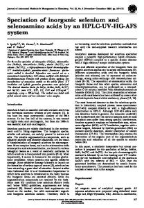
However, it is increasingly being recognized that speciation of dietary selenium is essential to the determination of the optimal food form and intake level: the negative outcome of recent prominent clinical trials [1] is in contrast to the majority of molecular, animal and human data showing a protective effect of selenium against prostate, colon and lung cancer incidence [2], and the form of the selenium species present in the nutritional supplement may partially explain the disparity [3]. Although speciation of selenium in foodstuffs appears critical to predicting biological outcome and potency, dietary selenium is also subject to extensive postprandial metabolic transformation that significantly alters its chemical properties and physiological effect [4, 5]. Thus, speciation of the forms of selenium actually present in tissues of subjects and animals, not just the forms of supplemental or background selenium present in the diet, is required to derive mechanistic insight from outcome-based experiments. The need for speciation is now increasingly being recognized in the design of clinical trials involving selenium supplementation [6]. A simplified version of the generally accepted selenium turnover is depicted in Fig. 1. The scheme is based on the generally accepted metabolism scheme first proposed by
Selenoproteins Excreted as Selenosugar
Genotoxicity
Grains/Legumes Selenomethionine
Transmethylation & Transulfuration
NH2
H
Hydrogen Selenide
Me CO2H
Se
Se H
Limited Interconversion β-elimination
Se Me
H
Methyl Selenol Selenite
Selenate
O
O
Se O
O O
Se O
O
Cruciferae/Allium Se-Methylselenocysteine NH2 Me
Se
Cell Cycle Arrest
CO2H
Fig. 1 Selenium from soil to cells. A simplified version of the generally accepted selenium turnover
Excreted as (Me)nSe
Redox Cycling
Surveying selenium speciation from soil to cell
Ganther [7]. Inorganic selenium absorbed by plants is metabolized to a variety of organic selenium compounds, the structures and amounts depending on the plant species, and these plant selenium metabolites are consumed by man and animals. Mammals further metabolize the ingested plant metabolites to multiple species, including hydrogen selenide (H2Se), which forms the basis for biosynthesis of essential selenoproteins. One of the products of mammalian metabolism of dietary selenium is methylselenol (MeSeH), a key metabolite in cancer prevention [8]. Excess selenium is excreted as volatile dimethylselenide (DMeSe) in breath or as a family of selenosugars in urine. However, there are many unanswered questions regarding selenium metabolism that have direct bearing on supplementation recommendations and public health policy. Many studies incorporating speciation methodology examine the presence of selenium forms within a food or tissue, or trace the metabolic conversion of selenium (generally via use of stable-isotope-labelled precursors or biochemical precedent). Terming these complementary analyses ‘horizontal speciation’ and ‘vertical speciation’, respectively, we propose that the most informative studies will combine aspects of both approaches. For horizontal speciation, this requires determination or prior knowledge of the basal diversity and relative quantities of selenium species in the food/tissue under study, and acknowledgement of this state in selenium interventions involving cells, animals or human subjects. For example, the translational machinery uniquely responsible for selenoprotein synthesis has shown discrimination towards the translation of individual selenoproteins, dependent upon adequacy of selenium status [9, 10]. Food forms of selenium can also determine foetal and postpartum selenium availability: inorganic forms are more readily able to increase the amount of the transport form of selenium, selenoprotein P (SEPP1), to increase the amount of foetal selenium, but organic-form selenium (as selenomethionine, SeMet) is better able to provide selenium via lactating [11]. Vertical speciation then is the process of detailing the dynamic conversions of selenium possible within a cell, tissue or organism, given the particular constraints on metabolism imposed by the genetic expression level of enzymes mediating selenium transformation and nutritional state. Thus, knowledge of the nutritional status of the system being studied is critical to accurately predicting the conversions to which dietary selenium will be subject. For example, the ability of SeMet to replete selenoprotein status in selenium-deficient animals is inversely correlated with the quantity of the amino acid methionine also present in the diet [12], and dietary methionine also affects the formation of methylated selenium [13]. Also, hepatic methylation capacity [14] and vitamin B6 status [15] influence the metabolism of dietary selenium to metabolically active forms. This variable conversion could poten-
1745
tially influence the effectiveness of clinical intervention with supplemental selenium, as methylated metabolites of selenium are much more potent anticancer agents than are non-methylated forms [8]. Clearly, the value of any vertical transformation inference rests on the quality of the horizontal survey of the species present, and, lacking some interpretation regarding metabolic conversion, simple surveys of selenium species in tissues do not provide sufficient predictive power for robust hypothesis generation. Horizontal speciation of the types and quantities of selenium present in dietary precursors and preintervention subjects as well as vertical speciation of the metabolic routes traced by ingested selenium are both critical to determining nutritional status and the potential for bioavailability or anticarcinogenicity. The aim of this review then is to evaluate the present knowledge of the selenium species available to the general population in the form of food and common supplements taking the horizontal approach regarding the selenium source. The presentation of the knowledge of selenium metabolism in mammals will include the horizontal approach as to which metabolites have been identified as well as the vertical approach describing attempts to elucidate metabolic pathways. Numerous metabolism studies and dose-effect studies are being performed analysing the impact of selenium on upregulation or downregulation of proteins, and correlating these effects with levels of added/supplemented selenium, or a determination of total selenium. Much more information could be deduced from these experiments if speciation analysis were included in addition to total selenium analysis. On the other hand, studies incorporating biological end points could inspire the analytical chemist to obtain new speciation insights. This review is an attempt to combine the knowledge from these two related but different research areas with the purpose of qualifying both areas. We also provide some perspectives on how recent developments within the general field of selenium research may present opportunities to contribute to speciation analysis and knowledge of selenium metabolism.
General analytical methods Liquid chromatography coupled with atomic and molecular mass spectrometry The present state of the art for selenium speciation is hyphenated techniques, where an efficient separation method is coupled with highly sensitive inductively coupled plasma (ICP) mass spectrometry (MS) detection. Detection limits in the low to sub microgram per litre range are easily obtainable with liquid chromatography (LC)–
1746
ICP-MS systems. As detection limits for direct infusion of selenium at the nanogram per litre level monitoring the 80Se isotope can be obtained with ICP-MS instruments prefaced with a collision/reaction cell, the limiting factor is most often the efficiency of the chromatographic system; optimization of the chromatography to obtain narrow well-defined peaks can improve the detection limits considerably. Several reviews on selenium speciation in biological material using ICP-MS detection have recently been given [16–18]. Productive improvements in selenium speciation are based on improved chromatographic separation systems and improved sample preparation procedures to obtain better recovery of the species present in a native sample. As an example, the original normal phase separation principle has found new applications following the introduction of hydrophilic interaction LC (HILIC) columns. The column material is polar, often based on silica, and is well suited for retention of hydrophilic compounds. The mobile phases for these columns are organic solvents, which is favourable for electrospray ionization (ESI) MS detection but not readily compatible with the ICP. This incompatibility, however, can be circumvented by use of microbore columns and correspondingly low flow rates, flow splitting or counterflows of aqueous solutions. As opposed to ESI-MS, where molecules are retained throughout the ionization process, ICP-MS is an atomic MS technique which obliterates analytes during ionization, stripping them of their structural information and exposing their constituent selenium. Thus, whereas ESI-MS allows massbased confirmation of the analyte structure, ICP-MS identification of metabolites is based on retention time comparison with standards. In the absence of molecular MS data, analysis of speciation data from atomic MS data alone has the potential for erroneous interpretation. In the absence of molecular MS, metabolites are typically assigned by using multiple, orthogonal chromatographic separations. Additionally, few standards for selenium speciation are commercially available, especially in stable-isotope-labelled form. The difficulties in molecular MS analysis, however, arise from poor ionization of metabolite leading to higher detection limits combined with ion suppression from the sample matrix demanding very clean extracts of the metabolite obtained by several chromatographic purification steps. Confident identification of a novel metabolite requires use of molecular MS, preferably synthesis of the proposed structure and demonstration of identical mass spectra and fractionation patterns of the synthesized standard and the purified metabolite. Thus, a current challenge for selenium speciation methods is to robustly incorporate molecular MS in parallel to atomic ICP-MS analysis. The MS instruments most often used are ion-trap instruments for tandem fragmentation (MSn) and time-of-flight (TOF) instruments providing accurate mass. Combinations
B. Gammelgaard et al.
of quadrupoles and TOF instruments (QTOF) can provide accurate mass together with structural information, and hybrid ion trap–TOF instruments are now available which allow tandem MSn and accurate mass analysis of parent and daughter fragments. The development of the Orbitrap, an electrostatic ion-trap mass analyser using fast Fourier transformation to obtain mass spectra with high resolution has opened up new possibilities [19]. The widening access to these instruments has resulted in fast identifications of large groups of new selenium metabolites in yeast [20, 21]. Use of molecular MS for selenium speciation has been reviewed [22] and critical evaluations of identification of selenium metabolites have been given as well [23–25] Additional analytical platforms Separation by capillary electrophoresis (CE), which gained temporary prosperity for speciation analysis, has only limited use. Since a review published in 2008 [26], only two applications have appeared; one for analysis of SeMet in yeast based on CE with electrochemiluminescence detection [27] and the latest CE with dynamic reaction cell–ICP-MS detection for analysis of a fish liver reference material and a dietary supplement [28]. Although interfaces for CE–ICP coupling have become commercially available, the technique is still complicated and has been more or less superseded by micro and capillary bore LC. An alternative electrophoretic technique is gel electrophoresis. Sodium dodecyl sulfate polyacrylamide gel electrophoresis (SDS PAGE) is still the most efficient technique for protein separation. SDS PAGE with laser ablation–ICP-MS detection has recently been used for identification of selenoproteins in yeast followed by QTOF MS/MS identification [29] and screening of selenium proteins in catfish [30]. The increasing interest in volatile selenium compounds has led to more intensified use of gas-chromatographic separations with MS detection, either by conventional electron impact or by the more sensitive ICP-MS detection. Gas chromatography (GC)–TOF MS has been applied for identification of DMeSe and dimethyldiselenide (DMeDSe) [31]. GC-ICP-MS is capable of detection limits of nanograms per litre, again at the expense of structure information loss. The major challenge in these analyses is loss of volatiles during sample processing and storage. The use of GC for amino acid analysis demands derivatization to volatilize the analytes; the most common reagents are silylation agents, fluorination agents and chloroformate [32]. Also for this technique, new developments are targeted towards improved sample collection and preparation, most often in the form of microfibre extractions [33]. X-ray absorption spectroscopy (XAS) has emerged as an alternative technique for selenium speciation at the oxida-
Surveying selenium speciation from soil to cell
tion state level. XAS uses synchrotron-radiation-based Xrays to probe local structural and electronic environments and can be used for fingerprint analysis of the different chemical forms of an element. The advantage is that the technique can be applied to bulk material, avoiding sample preparation and thus preventing artefactual transformations of selenium species. X-ray absorption near-edge spectroscopy (XANES) indicates the oxidation number and the coordination number of an element. Elemental selenium has the lowest edge energy, whereas selenate, in oxidation state six, has the highest edge energy. This technique was used in combination with multivariate data analysis to characterize yeast formulations [34] and revealed that SeMet was the principal species in the formulations. So far, this method has limited practicability in terms of identifying metabolite structures, but it may have future possibilities in screening methods, when used in combination with multivariate data analysis. In addition to qualitative assessment of selenium species having greatly differing chemical properties, the field of selenium analysis is increasingly incorporating a strict quantitative approach to selenium speciation, with isotopedilution-based approaches becoming the state of the art for ICP-MS-based analyses [35].
Speciating dietary selenium as a mammalian metabolic precursor
1747
derivative structures follow the core structures. Thus, selenocysteine (SEC) is considered a core structure and is followed by derivatives of this structure, e.g. Semethylselenocysteine (MeSEC). Selenolanthionine could also be regarded as such. Selenohomocysteine is the demethylated product of SeMet, although it has a free selenol group like SEC. Selenohomolanthionine is regarded a derivative of SeMet, whereas the asymmetric selenocystathionine can be regarded as a derivative of SEC as well as SeMet. The compounds containing a sulfur–selenium bond such as S-(selenomethyl)cysteine (MeSeCys) and the glutathione conjugates constitute another class. Hence, most of the selenium species are amino acids or small peptides based on a few selenoamino acids. Also the new compound selenoneine (2-selenyl-N, N,N-trimethyl-L-histidine) is an amino acid derivative. The forms of dietary selenium encountered can thus be grouped roughly into three categories: (1) inorganic forms such as selenite and selenate, (2) SEC-based forms and (3) SeMet. Generally, inorganic forms are relatively abundant in soils, but are found in low abundance in foodstuffs, whereas SEC-based species are variably produced by Allium species as seleno-analogues of their characteristic γ-glutamyl compounds. Generally, the various species exhibit different biological activities, albeit with significant overlap resulting from significant postprandial metabolic conversion. Horizontal overview of dietary selenium sources
Organization of molecular structures Few studies on selenium speciation in common food products have been published, but several studies on selenium-accumulating plants and selenized yeast have appeared. An overview of selenium species in these selenium sources is given in Table 1. For the purposes of this review, only species identified by the stringent metrics of ICP-MS in combination with molecular MS and/or nuclear magnetic resonance (NMR) spectroscopy are regarded as being unambiguously established in the given foodstuff and are signified in italics. These are indicated with references to the first published study only, but numerous confirmatory publications exist in some cases. Food products in which selenium species have been identified on the basis of chromatographic retention times only are signified in normal text and are not individually referenced. A compilation of the relevant references to these can be found in [56]. Consistent structures are only given once. Selenium content in food and beverages, including identification of selenium species by molecular MS, has recently been reviewed [23–25]. It appears that most species can be related to a few core molecular structures (Table 1). The table has been arranged so that
Selenium intake varies depending on the selenium content in the soil. Extreme values from below 10 μg/day up to 5 mg/day, both from China, have been reported [57]. In Western countries, such as Denmark, a European country, the daily intake is 38-47 μg, whereas on the North American continent, the daily intake in the USA is 106 μg/day and it is 98–224 μg/day in Canada [58]. The safe upper intake level is 400 μg "Se"/day, but intakes cannot readily be generalized across selenium species owing to their remarkably different biological effects at high levels. The intake required to observe adverse effects is more than double this amount, but the agreed limit leaves room for a safety margin necessary to take different bioavailability and toxicity of different selenium species into account [58]. Reported levels of selenium in food and beverages were recently reviewed [58]. Food forms contribute to overall human selenium intake to a much greater degree than drinking water. The selenium content in food products is highest in organ meats and seafood, followed by muscle meat, cereals, dairy products and vegetables. However, the selenium contents in these products are highly variable and dependent on the origin. In general, reported values for
1748 Table 1 Selenium species in food and supplementation sources
B. Gammelgaard et al.
Surveying selenium speciation from soil to cell Table 1 (continued)
1749
1750
B. Gammelgaard et al.
Table 1 (continued)
Species identified by the stringent metrics of inductively coupled plasma mass spectrometry in combination with molecular mass spectrometry and/or nuclear magnetic resonance spectroscopy are regarded as being unambiguously established in the given foodstuff and are signified in italics followed by a reference. Food products in which selenium species have been identified on the basis of chromatographic retention times only are signified in normal text. GSH glutathione
Surveying selenium speciation from soil to cell
beverages and most vegetarian food products such as cereals, bread and vegetables were below 100 μg/kg, whereas the selenium amounts in meat and marine products were higher and ranged from 100 to 800 μg/kg. Pork kidney showed the highest amount, up to 1,500 μg/kg. The food-based intake thus depends on the eating habits of the individual. As subjects with low plasma selenium levels (below 106 μg/L) benefited most from selenium supplementation in the Nutritional Prevention of Cancer trial [59], which used a selenium-enriched yeast-based supplement, and the average plasma levels in Europe are below this level, general selenium supplementation has been a matter of discussion [60]. Selenium supplementation in the form of functional foods such as selenium-fortified milk, broccoli, garlic, green onions, green tea and mushrooms has been proposed. The reason for suggesting these particular vegetables is that the Brassica species (rapeseed, broccoli, cabbage) and the Allium species (garlic, onion, ramps) are so-called secondary accumulators, producing a spectrum of methylated selenium species readily metabolizable by mammals to forms exhibiting anticarcinogenicity. Astragalus species and Brazil nuts are selenium accumulators; as they are not commercialized crops, readily volatilize selenium and are prone to accumulating undesirable metals, they have not been explored as dietary selenium sources outside traditional medicine. Finally, although cereal and pulse crops such as wheat and lentil, respectively are considered non-accumulators [58], they do accumulate selenium, largely in the form of proteinaceous SeMet; these plants provide the bulk of most people’s diets, and thus SeMet is the predominant form of ingested selenium in most cereal-consuming populations. Wheat in the Nawanshahr-Hoshiarpur region of Punjab, India, manifests some of the highest levels of recorded selenium in cereal grains. Cubadda et al. [61]. recently performed speciation analysis on wheat from this region and found that although SeMet formed the predominate selenium species, selenate and MeSEC constituted higher percentages of total selenium in wheat with the highest selenium levels. Thavarajah et al. have utilized the novel selenium speciation method based on XANES that can differentiate alkylselenides from inorganic selenium on the basis of the oxidation states [62]. After examining two individual cultivars of lentils grown in distinct North American regions, the authors concluded that selenium was present primarily as SeMet, although the presence of MeSEC and other organic selenides could not be ruled out. Intriguingly, up to 15% of total selenium was present as selenite and selenate, indicating the potential for lentils to supply a spectrum of benefits related to nutritional repletion; i.e. lentils might potentially provide immediate repletion of nutritional status with inorganic selenium and accrual of long-term stores in general body proteins as
1751
SeMet incorporated in general proteins. The authors undertook speciation analysis to define the organic selenides in a lentil cultivar and performed speciation by LCICP-MS with standard retention time matching . For the whole seed, the ratio of SeMet to (MeSEC + γ-glutamylSe-methylselenocysteine) to (selenate + selenite) was approximately 7:1:2 [63]. Another source of selenium is commercially available selenium supplements. The most common formulation is lyophilized, selenized yeast produced from Saccharomyces cerevisiae, but also tablets or capsules containing organic selenium in the form of SeMet or MeSEC and inorganic salts, often in combination with vitamins and minerals, are available. A capsule formulation containing a mixture of 50 μg SeMet, 50 μg sodium selenate, 25 μg selenodiglutathione and 75 μg MeSEC has also been described [64], and would be expected to provide ready substrates for multiple metabolic transformations involving selenoprotein synthesis and MeSeH production. Additionally, a product containing 2-hydroxy-4-methylselenobutanoic acid, which is suggested to be a SeMet precursor, is available (NutraSelen) [65], and with the increasing knowledge of selenium speciation and relevant metabolism, even more fanciful products will likely emerge. Acknowledging that species conversion within selenium dietary supplements might eliminate benefits, and potentially produce species with increased toxicity, Amoako et al. addressed the issue of selenium species stability in yeast-based and non-yeastbased supplements [64]. Combining the results of LCICP-MS after enzymatic hydrolysis of supplements with GC–atomic emission spectroscopy (AES) of extracts derivatized with ethyl chloroformate for amino acid analysis and headspace solid-phase microextraction (SPME) GC-MS for detection of volatiles, they showed that SeMet-containing supplements released DMeDSe in the headspace when heated at 100 °C for 1 day, whereas the amount of SeMet decreased and a relative increase in MeSeCys and selenomethionine oxide (SeOMet) was observed. The authors hypothesized that DMeDSe reacts with thiol groups or disulfide links in cysteine or cystine. Supplements containing only SeMet or MeSEC also released DMeDSe upon heating, but did not form MeSeCys. However, a supplement containing both amino acids together with selenate and selenodiglutathione formed MeSeCys as well. The work of Amoako et al. calls into question the utility of selenium supplements comprising multiple complex species, exposing the predisposition of selenium to interconvert at the expense of long-term stability. Much work on selenium metabolites in yeast has been performed by the Lobinski group. In the method typified by their group, hydrophilic compounds are extracted with water, followed by size-exclusion fractionation; fractions of interest, determined by HILIC-ICP-MS
1752
are selected for analysis by QTOF-MS/MS. Fifteen peaks were separated and 12 of these identified [42]. The same approach was improved by additional purification of the fractions containing large amounts of salt by anionexchange chromatography for identification of the anionic compounds [21]. Application of high-resolution MS to selenium speciation was characterized by identification of several glutathione conjugates of amino acids and selenium by Orbitrap analysis [20]. The compounds identified from these studies are included in Table 1. The new selenium compounds identified in yeast, however, are all largely derived from the previously mentioned core structures (Table 1) and may be metabolized via the same catabolic pathways. Analysis of volatiles in foods and supplements mainly has the focus of identifying volatiles for information on sample degradation or has the focus of sample purification by derivatization of non-volatile analytes into volatile species. As reviewed by Pyrzynska [66], common approaches for determination of volatile selenium species involve preconcentration via cryogenic trapping or SPME followed by thermal desorption or solvent extraction. The chromatographic method of choice is GC, with electron ionization (EI)–QTOF-MS and ICP-MS detection modes providing the best sensitivity and qualitative confidence. Campillo et al. [67]. addressed the microbial degradation of organoselenium compounds within beers, wines and spirits using headspace GC-AES with SPME sampling for the determination of DMeSe and DMeDSe along with related volatile organic sulfur compounds. The procedure was optimized for the analysis of milk and milk-related products such as yoghurt and infant formulas [68]. However, volatile selenium compounds were not detectable in 40 different alcoholic beverages or in 23 different milk and milk products. Related procedures were used for determination of volatiles in garlic, onion and their juices [33]. Methanol-desorbed SPME fibre extracts were analysed by GC-ICP-MS. DMeSe and DMeDSe were detected in garlic, onions and the juices. Some unidentified other volatile selenium species were observed as well. The amount of volatile species was determined; however, it was not discussed whether the volatile species comprised major or trace amounts of the total selenium accumulated in the plants and their juices. A method for trapping environmental volatile selenium species with some potential as a general method was recently developed. DMeSe and DMeDSe were trapped in concentrated nitric acid via N2 purging of the sample solution. DMeSe was trapped as non-volatile dimethyl selenoxide (verified by LC-ESI-MS) and DMeDSe was trapped as methylseleninic acid (MeSeA) (verified by spiking). The non-volatile trapped species was then quantified by anion-exchange chromatography ICPMS [69]. An example of sample purification is volatiliza-
B. Gammelgaard et al.
tion of selenite by ethylation with tetraethylborate. The volatile reaction product, diethylselenide, was extracted from the sample headspace by a coated SPME fibre and thermally desorbed in the following GC-EI-MS analysis in single ion monitoring mode. The advantage of this procedure is the efficient analyte extraction from the highsalt matrix of urine which may hamper the anion-exchange chromatography used for selenite determination. The method was proved valid for urine, saliva and milk matrices although no actual samples were analysed [70, 71]. Also the selenoamino acids SeMet and MeSEC are easily volatilized by ethylation with ethyl chloroformate. With use of hollow-fibre liquid-phase microextration, high enrichment factors and low carry-over of the amino acids were obtained. The organic extraction liquid was analysed by GC-ICP-MS using selenoethionine as an internal standard. The procedure was used for determination of SeMet and MeSEC in yeast (SELM-1), garlic, cabbage and mushroom [72]. The detection limits of the cited reports were in the 10–150 ng/L range. A novel selenium species named selenoneine was recently identified as the predominant chemical form of organic selenium in the deproteinized blood of bluefin tuna. The compound was identified by high-resolution MS and NMR spectroscopy after intensive purification by several chromatographic steps. The exact mass of the [M+H]+ ion of the isolated compound was 533.0562, with a molecular formula of C18H29N6O4Se2, which turned out to be the oxidized dimeric form of a selenium analogue of ergothioneine and was named selenoneine. The molecular peak at m/z 553 was converted to m/z 278 after reduction of the compound by dithiothretiol (DTT) or glutathione [53]. The monomeric compound was unstable and was readily oxidized to the dimeric diselenide form, which was stable at room temperature. Following its initial identification, its distribution in deproteinized tissue was determined by LC-ICP-MS in the supernatant from aqueous extraction from tissue homogenates using a hydrogel column. The presence of selenoneine and unidentified species was found in various animal tissues on the basis of standard comparison, whereas neither selenite, nor SEC nor SeMet was found; 98% of the selenium in the supernatant of the protein-precipitated tuna red muscle was present as selenoneine. Also spleen, liver, pancreas, heart white muscle and blood contained a large amount of the compound. It was suggested that selenoneine may be an important member of the redox cycle in animal cells as it has strong antioxidant capacity, binds to haem proteins and reacts with radicals and methylmercury [73]. Improved extraction method Although the online separation and detection of selenium forms is an indispensable aspect of selenium speciation, the
Surveying selenium speciation from soil to cell
initial extraction of the analytes from their biological matrix sets the stage for success at all subsequent steps of analysis. Thus, procedures must be adequate to efficiently extract the selenium species and sufficiently mild to avoid species conversion in the form of oxidation or volatilization. Extraction procedures are most often based on enzymatic degradation by use of proteolytic enzymes such as protease XIV and protein mixtures such as pronase E/lipase mixture. Acidic and basic hydrolysis with HCl or tetramethylammonium hydroxide (TMAH) is another possibility [18]. In this regard, several significant advancements have recently been brought to bear against retractable matrices, enabling a more comprehensive horizontal speciation of the precursors which form the starting materials of human selenium metabolism. One way to improve extraction efficiency is to accelerate the enzymatic digestion by microwave or ultrasonic assistance. Recently, a microwave-assisted enzymatic extraction (MAEE) procedure based on enzymatic extraction with a mixture of pronase E and lipase was demonstrated to provide robust extraction of selenium species in rice, allowing determination of approximately 100% of rice selenium as SeMet in a standard reference material and between 87 and 96% recovery as SeMet in various commercial varieties [74]. A sister report [75] determined that fish tissue was considerably more recalcitrant towards the MAEE procedure than rice. Analysis of canned tuna, shark and marlin showed that, again, SeMet was the major species detected in the fish tissues on the basis of comparison with retention times of standards, but this only accounted for between 9 and 56% of total selenium. The authors proposed that the balance of selenium is also in the SeMet form, but remains in peptides and proteins that were resistant to the protease employed. In the same study, enzymatic extraction was compared with the use of TMAH. The latter reagent showed full recovery but converted the selenium species to inorganic selenium. Besides the difference in efficiency of MAEE between rice and fish, it is intriguing that there is such variation in the extractability of SeMet from the different fish samples and standard reference materials. This indicates the potential for differential release of selenium from fish tissues upon enteric digestion. MAEE was shown to reduce the hydrolysis time from 40 to 1 h for extraction of SeMet from selenized yeast and it was demonstrated that the accelerated extraction did not compromise the accuracy and precision of the following quantification by isotope-dilution LC-ICP-MS [76]. With use of the same extraction method, 63% of watercress selenium could be accounted for as SeMet, MeSEC and selenate [45]. After quantification of the aforementioned selenium species by ICP-MS, LC-ESI-MS analysis confirmed the presence of SeMet and MeSEC by their characteristic MS transitions. The same authors addressed
1753
the issue of cooking procedures, contrasting prototypical boiling (mimicking common cooking techniques) with the MAEE technique (mimicking enteric digestive processes) [77]. All three major forms of selenium—inorganic selenate, SEC and SeMet—were found in both potato skin and flesh. The aspect of cooking was also addressed by Thavarajah et al., who discussed the commercial and cooking preparatory procedures that might impact the nutritional availability of selenium species from various seed fractions of lentils and how to maximize their nutritional impact, observing that traditional methods of preparation and consumption often provide the most available selenium [63]. Also sonication has been demonstrated as a fast alternative extraction method. An ultrasonic probe was employed for fast protein extractions from yeast [78]. The sample was extracted for 2 min in water containing DTT to cleave disulfide or selenosulfide bonds and phenylmethylsulfonyl fluoride as an inhibitor of protease activity on ice. After protein precipitation in acetone and reconstitution in water, proteins were separated by SDS PAGE, relevant spots were excised and analysed by nano-LC-ICP-MS and two proteins were subsequently identified by nano-LC-ESI-QTOF-MS/MS. Another improvement upon traditional extraction methods is the use of a complementary spectrum of focused extraction reagents. In recognition of the inadequacy of single or limited extractions for comprehensive speciation, the group of Szpunar and Lobinski developed a procedure for quantitative determination of SeMet and SEC from proteins in animal whole blood, tissues [79] and milk [80]. The method is based on protein unfolding by addition of urea, cross-linked sulfur/seleno bond breaking by DTT, followed by alkylation of cysteine and SEC residues with iodoacetamide (carbamidomethylation) and enzymatic digestion with protease. Samples were fractionated by sizeexclusion chromatography and the low molecular weight (LMW) fraction was analysed by ion pair reversed-phase LC. 77Se-labelled SeMet was used as an internal standard to correct for the SeMet derivatization yield. Analysis of chicken breast, and muscle, heart, liver and kidney from lamb showed the presence of SEC and SeMet. SEC was identified by spiking the sample with the SEC-containing selenoenzyme glutathione peroxidase 1 (GPX1; E.C. 1.11.1.9) prior to extraction. Both species were present in the samples, but in different ratios [79]. The method was applied for analysis of milk from cows supplemented by selenized yeast or selenite. However, chromatographic resolution of these species was not obtained and species were not identified by ESI-MS owing to inherently insufficient ionization in the intractable matrices. The same procedure was used for analysis of eggs [81]. Seleniumenriched eggs have recently been proposed as functional food [82]. Whereas egg white allowed direct analysis,
1754
analysis of yolk demanded defatting with cyclohexane and desalting was applied by protein precipitation with acetone and following reconstitution. Single peaks were obtained for the derivatized selenite and SEC, whereas the SeMet derivative resulted in two peaks. Derivatized standards of selenite and SeMet were analysed by ESI-QTOF-MS and showed spectra corresponding to the carbidomethylated compounds and the derivates produced were coeluted with the compounds in the egg extracts. A spectrum of the SEC derivative was not provided. The predominant form of selenium in yolk was SEC, whereas the predominant species in white was SeMet. Both yolk and white contained about 10% of selenite [81]. Along the same lines, a sample preparation platform for freeze-dried garlic was developed consisting of six separate manipulations, ultimately performing extraction-based speciation [83]. Sequentially, the researchers extracted water-soluble species followed by cell wall components (cellulases, chitinases, etc.), peptidyl SeMet and SEC (protease type XIV), inorganic selenium bound to organic components (HCl), elemental selenium (Na2SO3) and inorganic selenide (CS2). The authors used chromatographic matching to standards to identify species within these fractions with detection via ICP-MS. Also in this case they found QTOF-MS insufficiently sensitive to accommodate the analytical burden. From their report, it would seem that inorganic selenium composes a large portion of garlic cell wall, whereas SeMet is the primary species found after proteolysis. Isotope dilution: new standard in quantitative selenium analyses The quantitative aspect of selenium speciation has gained increasing interest and quantification by isotope dilution is an indispensable tool for precise and accurate measurements. The concepts of isotope dilution in general have been exhaustively treated in a recent tutorial [84] and specifically reviewed for selenium analysis [18]. An increasing number of reports on species-unspecific as well as species-specific applications in selenium speciation studies are emerging. An example of species-specific isotope dilution analysis is determination of ultratrace amounts of selenium species in garlic using an authentic standard produced in, and purified from, selenized yeast grown on isotopically enriched selenium [85]. As both allium species and selenized yeast produce clinically relevant methylated selenium-containing peptides, the purified enriched standard of γ-glutamyl-Se-methylselenocysteine from yeast was applied for quantification of the same compound in garlic. The increasing interest in species quantification augments the demand for standard reference materials proving the accuracy of the applied methods. So
B. Gammelgaard et al.
far, only one reference material for selenium speciation is available, the National Research Council Canada seleniumenriched yeast with a certificate for total selenium and SeMet. Alternatively, analytical speciation qualifications can be assessed by laboratory comparisons. An interlaboratory comparison of analysis of total selenium and SeMet in pharmaceutical yeast tablets was performed involving 15 laboratories, mainly national metrology institutes and selected expert laboratories, located in 12 countries throughout Europe, Africa, South America and North America [86]. A variety of techniques were used for sample preparation prior to SeMet speciation, including acid or enzymatic digestion, probe sonication and different variations of isotope dilution schemes. The coefficient of variation was 2.9% for total selenium and 7.9% for speciation of SeMet, demonstrating that speciation for this analyte—at least among skilled speciation laboratories—is reproducible across multiple sample preparation procedures and analysis platforms. It remains to be seen how other, more intractable selenium species hold up to this type of cross-platform study. Kitajima et al. have developed a mutant strain of S. cerevisiae that is capable of producing ectopically expressed recombinant proteins with up to 65% SeMet substitution for methionine [87]. This raises the possibility of producing recombinant standard reference materials for general body proteins having a known degree of SeMet substitution. This could then be used to assess the degree of SeMet substitution in a protein of interest found in a biological sample. To summarize the horizontal speciation of metabolic precursors, the volatile selenium compounds DMeSe and DMeDS are considered to be degradation products of the native selenium species initially present in food, beverages and nutritional supplements. The amino acid SeMet has been identified in rice, soybeans and wheat flour and is the far largest constituent of foods originating from plants as well as animals (although the latter acquire their selenium from diets of the former). SEC is also a major constituent of food originating from selenoproteins in animals, whereas plants do not contain selenoproteins and will thus have little if any proteinaceous SEC. However, molecular mass spectra of SEC of food origin have not yet been presented. The presence of selenite and selenate in food is intriguing from the standpoint of a species-balanced selenium intake and their role in selenium-rich foodstuffs is under active investigation. Despite MeSEC being the primary selenium compound in garlic [88] and γ-glutamyl-Se-methylselenocysteine being the predominant compound in seleniumfortified garlic [89] and the only selenium compound so far identified in onions [41], the contribution to total selenium intake from these compounds is of minor importance regarding the total selenium nutritional status for individ-
Surveying selenium speciation from soil to cell
uals with ordinary eating habits. However, the finding of the new selenium species selenoneine, a major compound in tuna and perhaps also other marine products, may require reconsideration of this assumption.
Horizontal survey of mammalian selenium species Compared with the number of reported identifications of selenium species in yeast and foods, relatively few metabolites have been identified by molecular MS as being present in mammalian fluids and tissues. This subgroup is summarized in Table 2. The primary focus in speciation of mammalian systems is currently identification of novel LMW selenium species and improvement of analytical methods for their determination, or speciation of seleniumcontaining proteins. Since the identification of the selenosugars, new unknown mammalian metabolites have not emerged. Except for the selenosugars and the glutathione– selenosugar conjugate, the metabolites in Table 2 were identified in in vitro systems. As in speciation analysis of the dietary selenium sources, sample preparation is important for analysis of mammalian target tissues. Tissues are most often homogenized and extracted with aqueous buffers. The extracts contain smallmolecule selenium species and soluble proteins, the latter can be precipitated either by heating or by adding precipitating agents such as organic solvents before separation and detection by LC-ICP-MS, which reveals the different species and their quantities. Identification with molecular MS demands further purification, often several steps, to reduce suppression of target analyte ionization by matrix components. Urinary metabolites Se-Methylseleno-N-acetylgalactosamine (SeGalNAc) is the major urinary metabolite and is excreted after ingestion of selenite, SeMet as well as yeast. The presence of this metabolite has been verified by molecular MS in rat urine as well as human urine and several subsequent studies have shown the presence of this species by comparison with standards. Also the glucose form, Se-methylseleno-Nacetylglucosamine, and the deacetylated form, Se-methylselenogalactosamine, were shown in subsequent studies after the molecular mass identification [98, 100]. The presence of trimethylselenonium (TMeSe) in human urine after ingestion of large amounts of SeMet (8,000 μg/ day for 7 days followed by 4,000 μg/day for 21 days) by five cancer patients was recently verified by molecular MS for the first time [98]. The concentration of SeGalNAc varied between 510 and 4,420 μg/L in the five patients, small amounts of the glucose form of the sugar were
1755
observed and levels between 36 and 258 μg/L of the deacetylated sugar were observed. Only one of the patients showed excretion of TMeSe in large amounts (332 μg/L), whereas all other TMeSe measurements were below 10 μg/ L. This implies that production of this metabolite is dependent on the individual and is not a detoxification pathway after ingestion of large amounts of selenium. Thus, human metabolism is probably different from rat metabolism in this respect. Hepatic metabolites The glutathione–sugar conjugate has been identified by the Suzuki group from the molecular MS spectrum for liver supernatant after selenite supplementation [101]. The presence of the major urinary selenosugar metabolite in porcine liver was recently demonstrated by selective reaction monitoring atmospheric pressure chemical ionization–MS/MS after meticulous purification by extraction, sequential centrifugation, preconcentration of the isolated cytosolic fraction, size-exclusion chromatography fractionation and tandem LC separation on a cation-exchange column and a reversed-phase column in series in an attempt to develop a quantification method. The selenosugar constituted less than 1% of the total selenium content of the liver [96]. MeSEC and SeMet were identified by molecular MS in isolated rat hepatocytes after incubation with MeSeA [92]. The amount of the metabolites, however, only constituted a very small percentage of the total selenium content [102]. This finding was preceded by the same observation from incubation of human lymphoma cells with MeSeA [31]. The finding of these selenoamino acids as products of MeSeA is highly surprising as mammal cells are not presumed to produce these species. Definite proof of this pathway by use of isotope labelling is desirable. Volatile metabolites Juresa et al. showed MS spectra of volatile species including DMeSe, DMeDSe and dimethylselenylsulfide (DMeSeS) collected by headspace SPME sampling in selenosugar-spiked urine samples after 8 weeks of storage [103]. Analysing blank urine by GC– microwave-induced plasma–AES only showed the presence of DMeSe and DMeSeS. Although GC-MS spectra for the blank urine samples were not given, the results render it very likely that the latter species are constituents of basal urine and that all three volatile species can be produced by selenosugar degradation [103]. Hydride generation by NaBH4 in aqueous solution is a component of the analytical procedure and produces DMeSe (and DMeDSe trace) from TMeSe and DMeDSe from SeMet, respectively [104]. Bueno and Pannier showed the presence of DMeSe and DMeDSe in
1756
B. Gammelgaard et al.
Table 2 Mammalian selenium species identified by molecular mass spectrometry
blank urine samples by GC-ICP-MS after headspace SPME and quantified these by standard addition [105]. The concentration of both volatiles was below 1 ng/L in the two urine samples analysed. Two unidentified seleniumcontaining compounds were detected; one of these was suggested to be DMeSeS. It was demonstrated that the urinary DMeSe signal did not increase after 21 days of storage. Hence, this study confirmed that urinary DMeSe was a constituent of basal urine and not a degradation product. Human breath has been collected after ingestion of 77Seenriched selenite in Tedlar bags and analysed for selenium by a combination of cryotrapping and cryofocusing
followed by thermal desorption and GC-ICP-MS [106]. The only volatile selenium metabolite detected in breath was DMeSe as identified by retention time matching with a DMeSe standard. Selenoproteins and selenium-containing proteins The human genome has been sequenced and many of the genetic parameters for selenoprotein expression have been determined; since then in silico analyses for the presence of novel selenoproteins have exposed the presence of all gene products likely to exist. With all human selenoproteins likely known, their physical–chemical properties (size, pI,
Surveying selenium speciation from soil to cell
etc.) are predictable from sequence data and thus the analysis of mammalian selenoproteins is focused on optimizing protein separations. Knowledge of exchange of dietary selenium to and from plasma proteins is important to understand how dietary selenium is transported and utilized in the body, and these proteins often serve as proximal biomarkers of nutritional status. Although much effort has been devoted to the simultaneous analysis of the two plasma selenoproteins SEPP1 and glutathione peroxidase 3 (GPX3) together with selenoalbumin (SeAlb; a form of human serum albumin which contains at least one cotranslationally inserted SeMet or selenium adducted to the Cys34 free thiol) significant challenges remain. The classic analytical procedure for the analysis of the three major selenium-containing proteins in plasma is based on affinity chromatography and consists of a two-step separation with selenium-selective detection such as ICPMS [107]. SEPP1 is selectively retained on heparinSepharose or immobilized metal-ion Sepharose stationary phases [108] and albumin is retained on blue Sepharose stationary phase. GPX3 and all other plasma proteins are not retained by any of the stationary phases. Separation of SEPP1, GPX3 and SeAlb is therefore achieved. The major drawback of this procedure is the lack of retention of GPX3; unretained salts provide spectral interference and the coelution of all other plasma proteins, which contain potentially large amounts of selenium as SeMet, artefactually contributes to the signal attributed to GPX3. Jitaru et al. addressed the issue of salt-based interferences by inclusion of a solid phase extraction clean-up step [109]. They used anion-exchange cartridges for effective removal of chlorine- and bromine-containing interferences from human serum as these elements impose spectral overlap on the most abundant selenium isotopes in ICPMS detection. They then coupled the sample clean-up step online with the separation procedure, leaving the procedure fully automated [110] and downscaled it for lower sample consumption by means of a microbore column set-up [111]. In the latter study, they evaded the spectral interferences from bromine and chlorine by use of high-resolution ICPMS and therefore omitted the sample clean-up step. Species-specific isotope dilution was use to validate the determination of proteolytically liberated SeMet from plasma and the authors addressed the concern of nonprotein SeMet as an artefact in proteinyl SeMet determination. It was argued that as 95% of serum selenium was protein-associated, interference from other unretained LMW selenium species possibly present in serum was negligible [112]. Xu et al. succeeded in developing an anion-exchange chromatography method for separation of SEPP1, GPX3 and SeAlb with ICP-MS detection [113]. They quantified
1757
the amount of selenium within these individual proteins by means of species-unspecific isotope dilution. Furthermore, they detected two unknown selenium species with molecular masses below 18 kDa determined by gel filtration. It should be noted that despite the promising analytical procedures, quantitative determination of selenoproteins is not a straightforward task. Selenoproteins, especially GPX3, are readily degraded in serum samples to produce selenite and apparently also volatile species [114] and in our experience even freeze-dried GPX3 is unstable. Modification of protein cysteine thiols by diselenide or selenosulfide metabolites of selenium is an effector of anticarcinogenicity, and is thought to result in functional [115] and structural [116] alterations. The direct analysis of proteinyl selenosulfides resulting from exposure to MeSeH congeners such as DMeDSe and Se-methylselenoglutathione has not yet been reported, but would likely aid structural and mechanistic interpretation of the biological effects of LMW selenium species. A recent publication described the utility of 77Se NMR spectroscopy in the analysis of peptidyl diselenides and aimed to elicit their use as proxies for disulfides [117]. In this light, 77Se NMR spectroscopy demonstrates the potential to determine specific methylselenyl adducts of critical cellular effectors; as sensitivity and sample size issues are addressed with NMR analysis, it might provide benefits towards the speciation of MeSeH adducts with mechanistic effector proteins that other, more traditionally employed, analytical platforms cannot. The potential for tagging as a speciation approach to analysis of selenoproteins is exhibited by a recent methodological publication describing the use of a nucleophilic tag to adduct the portion of the cellular GPX1 population whose single SEC residue has been irreversibly deselenated to dehydroalanine (DHA) [118], thus speciating native from deselenated GPX1. The immunoblot analysis was based on specific addition of biotin-conjugated cysteamine to DHA followed by detection after interaction with streptavidin. These authors also experienced conversion of the SEC residue during ageing of red blood cells in vivo as well as after exposure to oxidation, and suggested DHA–GPX1 as a biomarker for oxidative stress. A novel approach was taken by Tsopelas et al. to elucidate the pharmacokinetic parameters of eight selenium species on the basis of the chromatographic affinity of these species for immobilized human plasma proteins or artificial membranes designed to mimic the surface of biological membranes [119]. The retention factors (log tR,) of eight selenium compounds were determined by UV detection. The retention factors for the four biomimetic columns compared were quite similar, with retention decreasing in the order DMeDSe > DMeSe ~ selenocystamine > SeMet ~ SEC > selenate ~ selenite. The retention factors were used to estimate oral absorption and protein binding. This novel
1758
and creative chromatographic approach opens up new possibilities for elucidating interactions between selenium compounds and plasma proteins when coupled with a more specific and sensitive detection method.
Vertical speciation of selenium in mammalian metabolic pathways Enteric metabolism The conversion of selenium species in the gastrointestinal tract has previously been reviewed [23]. SeMet and other selenium species may be present in edible tissues that are resistant to enteric digestion and liberation of selenium for absorption in the small intestine (e.g. wheat bran). In this case, selenium might be expected to arrive intact in the colon and be subject to biotransformation by colonic microflora. To enable an analysis of the transformation of SeMet by colonic microflora, a high-performance LC (HPLC) assay with fluorescence detection involving derivatization with ortho-phthaldialdehyde and N-acetylcysteine was developed to detect SeMet in rat gut contents [120]. The authors recently expanded their investigation to examine the differences in biotransformation of inorganic (selenite) and organic (SeMet) selenium by gut contents from the jejunum, ileum, caecum and colon [121]. Incubations of selenite were analysed by hydride generation atomic absorption spectrometry and the volatiles were analysed by GC-MS. That caecum and colon contents were most metabolically active towards selenium species indicates the prominent role of microbial communities in selenium transformation, and stresses that enterohepatic reabsorption may be an overlooked route of selenium absorption. Selenite was transformed to DMeSe, whereas the predominant volatile produced during incubations with SeMet was DMeDSe. As both of these species are small, non-polar entities, they are expected to readily traverse the colonic lining to enter the systemic circulation. Whereas DMeSe is non-reactive and will be excreted via breath exhalation, DMeDSe has the potential to modify thiols and form selenosulfide adducts with plasma or cellular cysteines. It is unknown at this time how differing enterohepatic pharmacokinetic profiles impact the biological activity of selenium species. General mammalian selenium metabolism Mammalian selenium metabolism is most often described by different variations of a model originally proposed by Ganther [7]. An updated version of this scheme can be found in [25]. Once ingested, most available selenium is absorbed by the intestine and released in a form extracted by the liver with
B. Gammelgaard et al.
high efficiency [122]. In the liver, dietary selenium is metabolized to selenide and then incorporated as SEC into SEPP1, the physiological form of transport to peripheral tissues. Thus, all selenium incorporated into the selenoproteins has traversed a selenide pool. The other major plasma selenoprotein, GPX3, is synthesized in the kidneys [123]. Ingested SeMet, is either indiscriminately incorporated in any proteins in competition with methionine or transformed via the selenide pathway. Given the increasingly recognized role of methionine/ methionine sulfoxide redox couples in cell response and signalling [124], it is of interest to determine the degree of SeMet incorporation into plasma and cellular proteins after SeMet supplementation. Such information informs on the status of whole-body selenium pools, serving to assess the level of selenium present as recyclable, protein SeMet. SeMet metabolism SeMet is the largest source of dietary selenium and the compound that has been subject to most experiments regarding elucidation of metabolic pathways. A scheme of SeMet metabolism is presented in Fig. 2. Parts of this scheme appear from time to time in different versions of general selenium metabolism schemes together with new assumptions and suggestions. In the following, documentation of the pathways in Fig. 2 is briefly presented. SeMet is oxidized to SeOMet by complementary DNA expressed human flavin– containing monooxygenases purified from microsomes [125]. Interestingly, SeMet undergoes chemical oxidation with reaction rates that are 10–100-fold larger than those for the corresponding oxidation of methionine. The reverse reduction of SeOMet to SeMet proceeds spontaneously via oxidation of ubiquitously present thiols and antioxidants [126, 127], providing an antioxidant redox cycle. The transformation of SeMet by partially purified rat liver extracts containing S-adenosylmethionine synthetase and a variety of transmethylases has been shown the produce Seadenosylselenomethionine (AdoSeMet) and Se-adenosylselenohomocysteine in parallel to the metabolic pathway of methionine [128]. Transformation of AdoSeMet into SEC via selenocystathionine occurred by the enzymatic reactions of cystathionine β-synthase and cystathionine γ-lyase purified from rat liver [129]. Both liver and extrahepatic tissues have been predicted to metabolize SeMet to multiple end products with the potential for biological activity [5]. Recently, α-keto acid metabolites of SeMet and MeSEC have been shown to inhibit histone deacetylases, possibly contributing to their anticarcinogenicity in prostate cancer cells [130, 131]. The production of these α-keto metabolites is catalysed by transaminases and amino acid oxidases, detailed in a recent review [132]. Demonstrating the feasibility of analysis of these types of metabolites, Vacchina
Surveying selenium speciation from soil to cell
1759
Metabolism of Selenomethionine SelenomethionineSelenoxide Containing Proteins
SelenomethionineContaining Proteins
CO2H
CO2H
Me Se
O
tRNAMETSelenomethionine
Se
O
+
NH2
Ado
2-oxo-4-methyl Seleno-butyrate
Se-Adenosyl Selenomethionine CO2H
CO2H
Me
Ado Se
Me Se H
CO2H
Me
NH2
Selenomethionine
Methyl Selenol
CO2H Me Se
CO2H NH2
Se
NH2
Se-Adenosyl Selenohomocysteine
CO2H H Se
NH2
O
Selenomethionine Selenoxide
NH2
Selenohomocysteine
GSSG Me Se OH
2 GSH
Methyl Selenenic Acid
SeH2 Hydrogen Selenide
H2N
Se
H
CO2H
Selenocysteine
CO2H
H2N
Se
NH2
CO2H
Selenocystathionine
Fig. 2 Metabolism of selenomethionine
et al. developed an HPLC assay for simultaneous detection of SeMet and 2-hydroxy-4-methylselenobutanoic acid, the reduced congener of SeMet-derived α-keto acids [65]. They showed that the hydroxybutyric acid was transformed to SeMet by yeast by retention time matching. Interestingly, these authors envision α-keto acids of SeMet as precursors to SeMet, thus raising the intriguing possibility that selenium cycles into and out of a pool of α-keto metabolites of SeMet and MeSEC. Animal and cell selenium metabolism models Most of the reports on vertical selenium speciation have been provided by the group of Suzuki and have recently been reviewed by Ogra and Anan [4]. A large part of their contribution concerns the feeding of isotope-enriched selenium compounds to rats with subsequent speciation of body fluids and tissues by LC-ICP-MS. The isotopelabelled selenium compounds comprise selenate, selenite, SeMet, MeSEC and MeSeA. A typical set-up for these experiments is simultaneous oral ingestion of 25 μg Se/kg body weight of several selenium compounds labelled with
different isotopes by rats ingeniously depleted of selenium on a sustenance level of yet another selenium isotope, thus eliminating natural abundance signatures from the background in tissues [133]. Most separations were accomplished on two gel filtration columns of differing size exclusion, depending upon the sample matrix, with the identification of the selenium species based on coelution with standards. An overview of their results is given in Fig. 3. For small-molecule species, with supplemental selenate, unchanged selenate was observed in serum from 2–3 h after ingestion, SeGalNAc (selenosugar B) as well as its glutathione conjugate precursor (selenosugar A) appeared in liver from 1 to 12 h and both unchanged selenate (1–6 h) and SeGalNAc (2–48 h) appeared in urine. Considering protein selenium, large amounts of SEPP1 and small amounts of GPX3 were measured in serum during the whole time span. A small peak appearing as a shoulder on the SEPP1 peak was ascribed to SeAlb. Deduced from the chromatograms of the samples, the closely mapping retention times offer a potential to misidentify species that are determined solely on the basis of retention time mapping [133]. Ingestion of
1760 Fig. 3 Summary of vertical metabolism studies performed with isotope-labelled selenium compounds, mainly from [101, 133, 134]. SeMet selenomethionine, MeSeCys S-(selenomethyl)cysteine, MeSeA methylseleninic acid, SeGalNAc Se-methylseleno-Nacetylgalactosamine, GS-SeGalNAc glutathionylseleno-Nacetylgalactosamine, SePP1, selenoprotein P, GPX3, glutathione peroxidase 3, SeAlb selenoalbumin, DMeSe dimethylselenide, TMeSe trimethylselenonium
B. Gammelgaard et al.
SeO42-
HSeO3-
SeMet
MeSeCys
MeSeA
Liver SeO42-
Serum
Kidney
SeMet
SePP1
SeMet
MeSeCys
GPX3
MeSeCys
SeGalNAc
SeAlb
SeGalNAc
GS-SeGalNAc
Produced by all compounds
Breath
Urine
SeO42- origin
DMeSe
TMeSe
HSeO3- origin SeMet origin MeSeCys origin
DMeSe
TMeSe SeGalNAc
MeSeA origin
selenite and MeSeA resulted in the same picture in serum except the signal for selenate was missing, and in liver the same picture was seen. Interestingly, the selenate signal was not observed in liver, whereas the glutathionylselenoN-acetylgalactosamine (GS-SeGalNAc) peak was larger compared with the peaks from the MeSeA- and seleniteproduced signals. This was explained by the efficient selenate transformation in the liver. It could be argued, however, that this signal may be an unresolved peak of selenate and GS-SeGalNAc. Selenate was also found in lung and pancreas from animals treated with selenate as well as selenite. A reversed-phase column was used to compare metabolites of MeSEC, SeMet and selenite in liver and kidney supernatants of deproteinized samples. The presence of SeGalNAc was found in comparable amounts in liver as well as kidney supernatants from all three compounds. Intact SeMet and MeSEC were observed in liver and kidney from animals supplemented with these species [134]. The finding of unchanged SeMet and MeSEC in liver as well as kidney raises the question why these species are not present in serum—how are they transported to these organs? In this work no peaks were ascribed to the presence of albumin in serum. In a subsequent study, chromatograms of serum samples from MeSEC-supplemented rats showed the presence of GPX3 and SEPP1. Liver and kidney supernatants, however, only showed an unresolved peak corresponding to a species that was eluted at 12.9 min, which was ascribed to GPX1. In the kidney sample, a shoulder on the unresolved peak was ascribed to the glutathione sugar precursor although
an authentic standard was not available [101]. An important finding in this work was that 81Br1H nearly coeluted with selenosugar. Thus, the presence of the selenosugar could not be based on the retention time alone when 82Se was the labelling isotope [101]. Shigeta et al. used the more specific approach of retaining SEPP1 on a heparin affinity column, thereby separating this protein from other selenium-containing proteins and LMW compounds in plasma. When tissue and blood plasma samples from mice into which isotopelabelled selenite had been injected were analysed, most of the exogenous selenium was observed in the liver after 1 h. The plasma SEPP1 fraction peaked 6 h after injection, whereas the other fraction peaked after 1 h, indicating an initial transport via albumin or another compound to the liver, where SEPP1 was produced [135]. Volatile selenium forms were also speciated: DMeSe was observed in breath from rats supplied with labelled MeSEC and MeSeA [136]. Dimethylated and trimethylated compounds were primarily formed from methylated compounds such as MeSeA and MeSEC. These findings made the authors suggest urinary TMeSe and exhaled DMeSe as biomarkers for generation of cancer chemopreventive forms of Se [136]. In a recent study, Ohta and Suzuki studied the methylation and demethylation reactions of selenium in vitro, using labelled selenite, MeSeA and dimethyl selenoxide (DMeSeO) as precursors for selenide, MeSeH and DMeSe, respectively [137]. Precursors were reduced by glutathione in rat kidney homogenates and supernatants in the presence of various cofactors. Incubation was followed
Surveying selenium speciation from soil to cell
by oxidation and analysis by anion-exchange ICP-MS. The results showed that selenite (precursor of HSe−) produced monomethylated and dimethylated products, whereas MeSeA (precursor of MeSeH) produced dimethylated and demethylated products, and DMeDSe did not undergo noteworthy changes [137]. Thus, selenide was easily monomethylated and further dimethylated, but not trimethylated. On the other hand, monomethylated selenium was also demethylated to selenide, whereas DMeSe was not demethylated. Intriguingly, the experiments demonstrate that H2Se is not methylated to MeSeH with the same efficiency as MeSeH is demethylated, indicating a directional favourability in selenium metabolism away from production of methylated anticancer species. The results are also interesting as they question the production of TMeSe as a detoxification process for a large excess of selenium. Comparing the in vivo results with identified metabolites in isolated hepatocytes [102] and cancer cell lines [31], the production of MeSEC and SeMet from incubations with MeSeA was not observed in the rat liver from the in vivo experiments [133]. This could, however, be due to the poor resolution of the size-exclusion column for small metabolites. The formation of DMeSe exhaled by the rats [133] from MeSeA and MeSEC, but not from SeMet, was in accordance with results from incubation of these three species in Jurkat cells [99], where MeSeA and MeSEC produced DMeSe, but SeMet did not, and incubation of MeSeA in the lymphoma cell model, in which DMeSe was observed as well [31]. In both cell models, incubation with MeSeA produced DMeDSe as well. Formation of DMeDSe has been suggested to be an indication of MeSeH production as it was demonstrated that MeSeH produced from L-Met-γ-lyase spontaneously formed the dimer DMeDSe [138]. Surprisingly, treatment of MeSEC with the same enzyme resulted in MeSeH and then DMeDSe formation, although this process is expected to demand the presence of a β-lyase. When SeMet was incubated with isolated rat hepatocytes, it was hardly metabolized (93% of the dosed selenium was recovered as SeMet) and none of the metabolites in Fig. 2 were observed [102]. This indicates that the enzymes are not that abundant in the cultured hepatocytes and have to be purified from whole liver homogenates to obtain in vitro transformation. Suzuki et al. did not detect these metabolites in heat-treated liver homogenates of rats fed SeMet either [134]. However, a large selenium peak was observed in the column void volume and the possible presence of these metabolites in vivo cannot be precluded. Thus, the results of animal studies are often not in concordance with results from isolated cell cultures, probably because the cell culture model does not show the whole picture as, among other factors, metabolites can escape and the dynamics of the metabolite circulation in an animal is missing in the cell model.
1761
Conclusions Horizontal selenium speciation can be described as an improvement of analytical methods for analysis of selenoproteins and LMW metabolites together with identification of new metabolites in dietary selenium sources as well as mammalian tissue and body fluids. This area has a large focus and is steadily improving. Improved sample preparation methods applying multiple digestion agents and microwave assistance have appeared. New metabolites, identified by the combination of separation methods with atomic as well as molecular MS, are still emerging; the major contribution originates from selenium-enriched yeast and plants, whereas speciation of mammalian tissue is progressing somewhat more slowly. One reason for these achievements is the access to new more sensitive high-resolution MS instruments. Vertical selenium speciation, described as elucidation of metabolic pathways, is also progressing. Ingestion of isotope-labelled selenium compounds is a powerful approach for animal experiments and reports on such studies keep coming. Also studies on isolated cell cultures suggesting new pathways are appearing. The results of in vivo metabolism studies are in some cases not in accordance with those of in vitro studies in isolated cell cultures. Hence, samples from cell studies may be valuable for developing analytical procedures, whereas final establishment of metabolic pathways should be based on human or animal studies. More cooperation based on the analytical expertise obtained by the horizontal speciation studies combined with expertise on in vivo vertical experiments will probably qualify such a unified outcome of metabolism studies and provide more unambiguous answers to the questions posed.
References 1. Lippman SM, Klein EA, Goodman PJ, Lucia MS, Thompson IM, Ford LG, Parnes HL, Minasian LM, Gaziano JM, Hartline JA, Parsons JK, Bearden JD, Crawford ED, Goodman GE, Claudio J, Winquist E, Cook ED, Karp DD, Walther P, Lieber MM, Kristal AR, Darke AK, Arnold KB, Ganz PA, Santella RM, Albanes D, Taylor PR, Probstfield JL, Jagpal TJ, Crowley JJ, Meyskens FL, Baker LH, Coltman CA, Walsh PC (2009) J Urol 181:1686 2. Rayman MP (2005) Proc Nutr Soc 64:527 3. El-Bayoumy K (2009) Nutr Cancer 61:285 4. Ogra Y, Anan Y (2009) J Anal At Spectrom 24:1477 5. Jackson MI, Combs GF (2008) Curr Opin Clin Nutr Metab Care 11:718 6. Hurst R, Armah CN, Dainty JR, Hart DJ, Teucher B, Goldson AJ, Broadley MR, Motley AK, Fairweather-Tait SJ (2010) Am J Clin Nutr 91:923 7. Ganther HE (1986) J Am Coll Toxicol 5:1 8. Ip C, Thompson HJ, Zhu Z, Ganther HE (2000) Cancer Res 60:2882 9. Sunde RA, Paterson E, Evenson JK, Barnes KM, Lovegrove J, Gordon MH (2008) Br J Nutr 99:S37
1762 10. Sunde RA, Raines AM, Barnes KM, Evenson JK (2009) Biosci Rep 29:329 11. Anan Y, Ogra Y, Somekawa L, Suzuki KT (2009) Life Sci 84:888 12. Waschulewski IH, Sunde RA (1988) J Nutr 118:367 13. Ganther HE, Levander OA, Baumann CA (1966) J Nutr 88:55 14. Kobayashi Y, Ogra Y, Ishiwata K, Takayama H, Aimi N, Suzuki KT (2002) Proc Natl Acad Sci USA 99:15932 15. Yasumoto K, Iwami K, Yoshida M (1979) J Nutr 109:760 16. Polatajko A, Jakubowski N, Szpunar J (2006) J Anal At Spectrom 21:639 17. B'Hymer C, Caruso JA (2006) J Chromatogr A 1114:1 18. Pedrero Z, Madrid Y (2009) Anal Chim Acta 634:135 19. Perry RH, Cooks RG, Noll RJ (2008) Mass Spectrom Rev 27:661 20. Dernovics M, Lobinski R (2008) Anal Chem 80:3975 21. Dernovics M, Far J, Lobinski R (2009) Metallomics 1:317 22. McSheehy S, Mester Z (2003) Trends Anal Chem 22:210 23. Dumont E, Vanhaecke F, Cornelis R (2006) Anal Bioanal Chem 385:1304 24. Navarro-Alarcon M, Cabrera-Vique C (2008) Sci Total Environ 400:115 25. Gammelgaard B, Gabel-Jensen C, Stürup S, Hansen HR (2008) Anal Bioanal Chem 390:1691 26. Morales R, Lopez-Sanchez JF, Rubio R (2008) Trends Anal Chem 27:183 27. Deng BY, Shi AH, Li LQ, Xie F, Lu H, Xu QX (2009) Microchim Acta 165:279 28. Hsieh M, Liu C, Chen J, Jiang S (2010) Electrophoresis 31:2272 29. Tastet L, Schaumloffel D, Lobinski R (2008) J Anal At Spectrom 23:309 30. Pedrero Z, Madrid Y, Camara C, Schram E, Luten JB, Feldmann I, Waentig L, Hayen H, Jakubowski N (2009) J Anal At Spectrom 24:775 31. Infante H, Joel SP, Warburton E, Hopley C, Hearn R, Jüliger S (2007) J Anal At Spectrom 22:888 32. Pelaez MV, Montes Bayon M, Alonso J, Sanz-Medel A (2000) J Anal At Spectrom 15:1217 33. Duan J, Li X, Yu C, Hu B (2009) J Anal At Spectrom 24:297 34. Yu-Feng L, Xiaoyan W, Liming W, Bai L, Yuxi G, Chunying C (2010) J Anal At Spectrom 25:426 35. Iglesias HG, Sanchez M, Rodriguez-Castrillon JA, GarciaAlonso JI, Sastre JL, Sanz-Medel A (2009) J Anal At Spectrom 24:460 36. Montes-Bayón M, LeDuc DL, Terry N, Caruso JA (2002) J Anal At Spectrom 17:872 37. Dumont E, Ogra Y, Vanhaecke F, Suzuki KT, Cornelis R (2006) Anal Bioanal Chem 384:1196 38. Infante HG, O'Connor G, Rayman M, Wahlen R, Entwisle J, Norris P, Hearn R, Catterick T (1529) J Anal At Spectrom 19:1529 39. Infante HG, O'Connor G, Rayman M, Wahlen R, Spallholz JE, Hearn R, Catterick T (2005) J Anal At Spectrom 20:864 40. Kotrebai M, Birringer M, Tyson JF, Block E, Uden PC (2000) Analyst 125:71 41. Shah M, Kannamkumarath SS, Wuilloud CA, Wuilloud RG, Caruso JA (2004) J Anal At Spectrom 19:381 42. Far J, Preud'homme H, Lobinski R (2010) Anal Chim Acta 657:175 43. Block E, Glass RS, Jacobsen NE, Johnson S, Kahakachchi C, Kaminski R, Skowronska A, Boakye HT, Tyson JF, Uden PC (2004) J Agric Food Chem 52:3761 44. Chan QL, Afton SE, Caruso JA (2010) J Anal At Spectrom 25:186 45. Peachey E, Cook K, Castles A, Hopley C, Goenaga-Infante H (2009) J Chromatogr A 1216:7001
B. Gammelgaard et al. 46. Grant TD, Montes-Bayon M, LeDuc D, Fricke MW, Terry N, Caruso JA (2004) J Chromatogr A 1026:159 47. Ogra Y, Kitaguchi T, Ishiwata K, Suzuki N, Toida T, Suzuki KT (2009) Metallomics 1:78 48. Kotrebai M, Bird SM, Tyson JF, Block E, Uden PC (1999) Spectrochim Acta B 54:1573 49. Ogra Y, Kitaguchi T, Ishiwata K, Suzuki N, Suzuki KT Iwashita Y (2007) J Anal At Spectrom 22:1390 50. Dernovics M, Garcia-Barrera T, Bierla K, Preud'homme H, Lobinski R (2007) Analyst 132:439 51. Lindemann T, Hintelmann H (2002) Anal Chem 74:4602 52. Infante HG, O'Connor G, Rayman M, Hearn R, Cook K (2006) J Anal At Spectrom 21:1256 53. Yamashita Y, Yamashita M (2010) J Biol Chem 285:18134 54. Enzinar JR, Ruzik R, Buchmann W, Tortajada J, Lobinski R, Szpunar J (2003) Analyst 128:220 55. Dernovics M, Giusti P, Lobinski R (2007) J Anal At Spectrom 22:41 56. Rayman MP, Infante HG, Sargent M (2008) Br J Nutr 100:238 57. Combs GF (2001) Br J Nutr 85:517 58. Rayman MP (2008) Br J Nutr 100:254 59. Clark LC, Combs GF, Turnbull BW, Slate EH, Chalker DK, Chow J, Davis LS, Glover RA, Graham GF, Gross EG, Krongrad A, Lesher JL, Park HK, Sanders BB, Smith CL, Taylor JR (1996) JAMA 276:1957 60. Rayman MP (2002) Proc Nutr Soc 61:203 61. Cubadda F, Aureli F, Ciardullo S, D’Amato M, Raggi A, Acharya R, Reddy RAV, Prakash NT (2010) J Agric Food Chem 58:2295 62. Thavarajah D, Vandenberg A, George GN, Pickering IJ (2007) J Agric Food Chem 55:7337 63. Thavarajah D, Ruszkowski J, Vandenberg A (2008) J Agric Food Chem 56:10747 64. Amoako PO, Uden PC, Tyson JF (2009) Anal Chim Acta 652:315 65. Vacchina V, Moutet M, Yadan JC, de Baene F, Kudla B, Lobinski R (2010) J Chromatogr B 878:1178 66. Pyrzynska K (2009) Food Chem 114:1183 67. Campillo N, Penalver R, Lopez-Garcia I, Hernandez-Cordoba M (2009) J Chromatogr A 1216:6735 68. Campillo N, Penalver R, Hernandez-Cordoba M (2010) Talanta 80:1856 69. Winkel L, Feldmann J, Meharg AA (2009) Environ Sci Technol 44:382 70. Kapsimali DC, Zachariadis GA (2009) J Chromatogr B Anal Technol Biomed Life Sci 877:3210 71. Kapsimali DC, Zachariadis GA (2010) Talanta 80:1311 72. Duan J, Hu B (2009) J Mass Spectrom 44:605 73. Yamashita Y, Yamashita M (2010) World J Biol Chem 26:144 74. Mar JLG, Reyes LH, Rahman GAM, Kingston HMS (2009) J Agric Food Chem 57:3005 75. Reyes LH, Mar JLG, Rahman GMM, Seybert B, Fahrenholz T, Kingston HMS (2009) Talanta 78:983 76. Peachey E, McCarthy N, Goenaga-Infante H (2008) J Anal At Spectrom 23:487 77. Infante HG, Borrego AA, Peachey E, Hearn R, O'Connor G, Barrera TG, Ariza JLG (2009) J Agric Food Chem 57:38 78. Tastet L, Schaumloffel D, Bouyssiere B, Lobinski R (2008) Talanta 75:1140 79. Bierla K, Dernovics M, Vacchina V, Szpunar J, Bertin G, Lobinski R (2008) Anal Bioanal Chem 390:1789 80. Bierla K, Szpunar J, Lobinski R (2008) Anal Chim Acta 624:195 81. Lipiec E, Siara G, Bierla K, Ouerdane L, Szpunar J (2010) Anal Bioanal Chem 397:731 82. Fisinin VI, Papazyan TT, Surai PE (2009) Crit Rev Biotechnol 29:18 83. Mounicou S, Dernovics M, Bierla K, Szpunar J (2009) Talanta 77:1877
Surveying selenium speciation from soil to cell 84. Rodriguez-Gonzalez P, Marchante-Gayon JM, Alonso JIG, SanzMedel A (2005) Spectrochim Acta Part B Atom Spectrosc 60:151 85. Infante HG, Bendito MDO, Camara C, Evans L, Hearn R, Moesgaard S (2008) Anal Bioanal Chem 390:2099 86. Goenaga-Infante H, Sturgeon R, Turner J, Hearn R, Sargent M, Maxwell P, Yang L, Barzev A, Pedrero Z, Camara C, Huerta VD, Sanchez MLF, Sanz-Medel A, Emese K, Fodor P, Wolf W, Goldschmidt R, Vacchina V, Szpunar J, Valiente L, Huertas R, Labarraque G, Davis C, Zeisler R, Turk G, Rizzio E, Mackay LG, Myors RB, Saxby DL, Askew S, Chao W, Jun W (2008) Anal Bioanal Chem 390:629 87. Kitajima T, Yagi E, Kubota T, Chiba Y, Nishikawa S, Jigami Y (2009) FEMS Yeast Res 9:439 88. Dong Y, Lisk DJ, Block E, Ip C (2001) Cancer Res 61:2923 89. Larsen EH, Lobinski R, Burger-Meyer K, Hansen M, Ruzik R, Mazurowska L, Rasmussen PH, Sloth JJ, Scholten O, Kik C (2006) Anal Bioanal Chem 385:1098 90. Gabel-Jensen C, Gammelgaard B, Bendahl L, Stürup S, Jøns O (2006) Anal Bioanal Chem 384:697 91. Gabel-Jensen C, Lunoe K, Madsen KG, Cornett C, Bendix J, Stürup S, Hansen HR, Gammelgaard B (2008) J Anal At Spectrom 23:727 92. Gabel-Jensen C, Odgaard J, Skonberg C, Badolo L, Gammelgaard B (2009) J Anal At Spectrom 24:69 93. Cao TH, Cooney RA, Woznichak MM, May SW, Browner RF (2001) Anal Chem 73:2898 94. Ogra Y, Ishiwata K, Takayama H, Aimi N, Suzuki KT (2002) J Chromatogr B 767:301 95. Gammelgaard B, Madsen KG, Bjerrum J, Bendahl L, Jøns O, Olsen J, Sidenius U (2003) J Anal At Spectrom 18:65 96. Lu Y, Pergantis SA (2009) Metallomics 1:346 97. Bendahl L, Gammelgaard B (2004) J Anal At Spectrom 19:950 98. Kuehnelt D, Juresa D, Francesconi KA, Fakih M, Reid ME (2007) Toxicol Appl Pharmacol 220:211 99. Gabel-Jensen C, Lunoe K, Gammelgaard B (2010) Metallomics 2:167 100. Kuehnelt D, Kienzl N, Traar P, Le NH, Francesconi KA, Ochi T (2005) Anal Bioanal Chem 383:235 101. Tsuji Y, Suzuki N, Suzuki KT, Ogra Y (2009) J Toxicol Sci 34:191 102. Gabel-Jensen C, Gammelgaard B (2010) J Anal At Spectrom 25:414 103. Juresa D, Darrouzes J, Kienzl N, Bueno M, Pannier F, PotinGautier M, Francesconi KA, Kuehnelt D (2006) J Anal At Spectrom 21:684 104. Chatterjee A, Shibata Y, Yoneda M, Banerjee R, Uchida M, Kon H, Morita M (2001) Anal Chem 73:3181 105. Bueno M, Pannier F (2009) Talanta 78:759 106. Kremer D, Ilgen G, Feldmann J (2005) Anal Bioanal Chem 383:509 107. Koyama H, Omura K, Ejima A, Kasanuma Y, Watanabe C, Satoh H (1999) Anal Biochem 267:84 108. Sidenius U, Farver O, Jøns O, Gammelgaard B (1999) J Chromatogr B 735:85 109. Jitaru P, Prete M, Cozzi G, Turetta C, Cairns W, Seraglia R, Traldi P, Cescon P, Barbante C (2008) J Anal At Spectrom 23:402 110. Jitaru P, Cozzi G, Gambaro A, Cescon P, Barbante C (2008) Anal Bioanal Chem 391:661
1763 111. Jitaru P, Roman M, Cozzi G, Fisicaro P, Cescon P, Barbante C (2009) Microchim Acta 166:319 112. Jitaru P, Goenaga-Infante H, Vaslin-Reimann S, Fisicaro P (2010) Anal Chim Acta 657:100 113. Xu M, Yang LM, Wang QQ (2008) J Anal At Spectrom 23:1545 114. Palacios O, Lobinski R (2007) Talanta 71:1813 115. Gundimeda U, Schiffman JE, Chhabra D, Wong J, Wu A, Gopalakrishna R (2008) J Biol Chem 283:34519 116. Golombieski RM, Graichen DAS, da Rocha JBT, Valente VLD, Loreto ELD (2008) Genet Mol Biol 31:128 117. Mobli M, de Araujo AD, Lambert LK, Pierens GK, Windley MJ, Nicholson GM, Alewood PF, King GE (2009) Angew Chem Int Ed 48:9312 118. Rhee SG, Choo CS (2010) Methods Enzymol 474:23 119. Tsopelas F, Tsantili-Kakoulidou A, Ochsenkühn-Petropoulou M (2010) Anal Bioanal Chem 397:2171 120. Krittaphol W, McDowell A, Thomson CD, Tucker IG, Fawcett JP, Mikov M (2009) Biomed Chromatogr 23:1169 121. Krittaphol W, McDowell A, Thomson CD, Mikov M, Fawcett JP (2010) Biol Trace Elem Res. doi:10.1007/s12011-010-8653-x 122. Kato T, Read R, Rozga J, Burk RF (1992) Am J Physiol 262: G854 123. Avissar N, Ornt DB, Yagil Y, Horowitz S, Watkins RH, Kerl EA, Takahashi K, Palmer IS, Cohen HJ (1994) Am J Physiol 266: C367 124. Erickson JR, Joiner MLA, Guan X, Kutschke W, Yang JY, Oddis CV, Bartlett RK, Lowe JS, O'Donnell SE, Aykin-Burns N, Zimmerman MC, Zimmerman K, Ham AJL, Weiss RM, Spitz DR, Shea MA, Colbran RJ, Mohler PJ, Anderson ME (2008) Cell 133:462 125. Padmaja S, Squadrito GL, Lemercier JN, Cueto R, Pryor WA (1996) Free Radic Biol Med 21:317 126. Assmann A, Briviba K, Sies H (1998) Arch Biochem Biophys 349:201 127. Krause RJ, Elfarra AA (2009) Biochem Pharmacol 77:134 128. Kajander EO, Pajula RL, Harvima RJ, Eloranta TO (1989) Anal Biochem 179:396 129. Esaki N, Nakamura T, Tanaka H, Suzuki T, Morino Y, Soda K (1981) Biochemistry 20:4492 130. Lee JI, Nian H, Cooper AJL, Sinha R, Dai J, Bisson WH, Dashwood RH, Pinto JT (2009) Cancer Prev Res 2:683 131. Nian H, Bisson WH, Dashwood WM, Pinto JT, Dashwood RH (2009) Carcinogenesis 30:1416 132. Pinto JT, Lee J, Sinha R, MacEwan ME, Cooper AJL (2010) Amino Acids. doi:10.1007/s00726-010-0578-3 133. Suzuki KT, Ohta Y, Suzuki N (2006) Toxicol Appl Pharmacol 217:51 134. Suzuki KT, Doi C, Suzuki N (2006) Toxicol Appl Pharmacol 217:185 135. Shigeta K, Matsumura K, Suzuki Y, Shinohara A, Furuta N (2008) Anal Sci 24:1117 136. Ohta Y, Kobayashi Y, Konishi S, Hirano S (2009) Chem Res Toxicol 22:1795 137. Ohta Y, Suzuki K (2008) Toxicol Appl Pharmacol 226:169 138. Gabel-Jensen C, Bak S, Lauritsen F, Hansen H, Badolo L, Gammelgaard B (2009) J Anal At Spectrom 24:949