This material is protected by U.S. Copyright law. Unauthorized reproduction is prohibited. For reprints contact:
[email protected]
Original A rticle Temporal Expression Patterns and Corresponding Protein Inductions of Early Responsive Genes in Rabbit Bone Marrow–Derived Mesenchymal Stem Cells Under Cyclic Compressive Loading C.-Y. Charles Huang, Paul M. Reuben, Herman S. Cheung Research Service and Geriatrics Research, Education, and Clinical Center, Veterans Affairs Medical Center, Miami, Florida, USA; Department of Biomedical Engineering, University of Miami, Coral Gables, Florida, USA Key Words. Chondrogenesis • Adult bone marrow stem cells • TGF-β receptor • TGF-β1 Gene expression • Mesenchymal stem cell
Abstract Our recent study suggested that cyclic compressive loading may promote chondrogenesis of rabbit bone-marrow mesenchymal stem cells (BM-MSCs) in agarose cultures through the transforming growth factor (TGF)–β signaling pathway. It has been shown that the activating protein 1 (AP-1) (JunFos) complex mediated autoinduction of TGF-ß1 and its binding activity was essential for promoting chondrogenesis of mesenchymal cells, whereas Sox9 was identified as an essential transcription factor for chondrogenesis of embryonic mesenchymal cells. The objective of this study was to examine temporal expression patterns of early responsive genes (Sox9, c-Fos, c-Jun, and TGF-ß type Ι and II receptors) and induction of their corresponding proteins in agarose culture of rabbit BM-MSCs subjected to cyclic compressive loading. The rabbit BM-MSCs were obtained from the tibias and femurs of New Zealand White rabbits. Cell-agarose constructs were made
by suspending BM-MSCs in 2% agarose gel (107 cells/ml) for cyclic, unconfined compression tests performed in a custommade bioreactor. In the loading experiment, specimens were subjected to sinusoidal loading with a magnitude of 15% strain at a frequency of 1 hertz for 4 hours per day. Experiments were conducted for 2 consecutive days. This study showed that cyclic compressive loading promoted gene expressions of Sox9, cJun, and both TGF-ß receptors and productions of their corresponding proteins, whereas those gene expressions exhibited different temporal expression patterns among genes and between 2 days of testing. The gene expression of c-Fos was detected only in the samples subjected to 1-hour dynamic compressive loading. These findings suggest that the TGF-ß signal transduction and activities of AP-1 and Sox9 are involved in the early stage of BM-MSC chondrogenesis promoted by dynamic compressive loading. Stem Cells 2005;23:1113–1121
Introduction
passive motion enhanced the repair of full-thickness defects of articular cartilage with autogenous periosteal grafts containing MSCs [7]. A recent study repaired large, full-thickness cartilage defects of rabbit keen joints by transplanting BM-derived MSCs (BM-MSCs) and showed that 6 months after the implantation, different local mechanical environments resulted in substantial differences in mechanical properties of reparative tissues on the
When damage on articular cartilage extends into the subchondral bone, mesenchymal stem cells (MSCs) migrate from bone marrow (BM) to the injured area and form a new cartilage-like reparative tissue [1–6]. This clinical finding indicates that local stimuli at the injured site of articular cartilage can induce chondrogenic differentiation of MSCs. It has been demonstrated that continuous
Correspondence: Herman S. Cheung, Ph.D., Research Service, Miami VA Medical Center, 1201 NW 16th Street, Miami, Florida 33125, USA. Telephone: 305-575-3388; Fax: 305-575-3365; e-mail:
[email protected] Received August 17, 2004; accepted for publication April 11, 2005; first published online in Stem Cells EXPRESS June 13, 2005. ©AlphaMed Press 1066-5099/2005/$12.00/0 doi: 10.1634/stemcells.2004-0202
Stem Cells 2005;23:1113–1121 www.StemCells.com
1114
posterior and anterior aspects of the repair area [8]. These cartilage repair studies suggest that mechanical stimuli may affect chondrogenic differentiation of MSCs and/or cartilage-specific matrix formation of differentiated cells (i.e., chondrocyte). It has been well documented that compressive loading modulated the cartilage-specific macromolecule biosynthesis of mature chondrocytes [9–13], whereas compressive loading was shown to stimulate chondrogenic differentiation of chick and mouse embryonic mesenchymal cells [14–16]. Recently, Angele et al. showed that cyclic hydrostatic pressure enhanced the extracellular matrix deposition of human BM-MSCs, which underwent chondrogenesis in pellet cultures [17]. Our recent study found that cyclic compressive loading promoted gene expressions of chondrogenic markers (collagen type II and aggrecan) and transforming growth factor-β1 (TGF-ß1) in rabbit BM-MSCs in a serum-free media, suggesting that the TGF-ß signal pathway may be involved in BM-MSC chondrogenic differentiation stimulated by dynamic compressive loading [18]. In vitro studies have demonstrated that TGF-ß can induce chondrogenesis of BM-MSCs [19–24]. TGF-ß regulates a variety of cellular functions by signaling through a heteromeric receptor complex of two transmembrane serine-threonine kinases (receptor types I and II). The intracellular TGF-ß signal transduction is initiated by type I receptor (TßR-I) after its phosphorylation by ligand-bound type II receptor (TßR-II) [25]. Mizuta et al. found that the gene expressions of both receptors and TGF-ß1 ligand were upregulated during periosteal chondrogenesis induced by exogenous TGF-ß1 treatment wherein the temporal and spatial gene expression patterns of both receptors were consistent with those of the ligand itself [26], suggesting that TGF-ß1 regulated periosteal chondrogenesis. Therefore, the presence of both receptors is essential for chondrogenesis of BM-MSCs when TGF-ß signaling is involved. Activating protein 1 (AP-1), a transcription factor complex of the Jun and Fos nuclear oncoproteins, is one of the downstream targets for mitogen-activated protein kinase (MAPK) signaling pathway. It has been shown that MAPKs regulated chondrogenesis of chick embryo limb bud cells at postprecartilage condensation stages [27], whereas overexpression of c-Fos gene inhibited chondrocyte differentiation of ATDC5 chondrogenic cells [28]. Recent studies demonstrated that AP-1 binding activity is a requisite for promoting chondrogenesis of C3H10T1/2 mesenchymal cells [29, 30]. In addition, Kim et al. showed that induction of endogenous TGF-ß1 gene expression by TGF-ß1 treatment was mediated by binding of the AP-1 complex to the promoter region of TGF-ß1 gene, whereas both components (c-Fos and c-Jun) of the AP-1 complex were required for TGF-ß1 autoinduction [31]. Therefore, it is possible that activity of AP-1 may regulate BMMSC chondrogenesis induced by TGF-ß. Sox9, a member of Sry-type high-mobility-group box (Sox) genes, is identified as an essential transcription factor for chon-
Gene Expressions of BM-MSCs Under Compression drogenesis of mesenchymal cells and expressed in precartilaginous mesenchymal condensation and maturing cartilage [32–34]. It has been indicated that Sox9 can regulate expression of chondrocyte-specific collagen (i.e., type II, IX, and XI collagens) and aggrecan genes. Sox9 was able to promote type II collagen gene expression by binding directly to an enhancer element in the first intron of the collagen II gene [35–37] while it could bind to the promoter of type IX and XI collagen genes [38–40]. Sox9 also enhanced the gene promoter activity of aggrecan in the TC6 chondrocytic cell line [41]. Recently, it has been demonstrated that overexpression of Sox9 gene in mouse BM-MSCs promoted chondrogenesis in in vitro micromass culture and in vivo transplantation [42]. Because of its capability to regulate chondrogenic gene expressions, Sox9 may play an important role in regulation of chondrocyte differentiation of BM-MSCs. Our recent study suggested that dynamic compressive loading might promote chondrogenesis of rabbit BM-MSCs through the TGF-ß signaling pathway [18]. To advance our understanding of the mechanism behind this finding, our first step was to examine expressions of the early responsive genes that may regulate chondrogenesis and TGF-ß signal transduction. Therefore, the objective of this study was to examine the temporal expression patterns of c-Fos, c-Jun, Sox9, TGF-ß1, and TGF-ß receptors and induction of their corresponding proteins in agarose cultures of rabbit BM-MSCs under cyclic compressive loading.
Materials and Methods Preparation of Rabbit BM-MSC–Agarose Constructs The rabbit BM-MSCs were harvested from 3-month-old New Zealand White rabbits as previously described [18, 23]. Briefly, BM was obtained from the tibias and femurs by either aspirating or flushing out with a 16-gauge needle and a 10-ml syringe containing 1 ml of heparin (3,000 U/ml). After placing in a 50-ml tube containing 5 ml of low-glucose Dulbecco’s modified Eagle’s medium (DMEM) (GibcoBRL, Grand Island, NY, http://www. invitrogen.com/content.cfm?pageid=9371), the BM was centrifuged at 600g for 10 minutes to obtain cell pellet. Following removal of supernatant, cells were resuspended in 10 ml of lowglucose DMEM containing 10% fetal bovine serum (FBS) and 1% antibiotics, and then 105 cells were plated and cultured in 10-cm dishes at 37°C in a humidified atmosphere of 5% CO2 and 95% air. Nonadherent cells were removed by changing the culture medium after 5 days of culture. After 2 weeks of primary culture, each dish of cells was passaged into three 10-cm culture dishes every 7 days. After trypsinizing and cell counting, rabbit BM-MSCs of the fourth passage were suspended in a commercial serum-free medium, UltraCulture (Cambrex, East Ruthurford, NJ, http:// www.cambrex.com) supplemented with 1% antibiotics and 0.29 mg/ml L-glutamine, and then mixed with an equal volume of 4%
Huang, Reuben, Cheung (wt/vol) agarose solution at 37°C to produce mixtures of 107 cells per ml. The cell-agarose constructs (8 mm in diameter and 1.5 mm thick) were formed by casting the cell-agarose mixture in a customdesigned mold and gelling for 10 minutes at room temperature.
Dynamic Unconfined Compression Tests After construct preparation, specimens were cultured in 24-well culture plates containing a basic serum-free medium consisting of UltraCulture, 1% antibiotics, and 0.29 mg/ml L-glutamine. After a preliminary culture of 44–48 hours, specimens were divided into two groups: control (n = 20) and loading (n = 60) groups. Because our recent study demonstrated that chondrogenic gene expressions were upregulated in rabbit BM-MSCs subjected to 4-hour dynamic compressive loading each day for 3 days [18], this study examined the temporal expression patterns of early responsive genes in BMMSCs for 2 days under the same loading configuration. Briefly, specimens of the loading group were placed into the testing chamber of the bioreactor developed previously [18] and then preloaded with 5% static strain for 300 seconds. Following preloading, sinusoidal compressive loading was applied with a magnitude of 15% at a frequency of 1 hertz for 4 hours. After the unconfined compression test, specimens were unloaded and incubated for 20 hours to allow constructs to recover. Experiments were conducted for 2 consecutive days. Specimens of the control group were cultured in serum-free medium that was also used in the compression tests. The culture medium was changed every day after the compression test. For each day of testing, gene expressions of the loading group were analyzed at three time points (after 1, 2, and 4 hours of loading) during the 4-hour compression test and three time points (after 4, 8, and 20 hours of rest) during the recovery period. To examine protein induction, the samples, which were subjected to 2 and 4 hours of loading and recovered for 2 and 4 hours after 4-hour loading at the first day of testing, were used for protein analyses. Samples of the control group were analyzed at the beginning of the compression test and the recovery period.
RNA Extraction Total RNA was extracted from the cell-agarose constructs using the reagent, Trizol (Invitrogen Corporation, Carlsbad, CA, http:// www.invitrogen.com), according to the manufacturer’s instructions. The sample was homogenized in Trizol using a glass homogenizer, then incubated for 5 minutes at room temperature. After vigorous mixing with chloroform for 30 seconds, the mixture was separated into a lower phenol-chloroform phase and an upper aqueous phase by centrifuging at 12,000g for 15 minutes at 4°C. After the aqueous phase was transferred into a fresh tube, a pellet of RNA and agarose was formed by adding isopropyl alcohol for a 15-minute incubation at room temperature and centrifuging at 12,000g for 10 minutes at 4°C. After washing and freezing in 75% ethanol at –80°C overnight, the pellet was air-dried at room temperature and then homogenized in diethyl pyrocarbonate (DEPC)–treated
1115
water. Finally, the RNA solution was separated from agarose by centrifuging the homogenized pellet solution at 12,000g for 15 minutes at 4°C and then used for analysis of gene expression.
Reverse Transcription–Polymerase Chain Reaction Analysis Gene expressions of the specimens were analyzed using reverse transcription–polymerase chain reaction (RT-PCR) analysis, which was performed in a GeneAmp PCR system (2700; Applied Biosystems, Foster City, CA, http://www.appliedbiosystems. com) using the ThermoScript RT-PCR system (Invitrogen Corporation). The cDNA synthesis was performed over a 60-minute incubation at 50°C, with an avian RNase H-minus RT and Oligo(dT)20 primer, followed by enzyme inactivation at 85°C for 5 minutes. PCR amplifications for the resulting cDNA samples were carried out by denaturing at 95°C for 30 seconds, annealing at 58°C for 30 seconds, extending at 72°C for 45 seconds, with a final extension at 72°C for 10 minutes using the PCR primers shown in Table 1. The PCR products were analyzed by electrophoresis on a 2% agarose gel containing ethidium bromide and photographed with a low light image system (ChemiImager 4000; Alpha Innotech Corporation, San Leandro, CA, http://www. alphainnotech.com). Integrated density value (IDV) of each PCR product from the electrophoresis image was measured by the AlphaEase software (Alpha Innotech Corporation) and normalized with the IDV of the PCR product of the internal control gene, glyceraldehyde-3-phosphate dehydrogenase (GAPDH) gene.
Western Blot Analysis Western blotting was used to detect the proteins of c-Jun, Sox9, TGF-ß1, and TGF-ß receptors in the cell-agarose construct. The constructs were boiled in the sample buffer consisting of 0.125 M Tris-HCl, 5% sodium dodecyl sulfate (SDS), 10% 2-mercaptoethanol, 20% sucrose, and 0.04% bromphenol blue (100 μl samples buffer per construct) for 5 minutes. After the mixture was stored at –80°C overnight, the protein solution was separated by centrifuging the mixture at 12,000g for 10 minutes. Aliquots of the protein solution and positive controls were run on a 10% SDS-polyacrylamide gel and subsequently transferred onto a PVDF (polyvinylidene fluoride) membrane (Millipore Corporation, Bedford, MA, http://www.millipore.com) overnight at 4°C. After transfer, the membrane was incubated in the blocking buffer consisting of TBS-T (20 mM Tris, 137 mM sodium chloride, 0.1% polyoxyethylene sorbitan monolaurate) and 5% bovine serum albumin to eliminate nonspecific binding. After 30-minute incubation, the primary antibody for each protein was added separately to the blocking buffer, and the membrane was incubated for 1 hour. Following washing with TBS-T to remove the primary antibody, the membrane was incubated in the blocking buffer containing a monoclonal anti-mouse or anti-rabbit peroxidase-conjugated secondary antibody for 30 minutes. Finally, the membrane was
Gene Expressions of BM-MSCs Under Compression
1116 Table 1. Information of polymerase chain reaction primers Gene Collagen II (S) (As)
Sequence 5'-GCACCCATGGACATTGGAGGG-3' 5'-GACACGGAGTAGCACCATCG-3'
Size 366 bp
Cycle no. 32
Reference Genbank S83370
Sox9 (S) (As)
5'-TTCATGAAGATGACCGACGA-3' 5'-CACACCATGAAGGCGTTCAT-3'
326 bp
38
Genbank Z46629
c-Fos (S) (As)
5'-CGAGATTGCCAACCTGCTGAAG -3' 5'-TCTCTGTAATGCACCAGCTCGG -3'
628 bp
38
Genbank AB020214
c-Jun (S) (As)
5'-TCAACGCCTCGTTCCTGCAGA -3' 5'-TAGGGCGCGCACGAAGCCCT -3'
313 bp
30
Genbank AB020219
TGF-β1 (S) (As)
5'-CTTCCGCAAGGACCTGGG-3' 5'-CGGGGTTGTGCTGGTTGTAC-3'
142 bp
30
[26]
TGF-β (S) Receptor I (As)
5'-GAATCCTTCAAACGTGCTGACATC-3' 5'-CCATTGGCATACCAACATTCTCTC-3'
257 bp
32
[26]
TGF-β (S) Receptor II (As)
5'-CAACTGCAGCATCACGTCCATCT-3' 5'-TACAGGAACACATGAAGAAAGTCTC-3'
209 bp
32
[26]
GAPDH (S) (As)
5'-TCACAATCTTCCAGGAGCGA-3' 5'-CACAATGCCGAAGTGGTCGT-3'
293 bp
25
Genbank L23961
Abbreviations: As, antisense; S, sense.
washed in TBS-T, and the protein bands were detected using Western SuperSignal® West Pico Chemiluminescent Substrate (Pierce Biotechnology, Inc., Rockford, IL, http://www.piercenet.com). The following primary antibodies were used in the Western blotting: rabbit polyclonal antibodies against synthetic peptides of human TßR-I and TßR-II, recombinant protein of human Sox9, and a synthetic epitope of human c-Jun (TGF-ß RI [V-22], TGF-ß RII [C-16], Sox9 [H-90], c-Jun [H-79] from Santa Cruz Biotechnology, Inc., Santa Cruz, CA, http://www.scbt.com), and mouse monoclonal antibody against recombinant protein of human TGF−ß1 (R&D Systems, Inc., Minneapolis, http://www.rndsystems.com). Recombinant protein of mouse TßR-I (R&D Systems, Inc.), fusion protein of human TßR-II (Santa Cruz Biotechnology, Inc), nuclear extract of NIH/3T3 mouse fibroblasts treated with phorbol (Santa Cruz Biotechnology, Inc.), and cell lysates of human normal fibroblasts and rabbit chondrocytes were used as positive controls for TßR-I, TßR-II, c-Jun, TGF-ß1, and Sox9, respectively.
Day 1 of Testing During the 4-hour compression test, gene expressions of c-Jun, Sox9, type II collagen, TGF-ß1, TßR-I, and TßR-II for the loading group gradually increased and reached the peak after 2 hours of
Statistical Analysis Differences in gene expressions among different time points on the same testing day were analyzed statistically using a one-way analysis of variance (ANOVA) with Student-Newman-Keuls (SNK) comparison test. Significance was assumed for p < .05.
Results Typical Gene Expressions In general, the gene expressions of c-Fos, c-Jun, Sox9, type II collagen, TGF-ß1, and TßR-I and TßR-II were upregulated in the samples under dynamic compression, whereas the temporal expressions of those genes, except c-Fos, were different between the first and second days of testing (Figs. 1, 2).
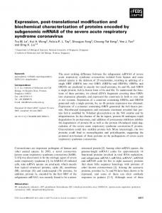
Figure 1. RT-PCR analysis of gene expressions of rabbit BM-MSCs on the first day of testing. Gene expressions of the loading group were analyzed after being subjected to 1 (L1), 2 (L2), and 4 (L4) hours of dynamic compressive loading and after 4 (R4), 8 (R8), and 20 (R20) hours of rest following 4-hour dynamic compressive loading. Gene expressions of the control group were analyzed at the beginning of the compression test. Total RNA was isolated and RT-PCR was performed on 1 μg of each sample using primers for c-Fos, c-Jun, Sox9, TßR-I, TßR-II, TGF-ß1, collagen II, and GAPDH as shown on the left. Bands shown are representatives of five independent experiments. Abbreviations: BM-MSC, bone-marrow mesenchymal stem cell; RT-PCR, reverse transcription–polymerase chain reaction.
Huang, Reuben, Cheung loading and then decreased from that peak after 4 hours of loading. Only the loading group exhibited a weak expression of c-Fos gene after 1 hour of loading (Fig. 1). After the 4-hour compression test, all gene expressions of the loading group decreased to a level similar to (i.e., c-Jun, type II collagen, TGF-ß1, and TßR-II) or slightly higher than (i.e., Sox9) those of the control group except that the expression of the TßR-I gene exhibited another peak after 8 hours of rest (Fig. 1).
Day 2 of Testing During the 4-hour compression test, all gene expressions of the loading group were upregulated. The gene expressions of c-Jun, Sox9, type II collagen, TßR-I, and TßR-II for the loading group gradually increased and reached the peak after 4 hours of loading, whereas the gene expression of TGF-ß1 quickly reached the highest level after 1 hour of loading (Fig. 2). Similar to the first day of testing, only weak expression of c-Fos gene was seen for the loading group after 1 hour of loading (Fig. 2). After the 4-hour compression test, gene expressions of c-Jun, Sox9, type II collagen, TGF-ß1, and TßR-I for the loading group gradually decreased to a level slightly higher than those of the control group, whereas TßR-I gene expression was similar to those of the control group (Fig. 2).
Figure 2. RT-PCR analysis of gene expressions of rabbit BM-MSCs on the second day of testing. Gene expressions of the loading group were analyzed after being subjected to 1 (L1), 2 (L2), and 4 (L4) hours of dynamic compressive loading and after 4 (R4), 8 (R8), and 20 (R20) hours of rest following 4-hour dynamic compressive loading. Gene expressions of the control group were analyzed at the beginning of the compression test. Total RNA was isolated and RT-PCR was performed on 1 μg of each sample using primers for c-Fos, c-Jun, Sox9, TßR-I, TßR-II, TGF-ß1, collagen II, and GAPDH as shown on the left. Bands shown are representatives of five independent experiments. Abbreviations: BM-MSC, bone-marrow mesenchymal stem cell; RT-PCR, reverse transcription–polymerase chain reaction.
1117
Statistical Analysis of Gene Expressions Day 1 of Testing Significant differences were found in all gene expressions between two experimental groups with the samples subjected to 2-hour dynamic compression having a higher level of expression than the control group (Figs. 3–5). After 4-hour dynamic compression, the loading group still exhibited significantly greater expression of type II collagen than the control (Fig. 5). Compared with the control group, the loading group also exhibited significantly higher levels of TßR-I and TßR-II gene expressions after 1-hour dynamic compression as well as higher expression of TßR-I gene after 8 hours of rest following 4 hours of dynamic compression (Fig. 3).
Day 2 of Testing Significant differences were found in all gene expressions between two experimental groups with the samples subjected to 4-hour dynamic compression having higher levels of expression than those of the control group (Figs. 3–5). For the gene expression of TGF-ß1, the samples subjected to 1- and 2-hour dynamic compression exhibited significantly higher levels of expression than the control group (Fig. 4). In addition, samples subjected to 2hour dynamic compression also exhibited a significantly greater
Figure 3. Statistical analysis of gene expressions of TGF-ß type I and type II receptors. The relative gene expressions of TGF-ß type I and type II receptors among six time points of 2 days of testing, as presented in Figures 1 and 2, were statistically analyzed using a one-way analysis of variance with Student-Newman-Keuls comparison test (n = 10 for control group; n = 5 for loading group). Gene expressions of the loading group were analyzed after being subjected to 1 (L1), 2 (L2), and 4 (L4) hours of dynamic compressive loading and after 4 (R4), 8 (R8), and 20 (R20) hours of rest following 4-hour dynamic compressive loading on each day of testing. Gene expressions of the control group were analyzed at the beginning of the compression test and the recovery period on each day of testing.
1118
expression of TßR-II gene than the control group (Fig. 3). However, no significant differences were found in all gene expressions between two experimental groups during the recovery period.
Gene Expressions of BM-MSCs Under Compression
Our recent study demonstrated that dynamic compression promoted expressions of chondrogenic markers and TGF-ß1 gene in rabbit BM-MSCs in a serum-free media, suggesting that the TGF-ß signaling pathway may be involved in this compressioninduced chondrogenesis [18]. To provide a better understanding of the mechanism linked to this finding, this study examined
the temporal expression patterns and the corresponding protein inductions of selected early responsive genes (c-Fos, c-Jun, Sox9, TßR-I, and TßR-II) that may be involved in chondrogenesis of BM-MSCs. It was found that the gene expressions of c-Fos, cJun, Sox9, TßR-I, and TßR-II were significantly upregulated, and the protein productions of c-Jun, Sox9, TßR-I, and TßR-II were induced in rabbit BM-MSCs by stimulation of dynamic compressive loading, whereas the temporal gene expression patterns differed among genes and between two testing days. Because TGF-ß signals through a heteromeric receptor complex of TßR-I and TßR-II, the presence of both receptors is an important indicator for TGF-ß signaling. The finding that dynamic compressive loading promoted gene expressions and protein productions of TßR-I and TßR-II in rabbit BM-MSCs suggests that TGF-ß signal transduction in BM-MSCs may be activated by dynamic compression. It also suggests that the upregulation of TGF-ß1, TßR-I, and TßR-II may result from an autocrine mechanism because TGF-ß1 is capable of regulating the expression of its ligand and receptors [31, 43, 44]. Furthermore, phosphorylated TßR-I protein can initiate the intracellular TGF-ß signal by activating Smad signaling [45], which may mediate chondrogenic differentiation of chondroprogenitor and mesenchymal cells [46]. This study found that the significant upregula-
Figure 4. Statistical analysis of gene expressions of TGF−ß1 and c-Jun. The relative gene expressions of TGF−ß1 and c-Jun among six time points of 2 days of testing, as presented in Figures 1 and 2, were statistically analyzed using a one-way analysis of variance with Student-Newman-Keuls comparison test (n = 10 for control group; n = 5 for loading group). Gene expressions of the loading group were analyzed after being subjected to 1 (L1), 2 (L2), and 4 (L4) hours of dynamic compressive loading and after 4 (R4), 8 (R8), and 20 (R20) hours of rest following 4-hour dynamic compressive loading on each day of testing. Gene expressions of the control group were analyzed at the beginning of the compression test and the recovery period on each day of testing.
Figure 5. Statistical analysis of gene expressions of Sox9 and type II collagen. The relative gene expressions of Sox9 and type II collagen among six time points of 2 days of testing, as presented in Figures 1 and 2, were statistically analyzed using a one-way analysis of variance with Student-Newman-Keuls comparison test (n = 10 for control group; n = 5 for loading group). Gene expressions of the loading group were analyzed after being subjected to 1 (L1), 2 (L2), and 4 (L4) hours of dynamic compressive loading and after 4 (R4), 8 (R8), and 20 (R20) hours of rest following 4-hour dynamic compressive loading on each day of testing. Gene expressions of the control group were analyzed at the beginning of the compression test and the recovery period on each day of testing.
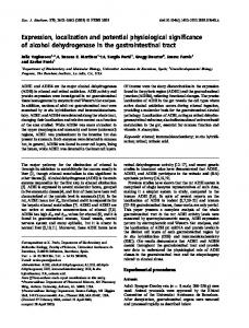
Typical Protein Induction In general, the protein inductions of c-Jun, Sox9, TGF-ß1, TßR-I, and TßR-II were seen in the samples subjected to 2- and 4-hour dynamic compression (Fig. 6). The protein inductions of c-Jun, Sox9, TßR-I, and TßR-II reached the highest levels in the loading group after 4 hours of loading and then decreased to a level similar to the control group after 4 hours of rest (Fig. 6). However, after the protein induction of TGF-ß1 in the loading group by the 2-hour dynamic compression, the protein level of TGF-ß1 was maintained at the similar level after 4-hour loading and during the 4-hour recovery period (Fig. 6).
Discussion
Huang, Reuben, Cheung tion of TßR-I and TßR-II gene expressions occurred earlier than that of type II collagen gene expression during the compression test. This finding supports the suggestion of our recent study [18] that TGF-ß signaling pathway may be involved in promoting chondrogenic gene expression of BM-MSCs under dynamic compressive loading. To our knowledge, this is also the first study to demonstrate the protein induction of TGF-ß1 in BM-MSCs by dynamic compression in a serum-free medium, suggesting that TGF-ß1 may play an important role in BM-MSC chondrogenesis promoted by dynamic compressive loading [18]. This study demonstrated that dynamic compressive loading upregulated gene expression and protein production of Sox9 in rabbit BM-MSCs. Takahashi et al. showed that static compressive loading promoted the expression of Sox9 gene in mouse embryonic limb bud mesenchymal cells [47]. Thus, Sox9 could be a responsive gene to mechanical loading for chondrogenic lineage. Furthermore, previous studies have demonstrated that Sox9 can bind directly to an enhancer element of the collagen II gene and enhance type II collagen gene expression [35–37] and also that mouse BM-MSC chondrogenesis can be promoted by overexpression of Sox9 gene [42]. Therefore, the upregulation of Sox9 gene expression and induction of its corresponding protein by dynamic compressive loading may contribute to the stimulation of type II collagen gene expression. c-Jun and c-Fos proteins can form a stable heterodimer (AP-1 complex) with a high affinity for the DNA target sequence. It has been shown that the AP-1 complex can positively regulate c-Jun [48, 49], whereas the binding of AP-1 complex to the promoter
Figure 6. Western blot analysis of corresponding protein inductions of c-Jun, Sox9, TGF-ß1, and TGF-ß receptors on the first day of testing. Protein inductions of the loading group were analyzed after being subjected to 2 (L2) and 4 (L4) hours of dynamic compressive loading and after 2 (R2) and 4 (R4) hours of rest following 4-hour dynamic compressive loading. Protein inductions of the control group were analyzed at the beginning of the compression test. Nuclear extract of NIH/3T3 mouse fibroblasts treated with phorbol, cell lysate of rabbit chondrocytes, recombinant protein of mouse TßR-I, fusion protein of human TßR-II, and cell lysate of human normal fibroblasts were used as positive controls (+) for c-Jun, Sox9, TßR-I, TßR-II, and TGF-ß1, respectively. Bands shown are representative of four independent experiments.
1119
regions of TGF-ß1 gene mediated autoinduction of TGF-ß1 [31]. This study found that the induction of c-Fos gene expression was detected earlier than the significant upregulation of c-Jun and TGF-ß1 gene expressions during the dynamic compression test. It may suggest that AP-1 complex was formed by dimerizing the newly synthesized c-Fos protein with the pre-existing c-Jun protein and then upregulated the expression of c-Jun and TGF-ß1 genes. Moreover, because the previous studies showed that TGFß1 treatment was able to activate gene expressions of c-Jun and c-Fos [49–51], TGF-ß1 upregulation by dynamic compression loading may be able to mediate transcription of c-Fos and c-Jun through feedback mechanism. Furthermore, because previous studies indicated that activity of the AP-1 complex may play an important role in regulating chondrocyte differentiation of chondrogenic cell lines [28, 30] and limb mesenchymal cells [29] as well as TGF-ß–induced type II collagen expression in chondrocytes [52], our finding also suggests that dynamic compressive loading may promote chondrogenic gene expressions of BMMSCs through activation of the AP-1 complex. Based on our findings, MAPK and Smad signal pathways may be activated in BM-MSCs by dynamic compressive loading. Firstly, c-Jun and c-Fos have been shown to be the downstream targets of stress-activated protein kinase/c-Jun N-terminal kinase (SAPK/JNK) and extracellular signal-regulated kinase (ERK) MAPK signal pathways, respectively [53]. SAPK/JNKs phosphorylated the transactivating domain of c-Jun protein that, in turn, regulated c-Jun expression [48, 53], whereas phosphorylation of ERK might cause induction of c-Fos [53]. The recent studies found that p38 and ERK MAPK pathways were involved in TGF-ß1–induced chondrogenesis of ATDC5 cells [54, 55], whereas Sox9 induction by fibroblast growth factors in chondrocytes and undifferentiated mesenchymal cells was mediated by the ERK MAPK pathway [56]. Moreover, it was recently demonstrated that the MAPK signal pathway was activated in cartilage by static compressive loading with a rapid induction of ERK and p38 MAPK pathways and a delayed stimulation of SAPK/JNK MAPK pathway [57]. Therefore, three major MAPK pathways could be activated in BM-MSCs by dynamic compressive loading. Secondly, the predominant downstream signaling protein of TGF-ß is Smad, which has been identified to relay the signal to the nucleus [45, 58]. The previous studies demonstrated that Smad proteins synergized in activating chondrogenic gene expression of mesenchymal and chondroprogenitor cells [46] and that Smad was involved in the regulation of cartilage-specific gene expression in chondrogenic ATDC5 cells after TGF-ß1 treatment [55]. Because our study suggests that dynamic compressive loading may induce chondrogenesis of BM-MSCs through TGF-ß signal transduction, Smad signaling may also play an important role in induction of chondrogenic gene expressions in BM-MSCs. This study found that the temporal expression patterns of the genes were different between two days of testing. For example,
1120
the expression of the genes reached the peak after the samples were subjected to 2-hour compressive loading on the first day of testing, whereas the peak expressions of the genes, except TGFß1, were found in the samples subjected to 4-hour compressive loading on the second day of testing. During the recovery period, most gene expressions in the loading group decreased from peak to the level that was similar to that of the control group on the first day of testing and, however, tended to be higher than that of the control group on the second day of testing. It may be possible that compressive loading on the first day of testing may change the initial condition of cells, such as deposition of extracellular matrix and protein accumulation of growth factors, receptors, and transcription factors, and then result in different responses of cells to compressive loading on the second day of testing. In summary, this study demonstrated temporal expression patterns of early responsive genes (Sox9, c-Fos, c-Jun, TßR−Ι, and TßR-II) and inductions of their corresponding proteins in
References 1
Gene Expressions of BM-MSCs Under Compression rabbit BM-MSCs with the stimulation of dynamic compressive loading, showing that the expressions of these genes were significantly upregulated by dynamic compressive loading. These findings suggest that the TGF-ß signal transduction and activities of AP-1 and Sox9 may be involved in the early stage of BM-MSC chondrogenesis promoted by dynamic compressive loading. The upregulation of these early responsive genes also indicates that dynamic compressive loading may activate MAPK and Smad signal pathways. Therefore, this study has established a basic model for future studies that will investigate mechanotransduction pathways in BM-MSCs in response to dynamic compressive loading.
Acknowledgments This work was supported by a National Institutes of Health grant (AR 38421) and a VA Merit Review Grant. The authors would like to thank Ms. Kristen Hagar for her technical assistance with RTPCR and cell culture.
articular cartilage through dynamic loading of chondrocyte-seeded agarose gels. J Biomech Eng 2000;122:252–260.
Cheung HS, Cottrell WH, Stephenson K et al. In vitro collagen biosynthesis in healing and normal rabbit articular cartilage. J Bone Joint Surg Am 1978;60:1076–1081.
13 Sah RL, Kim YJ, Doong JY et al. Biosynthetic response of cartilage explants to dynamic compression. J Orthop Res 1989;7:619–636.
2
Cheung HS, Lynch KL, Johnson RP et al. In vitro synthesis of tissue-specific type II collagen by healing cartilage. I. Short-term repair of cartilage by mature rabbits. Arthritis Rheum 1980;23:211–219.
14 Elder SH, Kimura JH, Soslowsky LJ et al. Effect of compressive loading on chondrocyte differentiation in agarose cultures of chick limb-bud cells. J Orthop Res 2000;18:78–86.
3
Friedman MJ, Berasi CC, Fox JM et al. Preliminary results with abrasion arthroplasty in the osteoarthritic knee. Clin Orthop 1984;182:200–205.
15 Elder SH, Goldstein SA, Kimura JH et al. Chondrocyte differentiation is modulated by frequency and duration of cyclic compressive loading. Ann Biomed Eng 2001;29:476–482.
4
Furukawa T, Eyre DR, Koide S et al. Biochemical studies on repair cartilage resurfacing experimental defects in the rabbit knee. J Bone Joint Surg Am 1980;62:79–89.
5
Johnson LL. Arthroscopic abrasion arthroplasty historical and pathologic perspective: present status. Arthroscopy 1986;2:54–69.
6
Levy AS, Lohnes J, Sculley S et al. Chondral delamination of the knee in soccer players. Am J Sports Med 1996;24:634–639.
7
O’Driscoll SW, Keeley FW, Salter RB. Durability of regenerated articular cartilage produced by free autogenous periosteal grafts in major full-thickness defects in joint surfaces under the influence of continuous passive motion. A follow-up report at one year. J Bone Joint Surg Am 1988;70:595–606.
8
9
Wakitani S, Goto T, Pineda SJ et al. Mesenchymal cell-based repair of large, full-thickness defects of articular cartilage. J Bone Joint Surg Am 1994;76:579–592. Buschmann MD, Gluzband YA, Grodzinsky AJ et al. Mechanical compression modulates matrix biosynthesis in chondrocyte/agarose culture. J Cell Sci 1995;108:1497–1508.
10 Guilak F, Meyer BC, Ratcliffe A et al. The effects of matrix compression on proteoglycan metabolism in articular cartilage explants. Osteoarthritis Cartilage 1994;2:91–101.
16 Takahashi I, Nuckolls GH, Takahashi K et al. Compressive force promotes sox9, type II collagen and aggrecan and inhibits IL-1beta expression resulting in chondrogenesis in mouse embryonic limb bud mesenchymal cells. J Cell Sci 1998;111:2067–2076. 17 Angele P, Yoo JU, Smith C et al. Cyclic hydrostatic pressure enhances the chondrogenic phenotype of human mesenchymal progenitor cells differentiated in vitro. J Orthop Res 2003;21:451–457. 18 Huang C-Y, Hagar CL, Frost LE et al. Effects of cyclic compressive loading on chondrogenesis of rabbit bone-marrow derived mesenchymal stem cells. Stem Cells 2004;22:313–323. 19 Huang C-Y, Reuben PM, D’Ippolito G et al. Chondrogenesis of human bone marrow-derived mesenchymal stem cells in agarose culture. Anat Rec 2004;278A:428–436. 20 Johnstone B, Hering TM, Caplan AI et al. In vitro chondrogenesis of bone marrow-derived mesenchymal progenitor cells. Exp Cell Res 1998;238:265–272. 21 Mackay AM, Beck SC, Murphy JM et al. Chondrogenic differentiation of cultured human mesenchymal stem cells from marrow. Tissue Eng 1998;4:415–428. 22 Pittenger MF, Mackay AM, Beck SC et al. Multilineage potential of adult human mesenchymal stem cells. Science 1999;284:143–147.
11 Lee DA, Bader DL. Compressive strains at physiological frequencies influence the metabolism of chondrocytes seeded in agarose. J Orthop Res 1997;15:181–188.
23 Solchaga LA, Johnstone B, Yoo JU et al. High variability in rabbit bone marrow-derived mesenchymal cell preparations. Cell Transplant 1999;8:511–519.
12 Mauck RL, Soltz MA, Wang CC et al. Functional tissue engineering of
24 Yoo JU, Barthel TS, Nishimura K et al. The chondrogenic potential of
Huang, Reuben, Cheung human bone-marrow-derived mesenchymal progenitor cells. J Bone Joint Surg Am 1998;80:1745–1757. 25 Wrana JL, Attisano L, Wieser R et al. Mechanism of activation of the TGF-beta receptor. Nature 1994;370:341–347. 26 Mizuta H, Sanyal A, Fukumoto T et al. The spatiotemporal expression of TGF-beta1 and its receptors during periosteal chondrogenesis in vitro. J Orthop Res 2002;20:562–574. 27 Oh CD, Chang SH, Yoon YM et al. Opposing role of mitogen-activated protein kinase subtypes, erk-1/2 and p38, in the regulation of chondrogenesis of mesenchymes. J Biol Chem 2000;275:5613–5619.
1121 stem cells. Biochem Biophys Res Commun 2003;301:338–343. 43 Inagaki M, Wang Z, Carr BI. Transforming growth factor beta 1 selectively increases gene expression of the serine/threonine kinase receptor 1 (SKR1) in human hepatoma cell lines. Cell Struct Funct 1994;19:305– 313. 44 Norgaard P, Spang-Thomsen M, Poulsen HS. Expression and autoregulation of transforming growth factor beta receptor mRNA in small-cell lung cancer cell lines. Br J Cancer 1996;73:1037–1043. 45 Massague J, Chen YG. Controlling TGF-beta signaling. Genes Dev 2000;14:627–644.
28 Thomas DP, Sunters A, Gentry A et al. Inhibition of chondrocyte differentiation in vitro by constitutive and inducible overexpression of the c-Fos proto-oncogene. J Cell Sci 2000;113:439–450.
46 Hatakeyama Y, Nguyen J, Wang X et al. Smad signaling in mesenchymal and chondroprogenitor cells. J Bone Joint Surg Am 2003;85(suppl 3):13– 18.
29 Tufan AC, Daumer KM, DeLise AM et al. AP-1 transcription factor complex is a target of signals from both WnT-7a and N-cadherin-dependent cell-cell adhesion complex during the regulation of limb mesenchymal chondrogenesis. Exp Cell Res 2002;273:197–203.
47 Takahashi I, Nuckolls GH, Takahashi K et al. Compressive force promotes sox9, type II collagen and aggrecan and inhibits IL-1beta expression resulting in chondrogenesis in mouse embryonic limb bud mesenchymal cells. J Cell Sci 1998;111:2067–2076.
30 Seghatoleslami MR, Tuan RS. Cell density dependent regulation of AP-1 activity is important for chondrogenic differentiation of C3H10T1/2 mesenchymal cells. J Cell Biochem 2002;84:237–248.
48 Angel P, Hattori K, Smeal T et al. The jun proto-oncogene is positively autoregulated by its product, Jun/AP-1. Cell 1988;55:875–885.
31 Kim SJ, Angel P, Lafyatis R et al. Autoinduction of transforming growth factor-β1 is mediated by the AP-1 complex. Mol Cell Biol 1990;10:1492– 1497.
49 Wong C, Rougier-Chapman EM, Frederick JP et al. Smad3-Smad4 and AP-1 complexes synergize in transcriptional activation of the c-Jun promoter by transforming growth factor beta. Mol Cell Biol 1999;19:1821– 1830.
32 Wright E, Hargrave MR, Christiansen J et al. The Sry-related gene Sox9 is expressed during chondrogenesis in mouse embryos. Nat Genet 1995;9:15–20.
50 Pertovaara L, Sistonen L, Bos TJ et al. Enhanced jun gene expression is an early genomic response to transforming growth factor beta stimulation. Mol Cell Biol 1989;9:1255–1262.
33 Zhao Q, Eberspaecher H, Lefebvre V et al. Parallel expression of Sox9 and Col2a1 in cells undergoing chondrogenesis. Dev Dyn 1997;209:377–386.
51 Okazaki R, Ikeda K, Sakamoto A et al. Transcriptional activation of cfos and c-jun protooncogenes by serum growth factors in osteoblast-like MC3T3-E1 cells. J Bone Miner Res 1992;7:1149–1155.
34 Bi W, Deng JM, Zhang Z et al. Sox9 is required for cartilage formation. Nat Genet 1999;22:85–89. 35 Bell DM, Leung KK, Wheatley SC et al. SOX9 directly regulates the typeII collagen gene. Nat Genet 1997;16:174–178. 36 Lefebvre V, Huang W, Harley VR et al. SOX9 is a potent activator of the chondrocyte-specific enhancer of the pro alpha1(II) collagen gene. Mol Cell Biol 1997;17:2336–2346. 37 Ng LJ, Wheatley S, Muscat GE et al. SOX9 binds DNA, activates transcription, and coexpresses with type II collagen during chondrogenesis in the mouse. Dev Biol 1997;183:108–121. 38 Bridgewater LC, Lefebvre V, de Crombrugghe B. Chondrocyte-specific enhancer elements in the Col11a2 gene resemble the Col2a1 tissue-specific enhancer. J Biol Chem 1998;273:14998–15006. 39 Liu Y, Li H, Tanaka K et al. Identification of an enhancer sequence within the first intron Required for cartilage-specific transcription of the α2(XI) collagen gene. J Biol Chem 2000;275:12712–12718. 40 Zhang P, Jimenez SA, Stokes DG. Regulation of human COL9A1 gene expression. Activation of the proximal promoter region by SOX9. J Biol Chem 2003;278:117–123. 41 Sekiya I, Tsuji K, Koopman P et al. SOX9 enhances aggrecan gene promoter/enhancer activity and is up-regulated by retinoic acid in a cartilagederived cell line, TC6. J Biol Chem 2000;275:10738–10744. 42 Tsuchiya H, Kitoh H, Sugiura F et al. Chondrogenesis enhanced by overexpression of sox9 gene in mouse bone marrow-derived mesenchymal
52 Miyazaki Y, Tsukazaki T, Hirota Y et al. Dexamethasone inhibition of TGF beta-induced cell growth and type II collagen mRNA expression through ERK-integrated AP-1 activity in cultured rat articular chondrocytes. Osteoarthritis Cartilage 2000;8:378–385. 53 Karin M. The regulation of AP-1 activity by mitogen-activated protein kinases. J Biol Chem 1995;270:16483–16486. 54 Nakamura K, Shirai T, Morishita S et al. p38 mitogen-activated protein kinase functionally contributes to chondrogenesis induced by growth/ differentiation factor-5 in ATDC5 cells. Exp Cell Res 1999;250:351–363. 55 Watanabe H, de Caestecker MP, Yamada Y. Transcriptional cross-talk between Smad, ERK1/2, and p38 mitogen-activated protein kinase pathways regulates transforming growth factor-beta-induced aggrecan gene expression in chondrogenic ATDC5 cells. J Biol Chem 2001;276:14466– 14473. 56 Murakami S, Kan M, McKeehan WL et al. Up-regulation of the chondrogenic Sox9 gene by fibroblast growth factors is mediated by the mitogen-activated protein kinase pathway. Proc Natl Acad Sci U S A 2000;97:1113–1118. 57 Fanning PJ, Emkey G, Smith RJ et al. Mechanical regulation of mitogen-activated protein kinase signaling in articular cartilage. J Biol Chem 2003;278:50940–50948. 58 Liberati NT, Datto MB, Frederick JP et al. Smads bind directly to the Jun family of AP-1 transcription factors. Proc Natl Acad Sci U S A 1999;96:4844–4849.