The osseous structures in the infratemporal fossa: foramen ovale, bony spurs, ossified ligaments and their contribution to the trigeminal neuralgia Konstantinos Natsis, Elpida Repousi, George Sofidis & Maria Piagkou
Acta Neurochirurgica The European Journal of Neurosurgery ISSN 0001-6268 Acta Neurochir DOI 10.1007/s00701-014-2197-3
1 23
Your article is protected by copyright and all rights are held exclusively by SpringerVerlag Wien. This e-offprint is for personal use only and shall not be self-archived in electronic repositories. If you wish to self-archive your article, please use the accepted manuscript version for posting on your own website. You may further deposit the accepted manuscript version in any repository, provided it is only made publicly available 12 months after official publication or later and provided acknowledgement is given to the original source of publication and a link is inserted to the published article on Springer's website. The link must be accompanied by the following text: "The final publication is available at link.springer.com”.
1 23
Author's personal copy Acta Neurochir DOI 10.1007/s00701-014-2197-3
LETTER TO THE EDITOR - NEUROSURGICAL ANATOMY
The osseous structures in the infratemporal fossa: foramen ovale, bony spurs, ossified ligaments and their contribution to the trigeminal neuralgia Konstantinos Natsis & Elpida Repousi & George Sofidis & Maria Piagkou
Received: 23 July 2014 / Accepted: 25 July 2014 # Springer-Verlag Wien 2014
Dear Editor, The etiology of the trigeminal neuralgia (TGN) varies. The paper of Liang et al. [2] was focused on the compression points of the trigeminal nerve intracranially. In addition, the entrapment of the mandibular nerve (MN) at the foramen ovale (FO) is a primary cause of the TGN. The incidence of TGN is higher on the right side [1, 4] and in females [1]. The FO is located in the infratemporal surface of the greater wing of the sphenoid bone, posterolateral to the foramen rotundum. It transmits the MN, the accessory meningeal artery, a venous plexus (connecting the cavernous sinus with the pterygoid venous plexus) and, sometimes, the lesser petrosal nerve [2, 9]. Occasionally, the FO may be compartmentalized by a bony spur situated antero-medially [5] or may be covered by bony bridges appearing after the partial or complete ossification of the pterygospinous (Pts) and pterygoalar (Pta) ligaments that are stretched between the lateral pterygoid plate and the greater wing of the sphenoid bone in the outer surface of the skull base [3] (Fig. 1). This study has not been presented to any congress K. Natsis : G. Sofidis Department of Anatomy, Medical School, Aristotle University of Thessaloniki, 54124 Thessaloniki, Greece K. Natsis e-mail:
[email protected] E. Repousi : M. Piagkou (*) Department of Anatomy, Medical School, National and Kapodistrian University of Athens, M. Asias 75, 11527 Athens, Greece e-mail:
[email protected] E. Repousi e-mail:
[email protected]
The FO morphometry and the ossified ligaments in the infratemporal fossa may cause pressure or mechanical irritation at the MN resulting in TGN [6]. With this letter we would like to share our experience regarding the FO morphometric details in 116 Greek adult dry (63 male and 53 female) skulls originated from the Departments of Anatomy of Medical Schools of Aristotle University (Thessaloniki) and National and Kapodistrian University (Athens). The antero-posterior length and transverse width of the FO were determined using a digital sliding caliper (0.01 mm precision). The surface area of the foramina was calculated using Radinsky’s formula (1967): (π×length×width) / 4. In addition the FO shape was classified into oval, almond, round and irregular types. All skulls were investigated in the infratemporal fossa for the existence of either an incomplete or complete Pts or Pta bar. The descriptive statistics are summarized in Table 1. The foramen was typically oval (62.1 % on the right and 49.1 % on the left), almond (24.1 % on the right and 14.7 % on the left), round (6.9 % on the right and 8.6 % on the left), and irregular (19.8 % on the right and 14.7 % on the left side). No correlation was found between the FO shape and the gender. Male skulls had a larger FO than the female (regarding the surface area, p=0.012 on the right and p = 0.04 on the left). The FO width was statistically significant smaller on the right side (p=0.02). Several authors mentioned that the right-sided foramina are narrower than the left-sided, but they found no statistically significant difference [5, 7]. Based on the topographic location of the structures passing through the FO, its width is the main parameter that affects the MN. The findings of our study contribute to the hypothesis
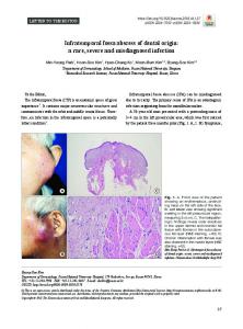
Author's personal copy Acta Neurochir Fig. 1 a The narrowing of the foramen ovale (FO) is apparent on the right side of the skull. b. The bony spur of the FO is apparent. c. The red arrow indicates the double bridging of the pterygoalar ligament’s ossification. d. The red arrow indicates the complete pterygospinous ligament’s ossification and the resulting pterygospinous foramen. LPP, lateral pterygoid plate
made by Neto et al. [4] according to which the TGN is more common on the right side, where the FO is narrower. Our finding regarding the narrowing of the foramen in female skulls (in which the width of the right-sided foramina is significant smaller than in male skulls, p=0.03) supports the fact that the TGN mainly affects women [1]. Few cases of TGN may appear as a result of the ossification of Pta and Pts ligaments [6] and the formation of FO bony spurs. In our sample, bony spurs were detected in 3.4 %. The complete Pta ligament ossification was detected in 4.9 % on the right and 1.8 % on the left side, while the incomplete ossified Pta ligament was observed in 20.7 % on the right and in 25 % on
Table 1 The descriptive statistics of the measured variables of the foramen ovale (FO)
FO dimensions
Length (in mm) Width (in mm) Surface area (in mm2)
the left side. The compression of the buccal, lingual, and auriculotemporal nerves by the ossified Pta ligament may result either in numbness, paresthesia, hypoesthesia, anaesthesia, and TGN [3]. In addition, the ossification of the Pts ligament (complete 1.8 % and incomplete 5.5 % on the right and 4.3 % on the left side) may result in TGN due to the entrapment of the MN branches [8]. Furthermore, these ossified ligaments may affect the blood vessels supplying the trigeminal ganglion leading to MN damage [6]. Thus, we estimate that the FO morphometry and the osseous structures in the infratemporal fossa should not be neglected in cases of TGN.
Mean±SD
Minimum
p-value
Maximum
Right
Left
Right
Left
Right
Left
7.2±1.2 3.9±0.7 22.1±5.6
7.0±1.1 4.1±0.8 22.9±5.6
5.0 3.0 12.0
4.0 2.0 10.0
13.0 6.0 48.0
10.0 7.0 37.0
0.106 0.02 0.093
Author's personal copy Acta Neurochir Conflict of interest None of the authors have any conflict of interest and any financial interest. 5.
References
6.
1. Bangash TH (2011) Trigeminal neuralgia: frequency of occurrence in different nerve branches. Anesth Pain 1:70–72 2. Liang L, Diao Y, Xu Q, Zhang M (2014) Transcranial segment of the trigeminal nerve: macro-/microscopic anatomical study using sheet plastination. Acta Neurochir (Wien) 156:605–612 3. Natsis K, Piagkou M, Skotsimara G, Totlis T, Apostolidis S, Panagiotopoulos NA, Skandalakis P (2014) The ossified pterygoalar ligament: an anatomical study with pathological and surgical implications. J Craniomaxillofac Surg 42:266–270 4. Neto HS, Camilli JA, Marques MJ (2005) Trigeminal neuralgia is caused by maxillary and mandibular nerve entrapment: greater inci-
7.
8.
9.
dence of right-sided facial symptoms is due to the foramen rotundum and foramen ovale being narrower on the right side of the cranium. Med Hypotheses 65:1179–1182 Ray B, Gupta N, Ghose S (2005) Anatomic variations of foramen ovale. Kathmandu Univ Med J (KUMJ) 3:64–68 Shaw JP (1993) Pterygospinous and pterygoalar foramina: a role in the etiology of trigeminal neuralgia? Clin Anat 6:173–178 Somesh MS, Sridevi HB, Prabhu LV, Swamy MS, Krishnamurthy A, Murlimanju BV, Chettiar GK (2011) A morphometric study of foramen ovale. Turk Neurosurg 21:378–383 Tubbs RS, May WR Jr, Apaydin N, Shoja MM, Shokouhi G, Loukas M, Cohen-Gadol AA (2009) Ossification of ligaments near the foramen ovale: an anatomic study with potential clinical significance regarding transcutaneous approaches to the skull base. Neurosurgery 65:60–64 Williams PL, Bannister LH, Berry MM, Collin P, Dyson M, Dussek JE, Ferguson MWJ (1995) Gray’s anatomy. Churchill Livingstone, New York