RESEARCH ARTICLE
Transient but Significant Visual Field Defects after Robot-Assisted Laparoscopic Radical Prostatectomy in Deep Trendelenburg Position Yukako Taketani1,2, Chihiro Mayama1*, Noriyuki Suzuki3, Akiko Wada4, Tatsuhiro Oka4, Kazuya Inamochi2,5, Yohei Nomoto2 1 Department of Ophthalmology, The University of Tokyo Graduate School of Medicine, Tokyo, Japan, 2 Department of Ophthalmology, Asahi General Hospital, Chiba, Japan, 3 Department of Urology, Asahi General Hospital, Chiba, Japan, 4 Department of Anesthesiology, Asahi General Hospital, Chiba, Japan, 5 Department of Ophthalmology, Tokyo Metropolitan Police Hospital, Tokyo, Japan *
[email protected]
Abstract Background OPEN ACCESS Citation: Taketani Y, Mayama C, Suzuki N, Wada A, Oka T, Inamochi K, et al. (2015) Transient but Significant Visual Field Defects after Robot-Assisted Laparoscopic Radical Prostatectomy in Deep Trendelenburg Position. PLoS ONE 10(4): e0123361. doi:10.1371/journal.pone.0123361 Academic Editor: Bang V Bui, University of Melbourne, AUSTRALIA Received: December 2, 2014 Accepted: March 3, 2015 Published: April 23, 2015 Copyright: © 2015 Taketani et al. This is an open access article distributed under the terms of the Creative Commons Attribution License, which permits unrestricted use, distribution, and reproduction in any medium, provided the original author and source are credited. Data Availability Statement: All relevant data are within the paper. Funding: The authors received no specific funding for this work. Competing Interests: The authors have declared that no competing interests exist.
Robot-assisted laparoscopic radical prostatectomy (RALP) is a minimally invasive surgical procedure for prostate cancer. During RALP, the patient must be in a steep Trendelenburg (head-down) position, which leads to a significant increase in intraocular pressure (IOP). The association of RALP with visual field sensitivity, however, has not been prospectively studied. The purpose of this study was to evaluate prospectively the visual field, retinal nerve fiber layer (RNFL) thickness, and optic disc morphology in 50 normal eyes of 25 male patients that underwent RALP.
Methods The subjects were 25 males among 33 consecutive patients who underwent uneventful RALP under general anesthesia in our hospital. Visual field tests using the Humphrey visual field analyzer 30-2 SITA-standard program were performed before, 7 days after, and 1-3 months after RALP. IOP was measured before, during, and after RALP; and ophthalmologic examinations, including slit-lamp, fundus examination, and optical coherence tomography (OCT), were scheduled before and 7 days after surgery.
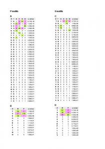
Results IOP was significantly increased during RALP up to 29.4 mmHg (P