tracking of the cross-sectional area of the IJV in ultrasound videos with different image ... and is actively deformed to minimize a pre-defined energy function.
Ultrasound Image Segmentation Techniques for Tracking and Measurement of the Internal Jugular Vein Ebrahim Karami1, Mohamed Shehata1, Peter Mcguire2, and Andrew Smith3 1
Faculty of Engineering and Applied Sciences, Memorial University, Canada 2 C-CORE, St. John’s, Newfoundland and Labrador, Canada 3 Faculty of Medicine, Memorial University, Canada for 2D medical images. AC models the desired contour as a time evolving curve and the segmentation process as an optimization over time of an adequate energy functional. An AC-based algorithm starts its operation with an initial contour and is actively deformed to minimize a pre-defined energy function. The minimum energy of the AC is supposed to be achieved at the desired border of the target object while neglecting gaps in the vessel wall which is a common feature in ultrasound imagery. The result of any AC algorithm is very dependent to the initial contour which is usually selected by an operator. In [10], AC technique and its combination with speckle tracking were proposed for the segmentation of the IJV images. In [10], the initial contour points for the first frame are manually selected by an experts and for the next frames, speckle tracking is used to move the control points of the AC (snake) along the motion of the edges of the IJV. This technique is efficient only if the deformation from one frame to the next one is negligible and consequently it requires a ultrasound device with a high frame rate. In this paper, we propose an AC-based algorithm which uses region growing technique to initialize the AC. Region growing is used to grow a region around some seed pixels and obtain an initial contour for the AC algorithm [11]. The initial seed pixel for the first frame is manually chosen by the operator and for the next frame, it is considered to be the centroid of the contour in the previous frame. Therefore, the only restriction on the proposed algorithm is that the centroid of the IJV must not have large displacement in two consecutive frames which is a very reasonable assumption. The rest of this paper is organized as follows: Section II presents the technical detail of the proposed algorithm. Section III reports experiments along with the results from the proposed algorithm and comparison of them with manual segmentation and the algorithms in [10]. Section IV concludes the paper.
Abstract--Estimation of circulating blood volume is important for the management of acute and chronic diseases. Both excessive and insufficient fluid administration have been shown to result increased morbidity and mortality. Studies have shown that the circulating blood volume is correlated to the cross-sectional area (CSA) of the internal jugular vein (IJV) which can be estimated from its ultrasound images. In this paper, an efficient active contour-based algorithm is proposed for segmentation of the IJV. In the proposed algorithm, region growing is used to obtain an initial contour for each frame and then an active contour is used for its fine adjustments. The proposed algorithm was applied for segmentation and tracking of the cross-sectional area of the IJV in ultrasound videos with different image quality and different IJV shapes and quality and results of the validation experiments showed that the algorithm performs very accurately compared to expert manual segmentation, as considered as ground truth, and efficiently tracks the changes in the cross-sectional area of the IJV. The results from the proposed algorithm are also compared to current state of the art algorithms.
I. INTRODUCTION Estimation and tracking of circulating blood volume is very important for assessment of the status of patients and is useful to prevent fluid overload [1-2]. Traditional methods of blood volume assessment are invasive and they are also recognized as poor predictors of fluid-responsiveness [3]. In recent year, ultrasound images of inferior vena cava (IVC) have been used to assess volume status and guide fluid administration [4]. However, this approach requires repeated measures over time by a skilled operator with image quality often limited by patient factors such as body habitus and bowel gas. Furthermore, Since IVC is located deep inside the chest; its ultrasound images do not have good quality. Ultrasound is extensively used for localization of the internal jugular vein (IJV) for replacement of the central venous catheter [5-6]. Recently, it has been shown that central venous pressure (CVP) is correlated with the cross-sectional area (CSA) of the IJV and hence the circulating blood volume can be estimated from the ultrasound images of the IJV [7-8]. Since the IJV is located in the neck, its ultrasound images are much clearer than the ones for the IVC and can provide more accurate results. Active Contour (AC), also called snakes, was proposed by Kass et al [9] and is a popular interactive segmentation method
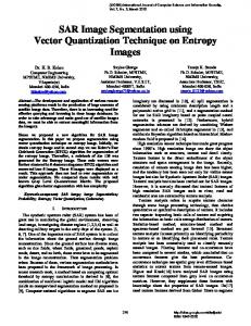
II. PROPOSED ALGORITHM The flow chart of the proposed algorithm is shown in Fig. 1. The proposed algorithm has the following steps: 1. Each frame of the captured video is preprocessed with a 7×7 median filter followed by a Gaussian filter to remove noises.
1
2. The initial seed pixel of the first frame is selected manually by the operator. For the next frames, the initial seed point is assumed to be the centroid of the contour in the previous frame. The centroid is calculated using first-order moments of the contour points as x c xI ( x, y ), x, yC
2
where A(C ) is the area of the contour which can be calculated as follows:
x, yC
A(C )
This approach is practical only if the relative motion between the ultrasound probe and the centroid of the region of interest is not significant, which is a reasonable assumption. 3. The quality of each frame is enhanced using an 8×8 adaptive histogram equalizer. This improves performance of the region growing. 4. A region is grown starting the initial seed point obtained from Step 2. Region growing is the simplest region-based image segmentation technique [11]. The concept of region growing algorithm is to choose an initial seed pixel and grow a region based on some feature such as intensity, gradient direction, or gradient magnitude. In this paper, we consider the intensity as the region growing feature. The region is iteratively grown by comparing all unallocated 8-connected neighboring pixels to the region such that if the difference between intensity of a candidate pixel and the mean intensity of the region is less than some threshold T, then we insert that candidate pixel to the region and continues the process until there are no unchecked candidate pixels. In this paper, we set the threshold as T 0.05( I max I min ) where I max and I min are maximum and minimum intensities of the frame, respectively. 5. Region growing provides a coarse contour with large number of contour points. In this step, we use cubic spline interpolation to obtain N =32 equidistant contour points. Without this resampling the performance of active contour algorithm will be very poor, because active contour do not work properly if the initial contour points are so close to each other. The contour generated by region growing is always underestimating the actual area of the IJV. 6. In this Step, the AC algorithm is used to move the contour points along the edges of the IJV and provide a continuous and smooth contour. The energy function in the proposed algorithm is defined as 1
1
0
0
E (C ) Einternal(C )dn Eimage (C )dn Econst (C ) ,
1 N ( x(n 1, t ) x(n 1, t ))( y(n 1, t ) y(n 1, t )) . (6) 2 n1
The energy function of the AC is minimized as x t (B I) 1 (x t 1 t f x (x t 1 , y t 1 ) wc Ax (Ct 1 )) y t (B I) 1 (y t 1 t f y (x t 1 , y t 1 ) wc Ay (Ct 1 ))
Image Capture
Preprocessing
1st frame
The initial seed pixel is the centroid of the previous frame
The operator selects the initial seed pixel by mouse clicking on center of the IJV
Adaptive equalization
Region growing
Contour points resampling
(2) Active contour
curve to the image edges and is defined as 2
I ( x, y) ,
,
(7)
where vectors x t and y t indicate x and y coordinates of the contour points at iteration t, subscripts x and y indicate gradient versus them, B is the penta-diagonal matrix defined in [9], 2000 , and t 0.98 t .
where Eimage (C ) is the energy term which is attracting the
Eedge
(4)
and Econst (C ) is the energy term related to any problem specific constraint and is defined as (5) Econst (C) 2( I ( x, y) 50) A(C) ,
(1)
y c yI ( x, y ).
2
Einternal (C ) 2 C (n) 2 C ' (n) ,
Figure 1: Block diagram of the proposed algorithm.
(3)
Einternal(C ) is the energy term which forces the contour to keep its shape as regular as possible and is defined as
2
Figure 2: Tracking of the IJV contour in different frames of the video captured at tilt angle 0 degree for manual segmentation, the proposed algorithm, and two algorithms proposed in [10].
IJV in the videos captured with an ultrasound machine with frame rate 30 fps1. Fig. 3 presents the DICE factor between the proposed algorithm and two algorithms in [10] with manual segmentation versus frame index for videos captured at tilt angles 0. One can see that the DICE coefficient of the proposed algorithm is always larger than 0.89 percent with mean value 0.93 while the algorithms in [10] only perform well at initial frames and they lose tracking after that. Fig. 4 presents the CSA of the IJV versus frame index for the manual segmentation, the proposed algorithm, and the algorithms in [10]. One can easily see that the proposed algorithm continuously follows the manual segmentation but always underestimates it. In other words, the relative variations in the CSA of the IJV estimated by the manual segmentation and the proposed algorithm are very close. On the other hand, the CSA estimated from the proposed algorithm seems smoother than the one from the manual segmentation which is due to the lack of manual error introduced by the natural fluctuations of the hand of the expert who extracts the manual segmentation. Figs. 5-7 present the same results when the head of the bed elevated at 30 degrees, where we expect lower CSA with larger fluctuations. From Fig. 5, one can see that the proposed algorithm efficiently follows the variations of the IJV contour while both algorithms presented in [10] lose tracking after initial frames. From Fig. 6, one can see that the minimum and average values for the DICE coefficient of the proposed algorithm are 0.88 and 0.95 while these values for the algorithms in [10] are even worse than the previous scenario. These results are also verified with Fig. 7. In both investigated scenarios, the proposed algorithm outperformed the algorithms in [10].
III. EXPERIMENT AND RESULTS The proposed algorithm was applied to track the area of two videos captured by ultrasound equipment Sonosite M-Turbo with a linear array transducer with frequency 6-15 MHz, frame rate 30 fps, and scan depth 6cm. Each video includes 450 frames corresponding to 15 seconds of data. Experimental data was collecting using healthy subjects supine and with head of the bed elevated at 0 and 30 degrees to simulate different volume status. The performance of the proposed algorithm is compared with the manual segmentation and the algorithms in [10]. Evaluation of extraction The accuracy of the proposed algorithm was evaluated by computing the DICE coefficient as the level of agreement between the areas extracted by the algorithm and the manual segmentation made by an expert. DICE coefficient S is defined as 2A M S , (8) A M where A and M are the CSA of the IJV estimated from the algorithm and the manual segmentation, respectively, and A M is intersection of them. Fig. 2. shows the contour obtained through different segmentation schemes and their tracking for different frame indices. From the second row of the Fig. 2, one can see that the proposed algorithm properly tracks the actual contour of the IJV, but it always underestimates the area. The third row of the Fig. 2 presents the result from the AC algorithm investigated in [10] which always overestimates the area and its error gradually increases over time. From the last row of Fig. 2, one can easily see that the speckle tracking-based AC algorithm presented in [10] cannot follow the variations in the
1 In [10], the frame rate is 69 fps and therefore the variations from one frame to the next one are less than our scenario.
3
Figure 4: The CSA of the IJV from the manual segmentation, the proposed algorithm, and the algorithms in [10] for ultrasound video captured at title angle 0 degree.
Figure 3: The DICE factor for the proposed algorithm and the algorithms in [10] for ultrasound video captured at title angle 0 degree.
Fig. 5: Tracking of the IJV contour in different frames of the video captured at tilt angle 30 degree for manual segmentation, the proposed algorithm, and two algorithms proposed in [10].
4
Figure 6: The DICE factor for the proposed algorithm and the algorithms in [10] for ultrasound video captured at title angle 30 degree.
Figure 7: The CSA of the IJV from the manual segmentation, the proposed algorithm, and the algorithms in [10] for ultrasound video captured at title angle 30 degree. venous pressure measurements improve the accuracy of leg raisinginduced change in pulse pressure to predict fluid responsiveness,” Intensive Care Med, vol. 36, no. 6, pp. 940-948, 2010. [4] J. Fields, P. Lee, K. Jenq, D. Mark, N. Panebianco and A. Dean, “The Interrater Reliability of Inferior Vena Cava Ultrasound by Bedside Clinician Sonographers in Emergency Department Patients,” Academic Emergency Medicine, vol. 18, no. 1, pp. 98-101, 2011. [5] B. Miller, “Locating the Right Internal Jugular Vein Using Ultrasound Is Different than Ultrasound Guidance or Ultrasound Confirmation of Right Internal Jugular Vein Cannulation,” Anesthesia & Analgesia, vol. 110, no. 3, pp. 974-975, 2010. [6] J. Ullman and R. Stoelting, “Internal Jugular Vein Location with the Ultrasound Doppler Blood Flow Detector,” Anesthesia & Analgesia, vol. 57, no. 1, p. 118, 1978. [7] U. Baumann, C. Marquis, C. Stoupis, T. Willenberg, J. Takala and S. Jakob, “Estimation of central venous pressure by ultrasound,” Resuscitation, vol. 64, no. 2, pp. 193-199, 2005. [8] J. Bailey, J. McCall, S. Smith and R. Kagan, “Correlation of Internal Jugular Vein/Common Carotid Artery Ratio to Central Venous Pressure,” Journal of Burn Care & Research, vol. 33, no. 1, pp. 89-92, 2012. [9] M. Kass, A. Witkin and D. Terzopoulos, “Snakes: Active contour models,” Int J Comput Vision, vol. 1, no. 4, pp. 321-331, 1988. [10] K. Qian, T. Ando, K. Nakamura, H. Liao, E. Kobayashi, N. Yahagi and I. Sakuma, “Ultrasound imaging method for internal jugular vein measurement and estimation of circulating blood volume,” Int J CARS, vol. 9, no. 2, pp. 231-239, 2013. [11] E. Karami, M. Shehata, P. McGuire, and A. Smith, “A semi-automated technique for internal jugular vein segmentation in ultrasound images using active contours,” in 2016 IEEE-EMBS International Conference on Biomedical and Health Informatics (BHI). IEEE, 2016, pp. 184–187.
IV. CONCLUSION In this paper, an active contour (AC) based segmentation algorithm was proposed for estimation and tracking of the cross-sectional area of the IJV from ultrasound images captured at rate 30 fps. In the proposed algorithm, the AC is initialized using a region growing algorithm which grows a region around an initial seed point and forms an initial but coarse contour for the AC algorithm. The proposed algorithm was applied to two ultrasound videos with frame rate 30 fps. The validation experiment showed that the proposed algorithm performs very close to the manual segmentation made by an expert and while the algorithms proposed in [10] lose tracking after initial frames. REFERENCES [1] T. Kudo, S. Suzuki and T. Iwabuchi, “Importance of monitoring the circulating blood volume in patients with cerebral vasospasm after subarachnoid hemorrhage”, Neurosurgery, vol. 9, no. 5, pp. 14-20, 1981. [2] Y. Sano, A. Sakamoto, Y. Oi and R. Ogawa, “Anaesthesia and circulating blood volume,” European Journal of Anaesthesiology, vol. 22, no. 4, pp. 258-262, 2005. [3] K. Lakhal, S. Ehrmann, I. Runge, D. Benzekri-Lefèvre, A. Legras, P. Dequin, E. Mercier, M. Wolff, B. Régnier and T. Boulain, “Central
5