Neuron
Article Using the Structure of Inhibitory Networks to Unravel Mechanisms of Spatiotemporal Patterning Collins Assisi,1,* Mark Stopfer,2 and Maxim Bazhenov1 1Department
of Cell Biology and Neuroscience, University of California, Riverside, Riverside, CA 92521, USA Institutes of Health, Eunice Kennedy Shriver National Institute of Child Health and Human Development, Bethesda, MD 20892-2425, USA *Correspondence:
[email protected] DOI 10.1016/j.neuron.2010.12.019 2National
SUMMARY
Neuronal networks exhibit a rich dynamical repertoire, a consequence of both the intrinsic properties of neurons and the structure of the network. It has been hypothesized that inhibitory interneurons corral principal neurons into transiently synchronous ensembles that encode sensory information and subserve behavior. How does the structure of the inhibitory network facilitate such spatiotemporal patterning? We established a relationship between an important structural property of a network, its colorings, and the dynamics it constrains. Using a model of the insect antennal lobe, we show that our description allows the explicit identification of the groups of inhibitory interneurons that switch, during odor stimulation, between activity and quiescence in a coordinated manner determined by features of the network structure. This description optimally matches the perspective of the downstream neurons looking for synchrony in ensembles of presynaptic cells and allows a low-dimensional description of seemingly complex high-dimensional network activity. INTRODUCTION Coordinated spiking of neuronal populations underlies the perception of sensory stimuli (Gray and Singer, 1989), the planning of movement (Bouyer et al., 1987), and the acquisition of new memories (Cheng and Frank, 2008). A number of studies have shown that such patterning, to a large extent, depends on the temporal and the spatial distribution of inhibition (Buzsa´ki and Chrobak, 1995). For example, in hippocampal networks, GABAergic interneurons form the hubs of a network that selectively synchronizes subpopulations of pyramidal cells (Bonifazi et al., 2009). The back-and-forth interaction between inhibitory granule cells and excitatory mitral cells in the mouse olfactory bulb leads to synchronous spiking in mitral cells (Schoppa, 2006). Synchrony extends across mitral cells that are electrically uncoupled and affiliated with different glomeruli. Feedback inhibition mediated by local interneurons synchronizes cortical pyramidal cells in the gamma frequency band underlying cogni-
tive processing and provides a mechanism for the temporal binding of sensory stimuli (Joliot et al., 1994; Llina´s and Ribary, 1993; Singer and Gray, 1995). In vertebrates (Grillner, 2003) and invertebrates (Marder and Bucher, 2007), coordinated movement is achieved by interneuron networks that work in concert to generate appropriate phases of spiking in motor neurons. The antennal lobe (AL), the insect equivalent of the olfactory bulb in mammals, provides an ideal system where the effects of inhibitory networks can be examined. Local inhibitory interneurons (LNs) extend extensive connections to each other and excitatory projection neurons (PNs) in the AL (Leitch and Laurent, 1996). Odor-driven activity of PNs in the AL evolves over multiple spatial and temporal scales (Laurent, 2002). The collective spiking activity of PNs generates an oscillatory local field potential (LFP) (Laurent and Davidowitz, 1994). The composition of synchronized groups of PNs contributing to the LFP oscillation changes on a cycle-by-cycle basis (Laurent and Davidowitz, 1994; Laurent et al., 1996; Wehr and Laurent, 1996). PNs receive input from local LNs. Blocking fast LN-mediated inhibition by the application of the GABA-activated chloride channel blocker picrotoxin leads to the desynchronization of PNs and consequently abolishes the oscillatory output of the AL (Ito et al., 2009; MacLeod and Laurent, 1996; Stopfer et al., 1997; Tanaka et al., 2009). Since dynamic changes in collective PN activity during odor stimulation exceed changes in input to the network, they must be attributed to the network interactions within the AL (Raman et al., 2010). Previously, we proposed that competitive inhibitory interactions between LNs generate alternately spiking groups of neurons. These groups entrain different populations of PNs, shaping their spike timings and generating a spatiotemporal representation of an odor (Bazhenov et al., 2001b). The basic cause of transient synchrony in PNs, recovery from concerted inhibition, is well understood in insect (MacLeod and Laurent, 1996; Stopfer et al., 1997) (Bazhenov et al., 2001b) and mammalian (Schoppa, 2006) olfactory circuits. However, a clear relationship between the global structure of the inhibitory network and the collective dynamics of PNs and LNs is not known. An understanding of structure-dynamics relationships in a network is often challenging because of the formidable dimensionality of the system and the inherent nonlinear properties of neurons and synapses that constitute the network. Recent approaches have been restricted to two broad classes (Boccaletti et al., 2006). The first class examines complex dynamics, albeit in relatively simple networks. These networks Neuron 69, 373–386, January 27, 2011 ª2011 Elsevier Inc. 373
Neuron Structure-Dynamics Relationship in Neural Networks
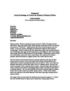
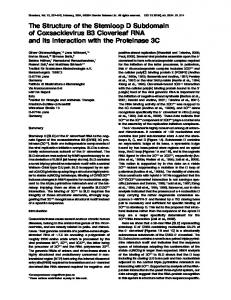
Figure 1. Clustering in Inhibitory Networks as a Function of Graph Coloring
B 50 mV
in vivo
model
100 ms
[Ca] 0.05 mM
A
1s
E
50 mV
000011111111 000011111111 000011111111 000011111111 111100011111 111100011111 111100011111 111111100000 111111100000 111111100000 111111100000 111111100000
10 neurons
D
C
500 ms
(A) A reciprocally connected pair of inhibitory neurons is an example of a graph with chromatic number two. Left traces: An alternating pattern of bursts is generated in response to a constant external stimulus to both neurons. A Ca2+-dependent potassium current (shown in red) causes spike frequency adaptation. Right traces: In the absence of a Ca2+-dependent potassium current, only one neuron produces spikes and the other is quiescent. Blue trace at the bottom: Spike frequency adaptation in a local inhibitory interneuron recorded in vivo from locust AL. (B) A coloring generated for a random network of 20 neurons with connection probability 0.5. (C) A graph with chromatic number three and its corresponding adjacency matrix. (D) Raster plot showing the activity of a network of 30 neurons with chromatic number three. Ten neurons are associated with each color. (E) The role of Ca2+ concentration on the timing of LN bursts. The bottom traces show the Ca2+ concentration in three LNs (top three traces) associated with three different colors. The neuron with the lowest concentration of Ca2+ tends to spike first.
[Ca]2+
0.1 mM
waves, a predictable and simple pattern where synchronous ensembles of excitatory projecting cells are successively recruited.
200 ms
(often consisting of a few [