computationally efficient vector field convolution to enhance ... The automated software Tree2Tree ... external force field to guide the active contour toward the.
VECTOR FIELD CONVOLUTION MEDIALNESS APPLIED TO NEURON TRACING Suvadip Mukherjee and Scott T. Acton Electrical & Computer Engineering, University of Virginia, Charlottesville, VA USA. ABSTRACT In this paper we propose a novel approach to the extraction of medial axis for grayscale objects. The method utilizes a computationally efficient vector field convolution to enhance the medialness feature. Local maxima of medialness are analyzed in scale space, yielding a robust medial axis for grayscale imagery. An important application of this work is the segmentation of neurons from noisy, cluttered microscopy images. Existing neuron segmentation methods depend heavily on accurate, noise-insensitive medial axis extraction. We propose the vector field convolution medialness operation as a first step in segmenting neurons. The proposed method requires no complex parameters or an initial binarization step. The efficacy of the method is demonstrated by a 60% reduction root mean squared error (2.9 pixels) as compared to an approach based on gradient vector flow. Index Terms— skeleton, VFC, neuron segmentation, microscopy 1. INTRODUCTION Equipped with state of the art imaging technology and adequate computational resources, biological image analysis is a field which is gaining popularity by the day. An important challenge in this field is to be able to trace neurons from light microscopy images. The typically low signal-to-noise ratio (SNR) obtained in confocal microscopy of the neurons poses difficulties, especially in achieving complete automation. The automated software Tree2Tree [2], [3] aims to create a neuronal morphology graph from confocal microscope images of single Drosophila neurons. Tree2Tree treats the segmentation problem in a graph theoretic framework, relying on an initial segmentation of the neuronal structures to create local subtrees. The connectivity between the subtrees is established by a specialized graph connectivity algorithm that connects the local medial trees (representing neurites, i.e., axons or dendrites or portions thereof) to create the global neuronal tree of a single neuron. The local medial trees are obtained by generating a medial graph from the medial axis of the disjoint connected components. Efficacy of the tracing
procedure thus largely depends on the initial segmentation and the quality of the obtained medial axis. The distance transform is a popular technique to extract the medial axis of binary objects [5]. The distance transform of an object calculates the minimum distance of an object point from the boundary. This method is sensitive to small perturbations on the object boundary, creating unwanted artifacts in the object skeleton. Methods based on electrostatic field theory [6] or Poisson solvers [7] avoid the above mentioned problem but depend heavily on proper segmentation of the foreground objects. The diffusion based method in [8] depends on a user selected parameter to control the diffusion. The “all path pruning” method by Peng et al. [1] traces a neuron by using a shortest path approach, but does not guarantee the traced path to be along the medial axis of the neuron structure. Consequently, the method requires manual editing to rectify the traced path to the medial axis. Gradient vector flow (GVF) [9] methods have been used to extract the medial axis of grayscale objects [10]. Wang et al. used GVF to drive an open curve snake to trace neurons [4]. Originally designed to be an external force to guide active contours, Gradient Vector Flow field can be used to extract the medial axis of objects. However, GVF suffers from significant computational cost and is sensitive to change in parameters. In this paper, we propose a novel method to compute the “medialness” function of a grayscale object using vector field convolution (VFC) [11], which is computationally efficient and robust to noise. We introduce VFC in Sec. 2, followed by the discussion of our algorithm in Sec. 3. Sec. 4 focuses on application in neuron segmentation, followed by experimental results in Sec. 5. 2. VECTOR FIELD CONVOLUTION A parametric active contour is an energy minimizing spline �(�), � ∈ [0,1] that attempts to minimize the following energy functional: �=
1 � � (�|�� (�)|� + �|��� (�)|� ) + ���� (�(�)) �� 2 �
(1)
The contour satisfies the Euler-Lagrange equation at the local minima which can be written down in terms of a force
balance equation involving an internal force ���� that regulates the smoothness and tautness of the active contour and an image dependent external force ���� : ���� ��(�)� + ���� ��(�)� = 0
(2)
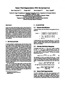
The external force pulls the contour toward the desired portions. VFC creates an external force field having large capture range with a reduced computational cost. For object segmentation, the edge-map of the object often provides the required feature of interest (FOI). The object edge-map convolved with the VFC kernel produces the desired external force field to guide the active contour toward the FOI. The vector field kernel (!, ") is so designed that the vectors point toward the center of the kernel (Fig. 1).
(a)
(b)
(c)
(d)
Figure 2: (a) A synthetic object; (b) the extracted medial axis in red; (c) the vector field obtained after convolving (a) with VFC kernel; (d) zoomed portion showing the medial axis.
Figure 1: A discrete VFC kernel.
3.1. Mathematical formulation
Formally, we define the kernel as (!, ") = $(!, ")%(!, ")
vector field is attracted to the feature of interest, if such a feature is within the capture range of the kernel. At the correct scale, convolution of an image with the VFC kernel produces a zero magnitude vector at the points of symmetry, since the vectors cancel each other. The points of symmetry of an object can be recognized by finding the local minima in the convolved image (assuming the object is brighter than the background). The vector field obtained by convolving the synthetic image in Figure 2(a) is shown in Figure 2 (c) and (d). The vector field has zero magnitude at the object medial axis, due to balancing of the vectors.
(3)
%(!, ") = ['!/), '"/)]* is the unit vector at position (x,y) pointing to the kernel origin and ) = +! � + " � . Two magnitude functions were introduced to create the VFC kernel. (4) $� (!, ") = ) ,$� (!, ") = exp(') � /1 � ) (5) Parameters 4 and 1 determine the vector magnitudes. VFC enjoys some advantages over popular methods like GVF [9] in terms of reduced computational expense, reduced sensitivity to noise and reduced dependence on paramaters. 3. VECTOR FIELD CONVOLUTION MEDIALNESS An object medial axis provides important information in image analysis, by providing a simple representation of its shape. In neuro-biological image analysis problems, skeleton extraction is often the initial step in establishing the morphology of neurons [1], [2], [4]. In this section, we introduce a novel medial axis extraction technique using VFC [11]. Our apporach satisfies the demands of a good quality medial axis extraction techniques in terms of low sensitivity to noise and low computational cost. A discrete VFC kernel is shown in Fig.1. The design of the kernel is such that a contour placed in the
(a)
(b)
(c)
Figure 3: (a) A simulated object; (b) corresponding VFC medialness image; (c) skeleton in red.
Let 5: 7 8 9 → ; be a digital grayscale image of dimension < 8 = (7 = >0, … , < ' 1@, 9 = >0, … , = ' 1@). We define the vector field obtained after performing vector field convolution as AB (!, ") = 5(!, ") ∗ FG
B (!, ")
(6)
where B (!, ") = exp E' G H %(!, ") . 1 is a scale selection B parameter which is discussed in the following sub-section. The magnitude of the vector field |AB | is low at the object medial axis. We now define a medialness function JB : 7 8 9 → [0,1]. |AB (!, ")| ' $LM|AB | JB (!, ") = 1 ' K O. (7) $N!|AB | ' $LM|AB | The medial points are thus charecterized by a higher magnitude of the medialness function (at proper scale). As
shown in Figure 3(b), the medialness image has locally maximal intensity at the medial axis of the object. 3.2. Scale selection Object thickness varies as in the case of neurites. Thus, we must capture the medial axis for each such branch. It is our hypothesis that a multiscale approach is required to obtain a scale invariant medialness function [12]. Let 1Q�� and 1QR� be the minimum and maximum thickness of the object of interest. In order to evaluate the correct medialness response over the scale space, we observe that the medialness response of a non-medial point diminishes with increasing scale, whereas, the response is significant for the medial points for each scale. This is illustrated in Figure 4.
(a)
(b)
(c)
Figure 4: (a) Simulated object; (b) VFC medialness response for S = T; (c) VFC medialness response for S = U.
Since the medial response decays with increasing scale for the non-medial portions, the appropriate medial response µ over the scale space Θ = >1Q�� , … , 1QR� @ is computed as J (!, ") =
1 W JB (!, "). |Θ| B∈X
(8)
This formulation ensures that we account for the medialness response of each pixel for all scales. The medial points can be extracted by finding the positions of local maxima of the scale space medial function J . 3.3. Connecting the disjoint components The extracted medial axis obtained from VFC medialness may be disconnected, producing disjoint components, which may be treated as local subgraphs. Connectivity between the disjoint components is analyzed using Tree2Tree’s graph connectivity algorithm [2]. Two disjoint components are connected depending on their orientation mismatch and Euclidian distance [2]. The global medial graph of the object is computed by intelligently connecting the local subgraphs, thus realizing a mathematical representation of its shape (Figure 5). 4. APPLICATION TO NEURON SEGMENTATION Medial axis extraction is an important preprocessing task in automated neuron tracing/segmentation. Tracing algorithms
(a)
(b)
(c)
(d)
Figure 5: (a) A broken component (circled), (b) connected by the green segment; (c), (d) fully connected trace of a neuron.
like [4] use active contour methods to use an open ended snake to trace the neuron fibers. Other methods like [1] use a shortest path approach to connect between seed points. Both these methods depend on a medialness function to perform the segmentation. The APP algorithm in [1] does not guarantee a trace that along the neuron medial axis since the medialness function is not defined. [4] uses a GVF based technique to obtain a medialness function for the neuron fibers. However, GVF is sensitive to the choice of parameters and presence of noise. Tree2Tree [2], [3] is an automated neuron tracing software that does not rely on user selected seed points to segment neurons. It treats the tracing process in a graph theoretic framework, depending on an initial segmentation technique to obtain disjoint neuronal components. The neuronal tree is represented as a collection of local subtrees, which are obtained by extracting the skeleton of each component using the distance transform [5]. This method suffers from two major disadvantages. First, it depends on the efficacy of the initial segmentation to obtain the binary components. This may result in neuron segments with nonsmooth boundary. Also, the obtained medial axis is sensitive to boundary perturbations. Both these problems result in unwanted artifacts in the neuron skeleton, thus introducing error in the subgraph connectivity process. Our method introduces a technique to combat these problems. The VFC medialness algorithm does not depend on an initial segmentation, and is robust to boundary undulation. Also, it creates a medialness function (8) that provides evidence of the medialness of a neuronal point, which can be used to guide an active contour along a neurite centerline. 4. RESULTS AND DISCUSSION The discussed VFC medialness algorithm is capable of extracting the medial axis of grayscale objects. Furthermore, the scale selection procedure yields a method that is robust to noise and boundary undulations. We have demonstrated our technique on synthetic images and also on light microscopy neuron images from the Diadem dataset [13].
4.1. Results on synthetic images First, we consider synthetic testing of the algorithm. Figure 6(a) shows the skeleton of a simulated neuron image. The image boundary reveals thorny peninsulas emerging from the main neurites. The scale selection procedure of VFC medialness avoids creating artifacts due to the small scale features, whereas traditional distance transform method produces artifacts in the form of fork-like projections from the medial axis (Fig. 6(b)). Similar results are obtained in Figure 6(c), where the VFC medialness method avoids the skeletal portions for the minor structures on the boundary, as seen in Figure 6(d).
algorithm is demonstrated by computing the RMSE of the obtained medial axis from the ground-truth data provided in Diadem challenge against GVF (Figure 9). It is observed that the proposed technique outperforms GVF, particularly in noisy images. Connectivity of the neuron centerline is established by the subgraph joining algorithm in [2]. This preserves the morphology and topology of the objects. The technique has produced impressive results in tracing neurons from light microscopy images with average RMSE of 2.9 pixels as opposed to 5.1 pixels for the GVF based technique. 5. CONCLUSION
(a)
(b)
(c)
(d)
Figure 6: (a) and (c): skeleton extracted using the medialness method; (b) and (d): skeleton using the distance transform.
In this paper we have proposed a novel medial axis extraction algorithm using a medialness function that is operable on grayscale images. The proposed method is robust to noise and boundary irregularities and demonstrates encouraging performance in a database of fifteen neurons and six noisy synthetic images.
Sample results on two simulated images (Figure 7(a)) via VFC medialness is shown in Figure 7(b). Figure 7(c) shows the medial axis extracted using GVF. The RMSE of our approach and the GVF based method for a set of six synthetic images with additive Gaussian noise (mean= 0, variance = 0.5) is shown in Figure 9.
(a)
(b)
(c)
Figure 8: (a) Maximum intensity projection of light microscopy neuron images; (b) medial axis extracted VFC medialness; (c) medial axis extracted using GVF. (a)
(b)
(c)
Figure 7: (a) noisy simulated images; (b) medial axis via VFC medialness; (c) medial axis by GVF.
4.2. Results on neuron images As mentioned earlier, medial axis computation is an important first step in neuron segmentation. We have used fifteen neurons for experimentation purposes from the Diadem dataset [13]. For the purpose of comparison, we have used the popular gradient vector flow based medial line extraction method [4], [10] on the 2D maximum intensity projections of the stacks. The extracted medial axes of two sample neurons in the database (out of fifteen) are shown in Figure 8(b). Figure 8(c) shows the medial axis extracted using GVF. It is observed that GVF is more sensitive to noise, creating unwanted artifacts. These artifacts are undesired, especially while constructing the global neuronal tree. Also, GVF depends on proper tuning of parameters and is computationally expensive due to the large number of differential equation updates required. Accuracy of our
(a) RMSE: synthetic images
(b) RMSE: neurons
Figure 9: RMSE (pixels) of our algorithm (red) and GVF based technique (green) computed against the ground truth.
The medialness function developed in this paper can be used as an external force field to guide an open ended active contour to trace the neuron filaments, or to guide a shortest path curve to join two components [3]. This would ensure a proper trace of the neurites along the medial axis. ACKNOWLEDGEMENT This work was supported in part by NSF under 1062433.
REFERENCES
[1] H. Peng, F. Long and G. Myers, "Automatic 3D neuron tracing using all-path pruning," Bioinformatics, vol. 27, pp. 239-247, 2011. [2] S. Basu, A. Aksel, B. Condron and S. T. Acton, "Segmentation and tracing of neurons in 3D," IEEE Transactions on Info. Tech. Biomedicine, in press,2012. [3] S. Mukherjee, S. Basu, B. Condron and S. T. Acton, "Tree2Tree2: Neuron tracing in 3D," in IEEE ISBI(accepted), 2013. [4] Y. Wang, A. Narayanaswamy, C.-L. Tsai and B. Roysam, "A Broadly Applicable 3-D Neuron Tracing Method Based on Open-Curve Snake," Neuroinform, pp. 193-217, 2011. [5] G. Borgefors, "Distance transformations in digital images," Computer Vision, Graphics, and Image Processing, pp. 344-371, 1986. [6] T. Grigorishin, G. Abdel-Hamid and Y. H. Yang, "Skeletonization: An Electrostatic Field-Based Approach," Pattern Analysis and Applications, vol. 1, pp. 163-177, 1998. [7] L. Gorelick, M. Galun, E. Sharon, R. Basri and A. Brandt, "Shape Representation and Classification Using the Poisson Equation," IEEE Transactions on PAMI, vol. 28, 2006.
[8] C. Direkoglu, R. Dahyot and M. Manzke, "Skeleton Extraction via Anisotropic Heat Flow," in British Machine Vision Conference, Aberystwyth, UK, 2010. [9] C. Xu and J. L. Prince, "Snakes, Shapes, and Gradient Vector Flow," IEEE Transactions on Image Processing, pp. 359-369, 1998. [10] M. S. Hassouna and A. A. Farag, "On the Extraction of Curve Skeletons using Gradient Vector Flow," in IEEE 11th International Conference on Computer Vision, 2007. [11] B. Li and S. T. Acton, "Vector Field Convolution for Image Segmentation using Snakes," in IEEE International Conference on Image Processing, Atlanta,GA, 2006. [12] C. Yang and S. T. Acton, "External forces for active contour via Multi-scale Vector Field Convolution," in IEEE International Conference on Image Processing, 2012. [13] K. M Brown et al. “The DIADEM data sets: representative light microscopy images of neuronal morphology to advance automation of digital reconstructions,” in Neuroinformatics, 9(2-3), 143-157, 2011.