FEMS Microbiology Letters 193 (2000) 123^127
www.fems-microbiology.org
Versatile biosensor vectors for detection and quanti¢cation of mercury Lars Hestbjerg Hansen, SÖren Johannes SÖrensen * Department of General Microbiology, University of Copenhagen, SÖlvgade 83 H, DK-1307 Copenhagen K, Denmark Received 26 May 2000 ; received in revised form 2 October 2000; accepted 3 October 2000
Abstract Three different whole cell biosensor constructs were made by fusing the mercury inducible promoter, Pmer , and its regulatory gene, merR, from transposon Tn21 with reporter genes luxCDABE, lacZYA, or gfp. In Escherichia coli these biosensor constructs responded to low levels of mercury by producing light, L-galactosidase or green fluorescent protein, respectively. Since the responses were quantitative, the constructs were used to quantify bioavailable mercury in different environments. The constructs were cloned into mini-Tn5 delivery vectors, thus enabling the transfer of the mer-lux, mer-lac or mer-gfp cassettes to a variety of Gram-negative bacteria. The mer-lux cassette was transferred to a Pseudomonas putida strain, which was used to quantify water-extractable mercury in contaminated soil. ß 2000 Federation of European Microbiological Societies. Published by Elsevier Science B.V. All rights reserved. Keywords : Mercury; Whole cell biosensor; Soil contamination ; lux; lacZ ; gfp
1. Introduction Many studies have used whole-cell biosensors to detect and measure the presence of metals in complex environments e.g. [1], many of which have focused on the detection of bioavailable mercury under both laboratory and environmental conditions [2^4]. Mercury, in the form of methyl mercury, is an environmental pollutant of great risk to public health. Measuring bioavailable mercury (Hg2 ) is essential for calculating methylation rates of mercury, and thereby predicting the bio-accumulation of methyl mercury in di¡erent environments [5]. All the mercury whole cell biosensors referred to above used a plasmid borne biosensor construct consisting of the mercury inducible promoter Pmer in combination with its regulatory gene merR, both obtained from the well described transposon Tn21 system [6]. However, some loss of sensitivity has been recorded when using plasmids instead of chromosomal inserts [7]. Furthermore, plasmids are sometimes lost from host organisms if selective pressure to maintain the plasmids is absent [8]. All mercury biosensor constructs so far use the host cell Escherichia coli. Paton et al. [9,10] found that di¡erent
* Corresponding author. Tel. : +45 (35) 32 20 53; Fax: +45 (35) 32 20 40; E-mail :
[email protected]
strains had remarkably di¡erent sensitivities to metals. It is therefore likely that some bacterial strains are better biosensor hosts than others. Furthermore it could be argued that E. coli is not the most ecologically relevant species to use as host, when examining the mercury content in environments like soil. We therefore developed three £exible biosensor vectors for the detection and quanti¢cation of low concentrations of mercury. They provide the choice of the three di¡erent reporter gene systems, luxCDABE, lacZYA and gfp, combined with Pmer . Furthermore, the constructs were placed in mini-Tn5 delivery vectors, thus providing a choice of Gram-negative bacteria to be used as biosensor host cells. 2. Materials, methods and results 2.1. Strains, plasmids and culture conditions All strains and plasmids are shown in Table 1. The E. coli strain MT102 was used as host strain in all DNA manipulation steps except for the ¢nal biosensor constructs using the mini-Tn5 plasmids : pUT-mer-lux, -merlac, -mer-gfp. These plasmids depend on the Z protein for replication [18], and were therefore transformed into strain E. coli MT102-PIR [12]. The MT102-PIR strains hosting the di¡erent biosensor plasmids were then in turn used as
0378-1097 / 00 / $20.00 ß 2000 Federation of European Microbiological Societies. Published by Elsevier Science B.V. All rights reserved. PII: S 0 3 7 8 - 1 0 9 7 ( 0 0 ) 0 0 4 6 2 - 6
FEMSLE 9681 16-11-00
124
L.H. Hansen, S.J. SÖrensen / FEMS Microbiology Letters 193 (2000) 123^127
donors in tri-parental ¢lter matings (using NF1815(RK600) as a mobilizing strain) to insert the biosensor cassettes into the chromosome of di¡erent Gramnegative bacteria. DNA, plasmids and fragments were analyzed, digested, manipulated, prepared and transformed by standard procedures [19] except in ¢lter matings. Strains of E. coli were grown in LB broth [20] at 37³C, unless otherwise stated. Other strains used were grown at 30³C. The pfu polymerase was used in all PCR reactions (purchased from Stratagene, La Jolla, CA, USA). Stock solutions of Hg2 were aqueous solutions of HgCl2 . All glassware in which the mer biosensor bacteria were used was washed, ¢rst in 0.1 M HNO3 , then rinsed in distilled H2 O, to avoid interference with induction of the biosensor construct by residual mercury. Tri-parental ¢lter matings were carried out to transfer the biosensor constructs to di¡erent Gram-negative bacteria (data not shown). This was done as described earlier [12], and biosensor transconjugants were selected on basal salts medium [21] containing 0.2% glucose and 50 Wg ml31 kanamycin. 2.2. Cloning of the mer-lux gene cassette The entire mer-lux gene cassette, containing merR, Pmer and part of merT in an operon fusion with the luxCDABE genes, was excised from plasmid pRB28 using EcoRI^PstI double digestion and cloned into pLOW2, yielding the plasmid pLOW2-mer-lux. This step was carried out in order to generate NotI ends on the fragment, which were used in the next cloning step. The biosensor cassette was then excised from pLOW2-mer-lux as a NotI fragment and
inserted into the unique NotI site of the mini-Tn5 delivery vector pUT-kn-res to generate pUT-mer-lux (Fig. 1). 2.3. Cloning of the mer-lac gene cassette Pmer was excised from pRB28 using EcoRI and BamHI and inserted into pRS415 (a pBR322-based plasmid) thereby creating an operon fusion between Pmer and the lacZYA genes. This plasmid was called pRSmer-lac. The mer-lac cassette was transferred from pRSmer-lac into pLOW2 as a PstI-SalI fragment in order to generate NotI ends on the biosensor cassette. The cassette was then transferred into the mini-Tn5 delivery vector pUTKn-res as a NotI fragment to generate pUT-mer-lac (Fig. 1). 2.4. Cloning of the mer-gfp gene cassette Due to the lack of suitable cloning sites in the GFP vector pAG408, a di¡erent cloning strategy was used to introduce restriction sites on the mer repressor/promoter fragment. This was done by PCR. The mer repressor/promoter was ampli¢ed from pRB28 using the following primers, Pmer NotI: 5P-GGCGGCGGCCGCGAATTCGAGCTCGCCC-3P and Pmer ClaI: 5P-GGGCATCGATGGATCCCCACTAGC-3P, to yield a 715bp fragment encoding the merR, Pmer and 29 codons of merT. These primers generate NotI and ClaI sites (underlined) at the ends of the mer repressor/promoter. After a double digest using NotI and ClaI, the resulting NotI^ClaI fragment was ligated into plasmid pAG408 to yield a plasmid containing the mercury-inducible Pmer in front of the gfp gene. This construct was called pAG-mer-gfp. A sec-
Table 1 Plasmids and strains Plasmid/strain Plasmids pAG408 pAG-mer-gfp pLOW2 pLOW2-mer-lac pLOW2-mer-lux pRB28 pRS415 pRS-mer-lac pUT-Kn-res pUT-mer-gfp pUT-mer-lac pUT-mer-lux RK600 Strains KT2440 KT2440 : :mer-lux MT102 MT102-PIR NF1815
Replicon/species
characteristics
Reference
R6K R6K p15A p15A p15A pMB1 pMB1 pMB1 R6K R6K R6K R6K pMB1
KmR , gfp KmR , Pmer -gfp KmR KmR , Pmer -lacZYA KmR , Pmer -luxCDABE KmR , ApR , Pmer -luxCDABE ApR , lacZYA ApR Pmer -lacZYA KmR , ApR , mini-Tn5 pUT-Kn-res-Pmer -gfp pUT-Kn-res-Pmer lacZYA pUT-Kn-res-Pmer -luxCDABE CmR
[11] This [12] This This [4] [13] This [14] This This This [15]
P. P. E. E. E.
glu+ KT2440 : :Pmer -luxCDABE, KmR leu pro thi SmR MT102 : :pir-aphA Z+ leu thi SmR
[16] This study [17] [12] N. Fiil
putida putida coli coli coli
ApR : ampicillin resistance, CmR : chloramphenicol resistance, KmR : kanamycin resistance, SmR : streptomycin resistance.
FEMSLE 9681 16-11-00
study study study
study study study study
L.H. Hansen, S.J. SÖrensen / FEMS Microbiology Letters 193 (2000) 123^127
125
(containing merR, Pmer and gfp) was then cloned into pUT-Kn-res to yield pUT-mer-gfp (Fig. 1). 2.5. Induction assays
Fig. 1. Construction of the three mercury biosensor cassettes and their insertion into pUT-Kn-res. (A) The mer-lux cassette, an operon fusion between merR-Pmer from Tn21 and the luxCDABE operon from Vibrio ¢sherii. (B) The Pmer fusion to the E. coli lacZYA operon. (C) The fusion of Pmer to the gfp gene from Aequorea victoria. Angled striped bars represent the merR gene and the crosshatched bar represents part of the cloning vector pRS415. Arrows below indicate transcription start and restriction endonuclease sites are shown above the cassettes.
ond PCR was then performed using primer Pmer NotI as forward primer and a new primer, gfp-revNotI: 5P-ATGGCGGCCGCATTCATTATTTGT-3P (also containing a NotI site) as reverse primer. The resulting NotI fragment
E. coli MT102-PIR(pUT-mer-lux) was grown overnight (ON) at 37³C in ABB1 media [22] containing 0.4% glycerol. Then, 1 ml of this culture was diluted into 40 ml of ABB1 media containing leucine and proline (no carbon source added). Three milliliter of this dilution was added to disposable plastic tubes (for luminometer counting). Mercury was added to obtain various total concentrations as shown in Fig. 2A. The tubes were mixed by gentle inversion and incubated at room temperature without shaking. Relative light units (RLU) per 30 s were measured at 80 min after incubation in a BG-P portable luminometer (MGM instruments, Hamden, USA), and plotted against mercury concentration (Fig. 2A). An ON culture of E. coli MT102-PIR(pUT-mer-lac) grown as above was diluted 100-fold into fresh ABB1 medium containing 0.2% glucose and various concentrations of mercury. The bacteria were incubated for 4 h at 37³C with shaking. 0.5 ml was transferred to 1.5-ml Eppendorf tubes and placed on ice. Ten microliter of toluene was added to each tube and the sample was vortexed for 10 s. The toluene was evaporated at 37³C for 30 min. A Lgalactosidase assay was then performed according to Miller [20]. L-Galactosidase levels were plotted against the concentration of mercury (Fig. 2B). Induction of E. coli MT102-PIR(pUT-mer-gfp) was carried out in a diluted and modi¢ed LB-type medium containing the following compounds: tryptone 1 g l31 yeast extract 0.5 g l31 and NaCl 4 g l31 (LB4.10). An ON culture of E. coli MT102-PIR(pUT-mer-gfp) (grown in LB4.10 medium) at 30³C, was diluted 100-fold into fresh LB4.10 medium containing various concentrations of mercury and grown for 16 h at 30³C. A 3-ml sample from each Hg2 concentration was washed twice and resuspended in 0.9% NaCl in order to minimize the background
Fig. 2. The quantitative response of the three mercury-biosensor constructs in the mini-Tn5 plasmid pUT-Kn-res, when situated in MT102-PIR. (A) Light production of MT102-PIR(pUT-mer-lux) in response to di¡erent HgCl2 concentrations after 80 min of induction. (B) The L-galactosidase activity of MT102-PIR(pUT-mer-lac) cells in the presence of increasing mercury concentrations after 4 h of induction. (C) Fluorescence of MT102-PIR(pUTmer-gfp) cells when grown in LB4.10 with di¡erent concentrations of mercury after 16 h of induction.
FEMSLE 9681 16-11-00
126
L.H. Hansen, S.J. SÖrensen / FEMS Microbiology Letters 193 (2000) 123^127
3. Discussion
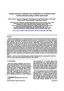
Fig. 3. Measurement of the water-extractable Hg2 vs. the total Hg2 concentration in arti¢cially contaminated agricultural soil. The extractable Hg2 was measured by the newly constructed biosensor strain P. putida KT2440: :mer-lux. 6 5: below detection limit (5 ng (g soil)31 ).
£uorescence of the LB media. The sample was then transferred to a luminescence spectrometer LS 50B (Perkin Elmer, Buckinghamshire, UK) and £uorescence was measured. The excitation wavelength was 395 nm and emission was measured at 509 nm. For each sample the optical density at 600 nm (OD600 ) was determined to ensure compatible cell numbers. Relative £uorescent units (RFU)/OD600 were plotted against the concentration of mercury (Fig. 2C). 2.6. Quanti¢cation of water-extractable mercury in soil Pseudomonas putida KT2440: :mer-lux was constructed by a tri-parental ¢lter mating between P. putida KT2440, E.coli MT102-PIR(pUT-mer-lux) and helper strain NF1815(RK600). These biosensor cells were used to determine the water-extractable concentration of mercury in extracts of arti¢cially contaminated soil. KT2440: :mer-lux was inoculated into LB4.10 media and grown for 8 h at 30³C by which time cells had reached stationary phase. The cells were then diluted in ABB1 media (no carbon source added) to 6.4U107 cells ml31 . Aliquots of 2.97 ml were then distributed into disposable plastic tubes for luminometer counting. The contaminated soil was made by adding 150 Wl of double distilled H2 O, containing 0, 2.5, 10 or 25 Wg of Hg2 , to 8-ml centrifuge tubes containing 1 g of sieved and dried agricultural soil [3]. After 8 h, the soil was mixed with 7 ml of H2 O and Hg2 was extracted for 1 h on a shaker at 25³C. The samples were centrifuged (15 000Ug for 10 min). A series of 10-fold dilutions were made to ensure mercury concentrations in the extracts within the linear range of the standard curve. 30 Wl of the soil extract dilutions was added to the biosensor cells. After 48 min, luminometer counts for cells with soil extracts were compared to luminometer counts from standard solutions (Fig. 3).
Three biosensor cassettes were each situated in a miniTn5 delivery vector. The biosensor constructs all responded in a quantitative manner to di¡erent concentrations of mercury. Adding to the versatility of these whole cell biosensors was the fact that they provide a choice of reporter genes. It appears that induction of the mer-lux and the mer-lac constructs were much more sensitive than induction of the mer-gfp construct (Fig. 1). However, the assay growth conditions were very di¡erent from one construct to another. Both the mer-lux and the mer-lac construct were grown in ABB1 media, compared to the modi¢ed LB media used for induction of E. coli MT102-PIR(pUT-mergfp), and the cell densities in the mer-gfp assay were much higher, compared to both mer-lux and mer-lac. Others studies have shown that both complex medium and high cell densities decrease the amount of bioavailable mercury in such assays [4,23]. The di¡erences in assay conditions re£ect the di¡erences between the three reporter genes. Detection of GFP required long incubation (16 h optimal) and detection of light gave £uctuating results, when cells containing the mer-lux construct were growing. Therefore the assay conditions were changed depending on which reporter gene was used. This complicates any comparison of sensitivity between the di¡erent biosensor constructs. However, the lux genes and lacZ are known as sensitive reporter genes in whole cell biosensors [24], whereas the gfp gene is known for its applications in in situ microbial ecology [25,26]. The mer biosensor constructs described in this study can be conjugated into a variety of Gram-negative bacteria, where integration into the host chromosome will ensure a stable construct, a constant copy number and maintenance even under non-selective conditions. All three miniTn5 constructs were successfully transferred into di¡erent Gram-negative bacteria such as Enterobacter aerogenes, Enterobacter chloacae, two strains of E. coli, Pseudomonas £uorescens and P. putida (data not shown). Soil bacteria like P. putida can readily be used to host biosensor constructs that are sensitive to mercury (Fig. 3). The results showed that only a small fraction of the mercury (less than 3%) was extracted from the soil. The waterextractable amounts mercury found in soil in this study, using KT2440: :mer-lux, are consistent with ¢ndings by Rasmussen et al. [3]. They found that approximately 50 ng of mercury was water-extractable from this soil spiked with 2.5 Wg g31 soil. In this study that number was 67 ng (Fig. 3). In the study by Rasmussen et al., E. coli hosted the plasmid-borne mer-lux construct. We are currently investigating the sensitivity and robustness of P. putida and several other potential host strains.
FEMSLE 9681 16-11-00
L.H. Hansen, S.J. SÖrensen / FEMS Microbiology Letters 193 (2000) 123^127
Acknowledgements We thank Pia Windel Kringelum for technical assistance and Lasse Dam Rasmussen for advice and discussion. This work was supported by the Danish Ministry of Food, Agriculture and Fisheries, Project MIL 96-2 and MIL 96-3.
[13]
[14]
[15]
References [1] Ramanathan, S., Ensor, M. and Daunert, S. (1997) Bacterial biosensors for monitoring toxic metals. Trends Biotechnol. 15, 500^ 506. [2] Lyngberg, O.K., Stemke, D.J., Schottel, J.L. and Flickinger, M.C. (1999) A single-use luciferase-based mercury biosensor using Escherichia coli HB101 immobilized in a latex copolymer ¢lm. J. Ind. Microbiol. Biotechnol. 23, 668^676. [3] Rasmussen, L.D., SÖrensen, S.J., Turner, R.R. and Barkay, T. (2000) Application of a mer-lux biosensor for estimating bioavailable mercury in soil. Soil Biol. Biochem. 32, 639^646. [4] Selifonova, O., Burlage, R. and Barkay, T. (1993) Bioluminescent sensors for detection of bioavailable mercury(II) in the environment. Appl. Environ. Microbiol. 59, 3083^3090. [5] Barkay, T., Turner, R.R., Rasmussen, L.D., Kelly, C.A. and Rudd, J.W.M. (1998) Luminescence facilitated detection of bioavailable mercury in natural waters. Methods Mol. Biol. 102, 231^246. [6] Condee, C.W. and Summers, A.O. (1992) A mer-lux transcriptional fusion for real-time examination of in vivo gene expression kinetics and promoter response to altered superhelicity. J. Bacteriol. 174, 8094^8101. [7] Vollmer, A.C., Belkin, S., Smulski, D.R., Van Dyk, T.K. and Larossa, R.A. (1997) Detection of DNA damage by use of Escherichia coli carrying recAP : :lux, uvrAP : :lux, or alkAP : :lux reporter plasmids. Appl. Environ. Microbiol. 63, 2566^2571. [8] Applegate, B.M., Kehrmeyer, S.R. and Sayler, G.S. (1998) A chromosomally based tod-luxCDABE whole-cell reporter for benzene, toluene, ethylbenzene, and xylene (BTEX) sensing. Appl. Environ. Microbiol. 64, 2730^2735. [9] Paton, G.I., Campbell, C.D., Glover, L.A. and Killham, K. (1995) Assessment of bioavailability of heavy metals using lux modi¢ed constructs of Pseudomonas £uorescens. Lett. Appl. Microbiol. 20, 52^56. [10] Paton, G.I., Palmer, G., Burton, M., Rattray, E.A.S., McGrath, S.P., Glover, L.A. and Killham, K. (1997) Development of an acute and chronic ecotoxicity assay using lux-marked Rhizobium leguminosarum Biovar trifolii. Lett. Appl. Microbiol. 24, 296^300. [11] Suarez, A., Gu«ttler, A., Stra«tz, M., Staendner, L.H., Timmis, K.N. and Guzma¨n, C.A. (1997) Green £uorescent protein-based reporter systems for genetic analysis of bacteria including monocopy applications. Gene 196, 69^74. [12] Hansen, L.H., SÖrensen, S.J. and Jensen, L.B. (1997) Chromosomal
[16]
[17]
[18]
[19]
[20] [21]
[22] [23]
[24]
[25]
[26]
127
insertion of the entire Escherichia coli lactose operon, into two strains of Pseudomonas, using a modi¢ed mini-Tn5 delivery system. Gene 186, 167^173. Simons, R.W., Houman, F. and Kleckner, N. (1987) Improved single and multicopy lac-based cloning vectors for protein and operon fusions. Gene 53, 85^96. Kristensen, C.S., Eberl, L., Sanchez-Romero, J.M., Givskov, M., Molin, S. and de Lorenzo, V. (1994) Site-speci¢c deletions of chromosomally located DNA segments with the multimer resolution system of broad-host-range plasmid RP4. J. Bacteriol. 177, 52^58. Kessler, B., de Lorenzo, V. and Timmis, K.N. (1992) A general system to integrate lacZ fusions into the chromosomes of Gram-negative eubacteria: regulation of the Pm promoter of the TOL plasmid studied with all controlling elements in monocopy. Mol. Gen. Genet. 233, 293^301. Bagdasarian, M., Lurz, R., Ruckert, B., Franklin, F.C.H., Bagdasarian, M.M., Frey, J. and Timmis, K.N. (1981) Speci¢c-purpose plasmid vectors II. Broad host range, high copy number, RSF1010-derived vectors, and a host-vector system for gene cloning in Pseudomonas. Gene 16, 237^247. Casabadan, M.J. and Cohen, S.N. (1980) Analysis of gene control signals by DNA fusion and cloning in Escherichia coli. J. Mol. Biol. 138, 179^207. Herrero, M., de Lorenzo, V. and Timmis, K.N. (1990) Transposon vectors containing non-antibiotic resistance selection markers for cloning and stable chromosomal insertion of foreign genes in Gram-negative bacteria. J. Bacteriol. 172, 6557^6567. Sambrook, J., Fritsch, E.F. and Maniatis, T. (1989) Molecular Cloning: A Laboratory Manual, 2nd edn., Cold Spring Harbor Laboratory, Cold Spring Harbor, NY. Miller, J.H. (1972) Experiments in Molecular Genetics, Cold Spring Harbor Laboratory Press, Cold Spring Harbor, NY. SÖrensen, S.J., Kroer, N., SÖrensen, E., SengelÖv, G. and Barkay, T. (1996) Conjugation in aquatic environments. In: Molecular Microbial Ecology Manual (Akkermans, A.D.L., van Elsas, J.D. and de Bruijn, F.L., Eds.), Kluwer Academic, Dordrecht. Clark, D.J. and MaalÖe, O. (1967) DNA replication and the division cycle of Escherichia coli. J. Mol. Biol. 23, 99^112. Rasmussen, L.D., Turner, R.R. and Barkay, T. (1997) Cell-densitydependent sensitivity of a mer-lux bioassay. Appl. Environ. Microbiol. 63, 3291^3293. Rouch, D.A., Parkhill, J. and Brown, N.L. (1995) Induction of bacterial mercury- and copper-responsive promoters ^ functional di¡erences between inducible systems and implications for their use in gene-fusions for in vivo metal biosensors. J. Ind. Microbiol. 14, 349^353. Normander, B., Hendriksen Niels, B. and Nybroe, O. (1999) Green £uorescent protein-marked Pseudomonas £uorescens: Localization, viability, and activity in the natural barley rhizosphere. Appl. Environ. Microbiol. 65, 4646^4651. Unge, A., Tombolini, R., Molbak, L. and Jansson Janet, K. (1999) Simultaneous monitoring of cell number and metabolic activity of speci¢c bacterial populations with a dual gfp-luxAB marker system. Appl. Environ. Microbiol. 65, 813^821.
FEMSLE 9681 16-11-00