Visual NetLogo-Based Simulation of Anti-SARS Immune System and Low-to-High Resolution Reconstruction of Sequence Medical CT Images Tao Gong, Lei Pei, Shangce Gao, Fang Han, Shuguang Zhao College of Information Science and Technology Engr. Research Center of Digitized Textile & Fashion Tech. for Ministry of Education, Donghua University Shanghai 201620, China E-mail:
[email protected] Abstract—In the immune responses against the SARS (Severe Acute Respiratory Syndromes), human immune systems are complex intelligent systems, which show good properties such as the self-organizing and adaptivity. Modeling the immune systems has important significance in both immunology and artificial immune system. In order to improve the visualization and readability of the anti-SARS immune system model, the visual tri-tier computational model of the anti-SARS immune system was simulated with NetLogo, which is a multi-agent-based tool. On the other hand, to fight against the SARS disease, the lowresolution medical CT (Computed Tomography) images should be transformed into the high-resolution ones for better SARS analysis. In order to obtain the high-resolution image from some low-resolution chest CT sequence images of a SARS patient, the low-to-high resolution reconstruction was designed and tested in this paper. First, the low-resolution medical images were preprocessed. Then the pretreated low-resolution medical images were registered with the sub-pixel-level image registration techniques. Finally, the POCS (Projections onto Convex Sets) image reconstruction algorithm was designed and tested. We obtained higher entropy and more detail information of the medical images with our approach than the Marcel method, especially for the rotated medical images in our experiments. Multiple-user browser-based experimental results show that the visual NetLogo-based simulation of the immune system is better to understand than the traditional mathematic equation model of the immune system. Keywords—visual immunization model; NetLogo; superresolution; medical image reconstruction; image registration
I.
INTRODUCTION
In the long history of mankind, human beings are always searching for the essence of life. In recent years, with the rapid development in medicine and physiology, some features of the immune system cause the attentions of researchers in other fields, and they begin to focus on the research and simulation of immune system. The research of immune mechanism has always been the focus point of studies home and abroad.
Zixing Cai, IEEE Senior Member College of Information and Engineering, Central South University Changsha, Hunan, China E-mail:
[email protected]
Inspired from the mechanism of the human immune system, new information processing system (i.e. the artificial immune system, AIS) can be established. Since 1997, an increasing number of international conferences have been organized on the AIS. Therefore, it is important to investigate the principle of the biological immune system and propose some mathematical models or engineering models of the immune system. First of all, according to the biological views, the development of the computer-based model of the immune system provides new way to test the theory of immunology, thus the model will help the immunologists to better understand and develop biological immunology. Besides, the visual simulation of the immune system has an important guiding significance in studying complex intelligent systems and proposing new intelligent methods. So an increasing number of researchers from different fields start to simulate the immune system with new techniques on computers. In the real industry procedure, when some visual images are obtained with the imaging system, the recording equipment, the transmission medium and some processing methods, the images are possibly blurred and deformed [1]. In these cases, these low-resolution images should be transformed into the high-resolution images. This problem becomes a hot issue of medical image processing. Though these single images have low resolutions, we can use the super-resolution reconstruction approach to break through the resolution limits of the medical images [2], in order to generate the high-resolution image for better disease analysis. SARS patients’ lungs show different degree of flake or patchy ground glass density shadow. The shadow shows a trend of progress, because in some cases a small shadow may quickly grows into a large shadow. The super-resolution reconstruction can help us to detect more shadows, so we can use this method for decrease early lesions. Why human beings can live healthy in this world with dangers and diseases? An important reason is that the nature has granted the mankind a natural defense system, i.e. immune system. With this system, human beings are able to survive
against the dangers and diseases. In medicine, the immunity refers to a physiological response when the body is exposed to the foreign substances named antigens [3]. The modern definition of the immunity is the reaction of the body against some exogenous substances. And the immulogical function is to identify the antigens and exclude the nonselfs from the body. Generally speaking, the reaction that maintains a stable state of the body is beneficial to the body, but it is also harmful under some certain conditions. There are great differences between the model of the natural immune system and the artificial immune system. These differences not only reflect in the application objects of the models, but also in the modeling approaches. So it’s difficult to establish an appropriate mapping between these two models. In order to investigate the immune system from the perspective of natural computing, the tri-tier visual model of the natural immune system includes the innate immune tier, the adaptive immune tier and the immune cell tier in this paper. II.
VISUAL IMMUNE MODEL AND SUPER-RESOLUTION RECONSTRUCTION OF MEDICAL CT IMAGES
The mapping relationship between the natural immune system and the computer- based artificial immune system is shown in Table I. TABLE I.
IMMUNE MAPPING FROM NATURE TO COMPUTERS
Biological immune system
Artificial immune system
Non-self antigen
Computer virus, faults
Self antigen
Normal component
Phagocyte
Object eliminator
Antibody
Immune Agent (IA)
Memory cell
Memory
Feature matching
Database query
Pattern recognition
Learning and reasoning
B cells, T cells etc.
Parallel computer
When the antigen enters body, the body is protected by the immune system. First of all, the system detect antigen by means of self/non-self identification mechanism, and then recognize the antigen by feature matching. When the antigen is identified as known antigen, it will be eliminated by phagocytes immediately; but if the antigen can’t be identified by innate immune tier, it will be handed to adaptive immune tier. Subsequently, a large number of immune cells begin to produce antibodies. Finally, the unknown antigen will be eliminated through pattern recognition and learning. After the antigen is identified by the antibody, the antigen becomes known antigen and be eliminated by phagocytes. Phagocytes are double-edged, and this is precisely the key of biological immune system [4]. When phagocytes work normally, they can combine with hazardous antigen and then elimination them; when phagocytes work anomaly, they may be combined with normal cells or molecules, and kill normal cells.
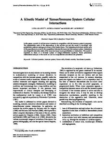
NetLogo is a programmable modeling environment, which is written with Java independent of the platforms such as Windows, Linux, Mac, etc. It is often used to simulate natural and social phenomena, and especially suited for modeling the complex systems, which evolved over time [5-6]. Researchers from different fields can create the relevant models according to their needs. Besides, a model library can be built to contain a number of simulation models. So the researchers can not only use them directly, but also use them after some modifications. These models cover many areas of nature and social science such as biology, medicine, physics, chemistry, computing mathematics, and computer science, etc [7]. There are two ways to view the simulation model with NetLogo: in the NetLogo client; in a remote web browser after the model is saved as Java Applet and embedded into a web page. Because of the complexity of biological systems and the limitations of technology, we are unable to simulate the immune system completely. Thus, we select an important immune mechanism named cellular immunity to simulate. The cellular immune mechanism is designed with the immune Dcells. The D cells have tree-root shapes under a microscope [8], and the D cells exist in the body's first line of defense (e.g. skin, mucous membranes). They are important for the innate immune system and the strongest professional antigenpresenting cells (pAPC) in the body. In addition, they are the main bearer of T cell activation. When the body encounters the invasion of pathogenic microorganisms, the D cells are activated through combining with pathogen, thus the antigen information is attained quickly. The immature D cells have a strong ability of migration. They can present the antigen information to the inferior immune system, also namely the adaptive immune system. The D cells that obtain sufficient antigen information move to the nearest lymph nodes through the lymphatic and blood circulation. These D cells get mature gradually, and then move to the T cell zones of the lymphoid organs with the function of chemokines. Mature D cells present information of the antigen to the T cells after they reach the T cell area. And then the T cells become into the effector T cells after the proliferation and differentiation. Meanwhile, a small portion of T cells become into the memory T cells. When the same kind of antigens intrude into body, the memory cells will proliferate and differentiate rapidly, the immune system can generate a large number of effector T cells, which could make stronger specific immune response. Thus, the mature D cells can stimulate the proliferation of the naive T cells effectively, which is the most prominent feature of the D cells. In fact, the D cells are the first link of the immune response. Whether the antigen presentation is effective or not, it is directly related to immune activation or induction of immune tolerance. Therefore, the D cells have a special status in immune response. And this is also one of important reasons for choosing the cellular immunity for the simulation. As shown in Fig. 1, the super-resolution medical image reconstruction has 5 steps: image preprocessing, image registration, image transformation, image reconstruction and inverse transformation. The medical image registration and the motion estimation model are both important for the superresolution reconstruction of the medical images.
Fig. 1. Super-resolution reconstruction of the medical images.
In addition, the higher are the accuracies of the image registration and the motion estimation, the better effect of the image reconstruction is made [8]. At first, the multi-frame low-resolution medical images should be made through the micro-displacement with each other, in order to get a highresolution image. The accuracy of the medical image registration is one of the main factors for the super-resolution image reconstruction, and this factor often affects the resolution of the reconstructed medical image [9]. After the image registration and the image recovery, the uniform sampling of the super-resolution image can be made. We register the medical image with the micro-displacement method, and the pre-processing of the medical images, the medical image registration, and the medical image reconstruction are based on the POCS super-resolution image reconstruction algorithm. III. MEDICAL IMAGE REGISTRATION WITH IMPROVED KEREN SUB-PIXEL-BASED REGISTRATION ALGORITHM The medical image registration is the seeking process of the one-to-one mapping between an image and another one, which is used to make the two images mapped to the coordinate position [11]. We designed the medical image registration with the Keren-pixel-based image registration method. The medical image registration can be roughly divided into four categories: multi-modal medical image registration, medical image template registration, medical image observation point registration and medical image time sequence registration. We used the time sequence registration method [12]. For example, if we use two medical images in the image registration, then the reference image of the two images is represented by I1(x, y), and the observed image of the two ones is represented by I2(x, y). Then the medical image registration is mathematically defined below.
I 2 ( x, y ) = g ( I1 ( f ( x, y )))
(1)
Here, f(x, y) is the coordinate transformation function, and g(·) denotes the 1-dimension gray-scale/radiation transformation function. For this reason, it is very crucial for the medical image registration to find the best space or geometric transformation [13]. This transformation can be made by the single-valued function I1(·) with the two variables fx and fy.
I 2 ( x, y ) = I1 ( f x ( x, y ), f y ( x, y ))
(2)
In the past few decades, an increasing number of scholars from different fields had investigated the problem of image registration from different angles and in different application backgrounds [14]. However, most of these methods are of the pixel-level accuracy, and this accuracy level is not enough for advanced processing of the medical images. For example, the remote sensing and the high-precision reconstruction of the medical image need the more accurate registration of the medical images, and the sub-pixel level registration of the medical images can make higher accuracy. Usually, in the medical image processing, only the sub-pixel level image registration can provide the possibility to detect the small differences between the sequence medical images. So the subpixel level registration method is a necessary foundation for this image reconstruction, and the Keren sub-pixel-based image registration method is used to process the medical sequence images. The Keren sub-pixel-based image registration algorithm uses the three-layered Gaussian pyramid to increase the speed, accuracy, and stability. After the Gaussian filtering and sampling, the original N × N images are transformed into the new two images, which have the N 2 × N 2 resolutions. When we repeat the above procedures, the final two N 4 × N 4 images are generated. This algorithm utilizes a coarse-to-fine resolution image pyramid, from the first coarse layer to strike X. According to X, the second layer is rotated and transformed, and then we get a new second layer with the interpolation. Then the new X is calculated again, and so we get the high accuracy registration parameters. However, the Keren sub-pixel-based image registration algorithm has the major drawback that this algorithm depends on the small angle Taylor series expansion. For the small angles (less than 6°), this algorithm is useful for the high registration accuracy. But the rotation angle of the medical sequence images sometimes causes relatively big errors by this Keren algorithm. To avoid these big errors, the following formula is used to improve this Keren algorithm with the successive approximation.
X k +1 = Ck−1Vk + X k
(3)
Here, k is the iteration sum of this Keren iterative algorithm.
In order to overcome the errors from the Taylor series, our improved medical image registration method embeds the rigid transformation model of the medical image into the simplified four-parameter affine transformation model.
x ' = x + a1 x + a2 y + a3 ⎫ ⎬ y ' = y + a1 y − a2 x + a4 ⎭
Step 2: Initial the iteration sum k, the rotation angle and the horizontal/vertical displacement all with zero. Step 3: Calculate C nk and K nk , and calculate X according to equation (3). Step 4: If K=k, then go to step 7. N
k
Step 5: According to X , resample Step 6: k=k+1, and return to step3. N
(4)
k
Step 7: If n