applied sciences Review
Wavefront Shaping and Its Application to Enhance Photoacoustic Imaging Zhipeng Yu, Huanhao Li
ID
and Puxiang Lai *
Department of Biomedical Engineering, The Hong Kong Polytechnic University, Hong Kong 999077, China;
[email protected] (Z.Y.);
[email protected] (H.L.) * Correspondence:
[email protected]; Tel.: +852-3400-8900 Received: 14 October 2017; Accepted: 30 October 2017; Published: 19 December 2017
Abstract: Since its introduction to the field in mid-1990s, photoacoustic imaging has become a fast-developing biomedical imaging modality with many promising potentials. By converting absorbed diffused light energy into not-so-diffused ultrasonic waves, the reconstruction of the ultrasonic waves from the targeted area in photoacoustic imaging leads to a high-contrast sensing of optical absorption with ultrasonic resolution in deep tissue, overcoming the optical diffusion limit from the signal detection perspective. The generation of photoacoustic signals, however, is still throttled by the attenuation of photon flux due to the strong diffusion effect of light in tissue. Recently, optical wavefront shaping has demonstrated that multiply scattered light could be manipulated so as to refocus inside a complex medium, opening up new hope to tackle the fundamental limitation. In this paper, the principle and recent development of photoacoustic imaging and optical wavefront shaping are briefly introduced. Then we describe how photoacoustic signals can be used as a guide star for in-tissue optical focusing, and how such focusing can be exploited for further enhancing photoacoustic imaging in terms of sensitivity and penetration depth. Finally, the existing challenges and further directions towards in vivo applications are discussed. Keywords: photoacoustic imaging; optical scattering; optical diffusion limit; penetration depth; wavefront shaping; iterative optimization; transmission matrix; Grueneisen effect; noninvasive internal guide star
1. Introduction High-resolution optical imaging has recently seen more and more biomedical applications, such as optical coherence tomography (OCT) for optometry [1], confocal microscopy for cytobiology research [2], two- or three-photon microscopy for deep tissue imaging [3–8], as well as stimulated emission depletion (STED) microscopy and photo-activated localization microscopy (PALM) for super-resolution imaging of single molecules [9,10]. In almost all of these applications, photons, whether illuminating or excited ones, need to propagate some distance in tissue. Therefore, the capability of focusing light plays an essential role, largely determining the sensitivity and spatial resolution of these techniques. It would be ideal if human tissues were as transparent as those of a jelly fish or X-ray fish. However, due to the inhomogeneities of the refractive index among different cells and organelles in tissue [11], photons are multiply scattered along their respective optical paths. As a result, an incident optical beam spreads out and becomes diffusive very quickly as it travels deep into tissue (typically ~1 mm beneath human skin for visible and near-infrared light). Beyond this so-called optical diffusion limit, optical focus can hardly be formed by using lenses or objects; instead, if the coherence length of light is sufficiently long, optical speckle patterns are developed within or behind a thick biological tissue sample. Moreover, because of the strong scattering, the in situ optical fluence also decays rapidly. These two folds pose a fundamental limitation to the aforementioned high-resolution techniques to be viable only within superficial depths (e.g., less than 100–200 micrometers). Appl. Sci. 2017, 7, 1320; doi:10.3390/app7121320
www.mdpi.com/journal/applsci
Appl. Sci. 2017, 7, 1320
2 of 17
Recently, photoacoustic (PA) imaging has been introduced to tackle the diffusion-induced resolution barrier in deep tissue [11–15]. This technique is based on the so-called PA effect [16–18], which mainly consists of four steps: first, tissue molecules in the region of interest (ROI) within the sample absorb pulsed light (no matter if it is in ballistic or diffusive form); the absorbed optical energy is converted to heat, causing local the temperature to increases; ultrasonic waves are generated due to thermal expansion; finally, these ultrasonic waves (typically around the order of MHz if the exiting light pulse width is several nanoseconds) can be detected by one or a series of ultrasound transducers mounted outside the sample. Since ultrasonic waves are scattered almost 1000 times less than light is in tissue (unless otherwise mentioned, we always mean soft tissue in the context), they can be used to reconstruct an image of the absorbing ROI with an acoustic resolution [19–27]. As the generation of such PA signals does not distinguish ballistic or diffusive exciting light formations, it allows for a penetration depth far exceeding the optical diffusion limit. On the other hand, if only shallow tissue depths (e.g., within one transport mean free path) are sought, where ballistic or quasi-ballistic photons dominate, the optical spot size can be much smaller than that of the ultrasonic focus, the resolution of PA imaging is determined by the optical—but not the acoustic—focal size. In this scenario, the technique is often referred to as optical-resolution photoacoustic imaging or microscopy [28,29]. As a hybrid modality, PA imaging takes advantage of the optical contrast and acoustic or optical spatial resolution at moderate tissue depth. Therefore, it has recently seen many fundamental and preclinical trials for the imaging or diagnosis of blood vessels [30], brain [31], breast tumor [32], sentinel lymph node (SLN) [33], oxygen saturation [34], sub-cellular structures [14], and small-animal whole body [35], among many other explorations. While promising, it must be noted that in PA imaging, only the detection of generated ultrasonic signals is “transparent” to tissue; the exciting photons experience the same strong scattering as in other optical microscopic techniques, and the optical fluence decays rapidly with tissue depth. This leads to limited practical depths of PA imaging: for optical resolution schemes, it is curbed to one transport mean free path (~1 mm) [36], beyond which diffusive light dominates and the imaging resolution is no longer determined by the light spot, but the ultrasonic focus; for acoustic resolution schemes, it is curbed to where the in situ optical fluence within the ultrasonic focus is too weak to generate reliable PA signals, usually being a couple of centimeters [13]. Therefore, it would be ideal if the diffusive light can be noninvasively refocused inside or behind thick scattering media. This actually has long been one of the holy grails in the community of biomedical optics. Traditional wisdom has treated multiple scattering of light a nuisance to be bypassed or eliminated. However, recently, researchers began to notice that the seemingly random scattering events and the resultant speckles are actually deterministic within a certain temporal window [37,38], and it is possible to reverse [39–41] or compensate for [42] the scattering-induced phase scrambling. To do so, researchers have developed several wavefront shaping (sometimes also referred to wavefront engineering) techniques, such as iterative wavefront optimization [23–26,28,42–51], measuring the transmission matrix of the scattering medium [21,22,52–56], and optical time reversal via phase conjugation [39,40,57–65]. Nevertheless, the goals of these implementations are identical, i.e., to make light wavelets traveling along different optical paths interfere coherently at a region of interest (ROI) and form a bright optical spot (focus) out of the much darker background. To create such an optical focus within or inside scattering media, which is more of biomedical interest, an internal guide star is needed to guide or provide a feedback for the wavefront shaping operations. For that purpose, researchers have proposed injected or embedded probes, such as small optical sensors [66,67], fluorescence particles [68,69], and nonlinear beads [57], ultrasound mediation [40,58,61,70,71], as well as absorption perturbation [22,25,46,48,72–76]. Among them, PA signal has been proved very attractive [22,25,28,47,48,53,55,73,74,76], as it is noninvasive, relatively deeply-penetrating, and it can pinpoint the focal spot accurately (the acoustic and optical foci overlap). Furthermore, with linear [22,25,28,53,73,74] or nonlinear [48] PA signals as the guide star, acousticand optical-diffraction limited optical focal spot can be achieved, respectively, benefiting from the nature of PA signal generation. In turn, during the process of such PA-guided wavefront optimization,
Appl. Sci. 2017, 7, 1320
3 of 17
the in situ optical fluence is gradually increased, and so is the strength of PA signals. Therefore, the signal-to-noise ratio (SNR) and the sensitivity of PA imaging can be greatly enhanced. Moreover, the formation of PA-guided optical focusing at depths in tissue paves the way to break the penetration depth limitation of current acoustic-resolution and optical-resolution PA imaging implementations. This technique, therefore, has the potential to impact the field. In this paper, we first briefly describe the propagation of light in tissue-like scattering media and the concept of wavefront shaping techniques. Then we discuss, in detail, how PA signals can be used as the guide star for wavefront shaping and how using wavefront shaping can enhance PA imaging in terms of sensitivity and resolution. Lastly, we discuss the existing limitations, challenges, as well as potential solutions in order to advance towards in vivo enhanced PA imaging at depth in tissue. 2. Exploiting Scattering of Light 2.1. Light Scattering in Complex Media and the Memory Effect In complex media, such as biological tissue samples, spatial inhomogeneities of the wavelength-scale refractive index cause light to be scattered once for every ~100 µm along its optical path. In general, such multiple-scattering feature, according to [19,20], can be described with specific parameters, such as ‘mean free path’ (MFP) and ‘transport mean free path’ (TMFP): the former represents the average distance between two consecutive scattering events, and the latter the effective MFP for forward-propagating light portion. Mathematically: l MPF = 1/µs
(1)
lTMFP = 1/[µs (1 − cos < θ >)]
(2)
where µs is the scattering coefficient of the medium, and θ is the deflection of angle of the scattered light. Typically, the TMFP of most biological tissues varies from 0.1 to 1 mm. Note that the TMFP of biological tissues is wavelength dependent. Some near-infrared (NIR) light can reach a depth of up to several millimeters [15]; visible light that is widely used in many biomedical techniques only has a shallow penetration depth of hundreds of micrometers inside biological tissue, while a good resolution is sought. Therefore, traditionally scattering events have been regarded as an obstacle, and the speckle patterns generated by the coherent light propagation in tissue has been seen as one of the major noise sources in biomedical optical imaging [77,78]. However, recently, pioneering scientists have begun to explore the feasibility of exploiting scattering and the corresponding speckle patterns based on their deterministic feature within the medium’s temporal correlation window [40,63–65]. 2.2. Wavefront Shaping Wavefront shaping is one such technique that scientists have developed. The basic principle is to spatially modulate light incident onto the turbid medium in a way so that scattering-induced phase distortions between the point of incidence and the ROI can be pre-compensated. This technique was first introduced by Vellekoop et al. in 2007 [42]. They used a liquid-crystal-on-silicon (LCoS)-type spatial light modulator (SLM) as the phase/wavefront modulator, with up to 3228 independently controlled pixels to control the phase of every input mode individually. A charge coupled device (CCD) camera was placed to monitor the light intensity at a target position behind a scattering medium. Light intensity reading from the specific CCD pixels was used as the feedback for an optimization algorithm to program the phase patterns on the SLM. In order to maximize the intensity of a chosen area (output mode) recorded by the camera pixels, the phase of each input mode (SLM pixel) was gradually tuned from 0 to 2π. The one that yielded highest feedback was assigned as the optimum phase for that specific SLM pixel. Such iterative optimization process was performed for each SLM pixel. After the optimization, a strong optical focus behind an opaque ground glass diffuser is obtained
Appl. Sci. 2017, 7, 1320
4 of 16
Appl. Sci. 2017, 7, 1320
4 of 17
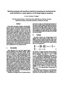
is obtained (Figure 1b). In comparison, if wavefront shaping is not employed, a fully-randomized speckle pattern is seen (Figure 1a), which is an indicator of sufficient optical diffusion from the (Figure 1b). In comparison, if wavefront shaping is not employed, a fully-randomized speckle pattern medium. is seen (Figure 1a), which is an indicator of sufficient optical diffusion from the medium.
Figure 1. optical speckle pattern behind a ground glass diffuser withoutwithout a shapeda incident Figure 1. (a) (a)AAtypical typical optical speckle pattern behind a ground glass diffuser shaped wavefront; (b) Optical(b) pattern of the regionofwith optimized wavefront compensation by incident wavefront; Optical pattern theanregion withincident an optimized incident wavefront using wavefront shaping. A lot of energy of the multiply-scattered light is focused to a spot that compensation by using wavefront shaping. A lot of energy of the multiply-scattered light is focused is 1000 times speckle grains. size of the optical focus determined by the to a spot thatbrighter is 1000 than timesother brighter than otherThe speckle grains. The size of isthe optical focus is sensitivity area of the feedback signal (Figure reproduced from [42]). determined by the sensitivity area of the feedback signal (Figure reproduced from [42]).
Since the initial implementation, wavefront shaping has seen many exciting advancements and has been explored for wide applications, which has been well reviewed [19,79–81] and, thus, will not be reiterated reiteratedherein. herein.However, However, it must be pointed out most that efforts most efforts in the community have it must be pointed out that in the community have centered centered on threetoaspects: improve the focal intensity enhancement (R),the tooptimization shorten the on three aspects: improvetothe focal intensity enhancement ratio (R), to ratio shorten optimization process, and to seeknoninvasive appropriateguide noninvasive guide star producesignal. the feedback signal. process, and to seek appropriate star to produce thetofeedback Although the Although the focal enhancement R at theand ROIafter before after the optimization maycase vary focal enhancement ratio R at the ratio ROI before theand optimization may vary from tofrom case case (e.g., the working principle of the wavefrontitmodulator), it is overalltoproportional to α × (e.g.,to thecase working principle of the wavefront modulator), is overall proportional α × (N/M), where (N/M), N is number of independently controlled elements M of is the number of N is thewhere number ofthe independently controlled elements on the SLM, Mon is the SLM, number output modes output modes (speckle grains) within the ROI, and α is a constant depending on the type of wavefront (speckle grains) within the ROI, and α is a constant depending on the type of wavefront modulator modulator (Table 1). As one caninimagine, order to achieve high N R, and/or a large N and/or small M are (Table 1). As one can imagine, order toin achieve a high R, aalarge small M are required: required: is determined by the technical specification of the wavefront modulator, and the the formerthe is former determined by the technical specification of the wavefront modulator, and the latter by latter by the sensitivity zone of the feedback signal withto respect to the grain speckle grain size. That said,N a the sensitivity zone of the feedback signal with respect the speckle size. That said, a large large N requires usually requires longer optimization process [43], which is also significantly by the usually a longer aoptimization process [43], which is also significantly affected byaffected the refreshing refreshing of the modulator, wavefront modulator, as speed well asofthe speed of data andbetween processing rate of the rate wavefront as well as the data transfer and transfer processing the between thewavefront sensor, the wavefrontand modulator, and the processor (usually a personal computer [56]). sensor, the modulator, the processor (usually a personal computer [56]). Table 1 lists three types of commercial commercial wavefront modulators, i.e., LCoS-type spatial spatial light modulator (SLM), deformable mirror (DM), and digital micromirror device (DMD), that modulator (SLM), deformable mirror (DM), and digital micromirror device (DMD), that have been popularly used usedininwavefront wavefront shaping. Each its own advantages and disadvantages; is shaping. Each hashas its own advantages and disadvantages; none isnone perfect. perfect. For example, DMD can respond but its modulation relies the of tilting of the microFor example, DMD can respond rapidly,rapidly, but its modulation relies on the on tilting the micro-mirror mirror and only (i.e., binary (i.e.,ON either ON orintensity OFF) intensity modulation is supported byFor DMD. arrays, arrays, and only binary either or OFF) modulation is supported by DMD. the For the other twofiner types, finer steps modulation of phase modulation are usually supported. Onhand, the other hand, other two types, steps of phase are usually supported. On the other LCoS-SLM LCoS-SLM slow, and DM is expensive supports small pixel numbers. choosing is slow, andis DM is expensive and supportsand small pixel numbers. Therefore, the Therefore, choosing ofthe a wavefront of a wavefront modulator is usually a trade-off between the speed,efficiency, the modulation the modulator is usually a trade-off between the speed, the modulation the pixelefficiency, number, and pixel number, and the cost, many other from the specific application. the cost, among many otheramong requirements from requirements the specific application.
Appl. Sci. 2017, 7, 1320
5 of 17
Table 1. Representative commercial wavefront modulators 1 (modified from [79]). Character
LCoS-SLM
DM
DMD
Working principle
Electrically controlled liquid crystal arrays
Piezoelectric arrays and flexible reflective surface
Tilting of micro-mirror arrays
Max pixel number
High (1920 × 1080)
Low (10–1000)
High (3000 × 2000)
Refreshing rate
Slow (10–100 Hz)
Fast (1–10 kHz)
Fast (up to 23 kHz)
Diffraction efficiency
High (60–90%)
High (~100%)
Low (100,000, with an optimization process slightly longer than one hour, even though a much higher N was employed. This will not be possible without the use of a much faster-responding modulator DMD, even though the full power of the DMD has not been released yet and the focusing speed can be further improved. In conclusion, using photoacoustic signal as a virtual internal guide star allows for acousticor optical-diffraction limited optical focusing with an intensive focal-to-background ratio within biological tissue or tissue-like media. With further engineered, PAWS can be a promising tool to bring the sensitivity, the resolution, and potentially the penetration depth of existing photoacoustic imaging implementations to a new level. Acknowledgments: The work has been supported by the Hong Kong Research Grant Council (no. 252044/16E) and the National Natural Science Foundation of China (no. 81671726 and no. 81627805) Author Contributions: Zhipeng Yu initiated the manuscript. All authors participated in the revision of the manuscript. Puxiang Lai provided overall supervision. Conflicts of Interest: The authors declare no conflict of interest.
References 1. 2. 3. 4. 5.
Huang, D.; Swanson, E.A.; Lin, C.P.; Schuman, J.S.; Stinson, W.G.; Chang, W.; Hee, M.R.; Flotte, T.; Gregory, K.; Puliafito, C.A.; et al. Optical coherence tomography. Science 1991, 254, 1178–1181. [CrossRef] [PubMed] Pawley, J.B. Handbook of Biological Confocal Microscopy, 3rd ed.; Springer: Berlin, Germany, 2006. Beaurepaire, E.; Oheim, M.; Mertz, J. Ultra-deep two-photon fluorescence excitation in turbid media. Opt. Commun. 2001, 188, 25–29. [CrossRef] Theer, P.; Hasan, M.T.; Denk, W. Two-photon imaging to a depth of 1000 µm in living brains by use of a Ti: Al2 O3 regenerative amplifier. Opt. Lett. 2003, 28, 1022–1024. [CrossRef] [PubMed] Helmchen, F.; Denk, W. Deep tissue two-photon microscopy. Nat. Methods 2005, 2, 932–940. [CrossRef] [PubMed]
Appl. Sci. 2017, 7, 1320
6.
7. 8.
9.
10. 11. 12. 13. 14. 15. 16. 17. 18. 19. 20. 21.
22. 23. 24. 25. 26. 27. 28.
29. 30.
14 of 17
Sakadži´c, S.; Demirbas, U.; Mempel, T.R.; Moore, A.; Ruvinskaya, S.; Boas, D.A.; Sennaroglu, A.; Kartner, F.X.; Fujimoto, J.G. Multi-photon microscopy with a low-cost and highly efficient Cr: LiCAF laser. Opt. Express 2008, 16, 20848–20863. [CrossRef] [PubMed] Kobat, D.; Horton, N.G.; Xu, C. In vivo two-photon microscopy to 1.6 mm depth in mouse cortex. J. Biomed. Opt. 2011, 16, 106014. [CrossRef] [PubMed] Horton, N.G.; Wang, K.; Kobat, D.; Clark, C.G.; Wise, F.W.; Schaffer, C.B.; Xu, C. In vivo three-photon microscopy of subcortical structures within an intact mouse brain. Nat. Photonics 2013, 7, 205–209. [CrossRef] [PubMed] Betzig, E.; Patterson, G.H.; Sougrat, R.; Lindwasser, O.W.; Olenych, S.; Bonifacino, J.S.; Davidson, M.W.; Lippincott-Schwartz, J.; Hess, H.F. Imaging intracellular fluorescent proteins at nanometer resolution. Science 2006, 313, 1642. [CrossRef] [PubMed] Willig, K.I.; Rizzoli, S.O.; Westphal, V.; Jahn, R.; Hell, S.W. STED microscopy reveals that synaptotagmin remains clustered after synaptic vesicle exocytosis. Nature 2006, 440, 935–939. [CrossRef] [PubMed] Tuchin, V.V.; Tuchin, V. Tissue Optics: Light Scattering Methods and Instruments for Medical Diagnosis; SPIE Press: Bellingham, WA, USA, 2007. Ntziachristos, V.; Razansky, D. Molecular imaging by means of multispectral optoacoustic tomography (MSOT). Chem. Rev. 2010, 110, 2783–2794. [CrossRef] [PubMed] Beard, P. Biomedical photoacoustic imaging. Interface Focus 2011, 1, 602–631. [CrossRef] [PubMed] Wang, L.V.; Hu, S. Photoacoustic tomography: In vivo imaging from organelles to organs. Science 2012, 335, 1458–1462. [CrossRef] [PubMed] Wang, L.V.; Wu, H.I. Biomedical Optics: Principles and Imaging; John Wiley & Sons: Hoboken, NJ, USA, 2012. Kruger, R.A. Photoacoustic ultrasound. Med. Phys. 1994, 21, 127–131. [CrossRef] [PubMed] Karabutov, A.; Podymova, N.; Letokhov, V. Time-resolved laser optoacoustic tomography of inhomogeneous media. Appl. Phys. B 1996, 63, 545–563. [CrossRef] Oraevsky, A.A.; Jacques, S.L.; Tittel, F.K. Measurement of tissue optical properties by time-resolved detection of laser-induced transient stress. Appl. Opt. 1997, 36, 402–415. [CrossRef] [PubMed] Mosk, A.P.; Lagendijk, A.; Lerosey, G.; Fink, M. Controlling waves in space and time for imaging and focusing in complex media. Nat. Photonics 2012, 6, 283–292. [CrossRef] Popoff, S.; Lerosey, G.; Fink, M.; Boccara, A.C.; Gigan, S. Image transmission through an opaque material. Nat. Commun. 2010, 1, 81. [CrossRef] [PubMed] Popoff, S.; Lerosey, G.; Carminati, R.; Fink, M.; Boccara, A.; Gigan, S. Measuring the transmission matrix in optics: An approach to the study and control of light propagation in disordered media. Phys. Rev. Lett. 2010, 104, 100601. [CrossRef] [PubMed] Chaigne, T.; Katz, O.; Boccara, A.C.; Fink, M.; Bossy, E.; Gigan, S. Controlling light in scattering media non-invasively using the photoacoustic transmission matrix. Nat. Photonics 2014, 8, 58–64. [CrossRef] Conkey, D.B.; Brown, A.N.; Caravaca-Aguirre, A.M.; Piestun, R. Genetic algorithm optimization for focusing through turbid media in noisy environments. Opt. Express 2012, 20, 4840–4849. [CrossRef] [PubMed] Conkey, D.B.; Caravaca-Aguirre, A.M.; Piestun, R. High-speed scattering medium characterization with application to focusing light through turbid media. Opt. Express 2012, 20, 1733–1740. [CrossRef] [PubMed] Kong, F.; Silverman, R.H.; Liu, L.; Chitnis, P.V.; Lee, K.K.; Chen, Y.C. Photoacoustic-guided convergence of light through optically diffusive media. Opt. Lett. 2011, 36, 2053–2055. [CrossRef] [PubMed] Conkey, D.B.; Caravaca-Aguirre, A.M.; Dove, J.D.; Ju, H.Y.; Murray, T.W.; Piestun, R. Super-resolution photoacoustic imaging through a scattering wall. Nat. Commun. 2015, 6, 1. [CrossRef] [PubMed] Tay, J.W.; Lai, P.X.; Suzuki, Y.; Wang, L.V. Ultrasonically encoded wavefront shaping for focusing into random media. Sci. Rep. 2014, 4, 5. [CrossRef] [PubMed] Caravaca-Aguirre, A.M.; Conkey, D.B.; Dove, J.D.; Ju, H.; Murray, T.W.; Piestun, R. High contrast three-dimensional photoacoustic imaging through scattering media by localized optical fluence enhancement. Opt. Express 2013, 21, 26671–26676. [CrossRef] [PubMed] Yao, J.; Wang, L.V. Photoacoustic Microscopy. Laser Photonics Rev. 2013, 7. [CrossRef] [PubMed] Hoelen, C.; de Mul, F.; Pongers, R.; Dekker, A. Three-dimensional photoacoustic imaging of blood vessels in tissue. Opt. Lett. 1998, 23, 648–650. [CrossRef] [PubMed]
Appl. Sci. 2017, 7, 1320
31.
32.
33.
34.
35. 36. 37. 38. 39. 40.
41. 42. 43. 44. 45.
46. 47. 48. 49. 50. 51. 52. 53.
15 of 17
Wang, X.; Pang, Y.; Ku, G.; Xie, X.; Stoica, G.; Wang, L.V. Noninvasive laser-induced photoacoustic tomography for structural and functional in vivo imaging of the brain. Nat. Biotechnol. 2003, 21, 803–806. [CrossRef] [PubMed] Xi, L.; Grobmyer, S.R.; Wu, L.; Chen, R.; Zhou, G.; Gutwein, L.G.; Sun, J.; Liao, W.; Zhou, Q.; Xie, H. Evaluation of breast tumor margins in vivo with intraoperative photoacoustic imaging. Opt. Express 2012, 20, 8726–8731. [CrossRef] [PubMed] Song, K.H.; Kim, C.; Cobley, C.M.; Xia, Y.; Wang, L.V. Near-infrared gold nanocages as a new class of tracers for photoacoustic sentinel lymph node mapping on a rat model. Nano Lett. 2009, 9, 183–188. [CrossRef] [PubMed] Zhang, H.F.; Maslov, K.; Sivaramakrishnan, M.; Stoica, G.; Wang, L.V. Imaging of hemoglobin oxygen saturation variations in single vessels in vivo using photoacoustic microscopy. Appl. Phys. Lett. 2007, 90, 053901. [CrossRef] Xia, J.; Wang, L.V. Small-animal whole-body photoacoustic tomography: A review. IEEE Trans. Biomed. Eng. 2014, 61, 1380–1389. [PubMed] Maslov, K.; Zhang, H.F.; Hu, S.; Wang, L.V. Optical-resolution photoacoustic microscopy for in vivo imaging of single capillaries. Opt. Lett. 2008, 33, 929–931. [CrossRef] [PubMed] Freund, I.; Rosenbluh, M.; Feng, S. Memory effects in propagation of optical waves through disordered media. Phys. Rev. Lett. 1988, 61, 2328–2331. [CrossRef] [PubMed] Feng, S.; Kane, C.; Lee, P.A.; Stone, A.D. Correlations and fluctuations of coherent wave transmission through disordered media. Phys. Rev. Lett. 1988, 61, 834. [CrossRef] [PubMed] Ma, C.; Zhou, F.; Liu, Y.; Wang, L.V. Single-exposure optical focusing inside scattering media using binarized time-reversed adapted perturbation. Optica 2015, 2, 869–876. [CrossRef] Liu, Y.; Lai, P.; Ma, C.; Xu, X.; Grabar, A.A.; Wang, L.V. Optical focusing deep inside dynamic scattering media with near-infrared time-reversed ultrasonically encoded (TRUE) light. Nat. Commun. 2015, 6, 5904. [CrossRef] [PubMed] Yaqoob, Z.; Psaltis, D.; Feld, M.S.; Yang, C. Optical phase conjugation for turbidity suppression in biological samples. Nat. Photonics 2008, 2, 110–115. [CrossRef] [PubMed] Vellekoop, I.M.; Mosk, A. Focusing coherent light through opaque strongly scattering media. Opt. Lett. 2007, 32, 2309–2311. [CrossRef] [PubMed] Vellekoop, I.M.; Mosk, A.P. Phase control algorithms for focusing light through turbid media. Opt. Commun. 2008, 281, 3071–3080. [CrossRef] Akbulut, D.; Huisman, T.J.; van Putten, E.G.; Vos, W.L.; Mosk, A.P. Focusing light through random photonic media by binary amplitude modulation. Opt. Express 2011, 19, 4017–4029. [CrossRef] [PubMed] Chaigne, T.; Gateau, J.; Katz, O.; Boccara, C.; Gigan, S.; Bossy, E. Improving photoacoustic-guided optical focusing in scattering media by spectrally filtered detection. Opt. Lett. 2014, 39, 6054–6057. [CrossRef] [PubMed] Deán-Ben, X.L.; Estrada, H.; Razansky, D. Shaping volumetric light distribution through turbid media using real-time three-dimensional opto-acoustic feedback. Opt. Lett. 2015, 40, 443–446. [CrossRef] [PubMed] Tay, J.W.; Liang, J.; Wang, L.V. Amplitude-masked photoacoustic wavefront shaping and application in flowmetry. Opt. Lett. 2014, 39, 5499–5502. [CrossRef] [PubMed] Lai, P.; Wang, L.; Tay, J.W.; Wang, L.V. Photoacoustically guided wavefront shaping for enhanced optical focusing in scattering media. Nat. Photonics 2015, 9, 126–132. [CrossRef] [PubMed] Wang, L.; Zhang, C.; Wang, L.V. Grueneisen relaxation photoacoustic microscopy. Phys. Rev. Lett. 2014, 113, 174301. [CrossRef] [PubMed] Katz, O.; Small, E.; Guan, Y.; Silberberg, Y. Noninvasive nonlinear focusing and imaging through strongly scattering turbid layers. Optica 2014, 1, 170–174. [CrossRef] Tang, J.; Germain, R.N.; Cui, M. Superpenetration optical microscopy by iterative multiphoton adaptive compensation technique. Proc. Natl. Acad. Sci. USA 2012, 109, 8434–8439. [CrossRef] [PubMed] Katz, O.; Ramaz, F.; Gigan, S.; Fink, M. Controlling light in complex media beyond the acoustic diffraction-limit using the acousto-optic transmission matrix. arXiv 2017. Chaigne, T.; Gateau, J.; Katz, O.; Bossy, E.; Gigan, S. Light focusing and two-dimensional imaging through scattering media using the photoacoustic transmission matrix with an ultrasound array. Opt. Lett. 2014, 39, 2664–2667. [CrossRef] [PubMed]
Appl. Sci. 2017, 7, 1320
54.
55. 56. 57.
58.
59. 60. 61. 62.
63. 64.
65. 66. 67.
68. 69. 70. 71. 72.
73. 74.
75.
16 of 17
Drémeau, A.; Liutkus, A.; Martina, D.; Katz, O.; Schülke, C.; Krzakala, F.; Gigan, S.; Daudet, L. Reference-less measurement of the transmission matrix of a highly scattering material using a DMD and phase retrieval techniques. Opt. Express 2015, 23, 11898–11911. [CrossRef] [PubMed] Abe, H.; Shiina, T. Visualization of photoacoustic images in a limited-View measuring system using eigenvalues of a photoacoustic transmission matrix. Photoacoustics 2017, 8, 1–7. [CrossRef] [PubMed] Yu, H.; Lee, K.; Park, Y. Ultrahigh enhancement of light focusing through disordered media controlled by mega-pixel modes. Opt. Express 2017, 25, 8036–8047. [CrossRef] [PubMed] Hsieh, C.L.; Pu, Y.; Grange, R.; Laporte, G.; Psaltis, D. Imaging through turbid layers by scanning the phase conjugated second harmonic radiation from a nanoparticle. Opt. Express 2010, 18, 20723–20731. [CrossRef] [PubMed] Judkewitz, B.; Wang, Y.M.; Horstmeyer, R.; Mathy, A.; Yang, C.H. Speckle-scale focusing in the diffusive regime with time reversal of variance-encoded light (TROVE). Nat. Photonics 2013, 7, 300–305. [CrossRef] [PubMed] Ma, C.; Xu, X.; Liu, Y.; Wang, L.V. Time-reversed adapted-perturbation (TRAP) optical focusing onto dynamic objects inside scattering media. Nat. Photonics 2014, 8, 931–936. [CrossRef] [PubMed] Zhou, E.H.; Ruan, H.W.; Yang, C.H.; Judkewitz, B. Focusing on moving targets through scattering samples. Optica 2014, 1, 227–232. [CrossRef] [PubMed] Ruan, H.W.; Jang, M.; Yang, C.H. Optical focusing inside scattering media with time-reversed ultrasound microbubble encoded light. Nat. Commun. 2015, 6, 8. [CrossRef] [PubMed] Shen, Y.; Liu, Y.; Ma, C.; Wang, L.V. Focusing light through biological tissue and tissue-mimicking phantoms up to 9.6 cm in thickness with digital optical phase conjugation. J. Biomed. Opt. 2016, 21, 085001. [CrossRef] [PubMed] Wang, D.; Zhou, E.H.; Brake, J.; Ruan, H.; Jang, M.; Yang, C. Focusing through dynamic tissue with millisecond digital optical phase conjugation. Optica 2015, 2, 728–735. [CrossRef] [PubMed] Jang, M.; Ruan, H.; Vellekoop, I.M.; Judkewitz, B.; Chung, E.; Yang, C. Relation between speckle decorrelation and optical phase conjugation (OPC)-based turbidity suppression through dynamic scattering media: A study on in vivo mouse skin. Biomed. Opt. Express 2015, 6, 72–85. [CrossRef] [PubMed] Liu, Y.; Ma, C.; Shen, Y.; Shi, J.; Wang, L.V. Focusing light inside dynamic scattering media with millisecond digital optical phase conjugation. Optica 2017, 4, 280–288. [CrossRef] [PubMed] Thompson, J.V.; Throckmorton, G.A.; Hokr, B.H.; Yakovlev, V.V. Wavefront shaping enhanced Raman scattering in a turbid medium. Opt. Lett. 2016, 41, 1769–1772. [CrossRef] [PubMed] Suzuki, Y.; Tay, J.W.; Yang, Q.; Wang, L.V. Continuous scanning of a time-reversed ultrasonically encoded optical focus by reflection-mode digital phase conjugation. Opt. Lett. 2014, 39, 3441–3444. [CrossRef] [PubMed] Vellekoop, I.M.; Cui, M.; Yang, C. Digital optical phase conjugation of fluorescence in turbid tissue. Appl. Phys. Lett. 2012, 101, 081108. [CrossRef] [PubMed] Wang, Y.M.; Judkewitz, B.; DiMarzio, C.A.; Yang, C. Deep-tissue focal fluorescence imaging with digitally time-reversed ultrasound-encoded light. Nat. Commun. 2012, 3, 928. [CrossRef] [PubMed] Xu, X.; Liu, H.; Wang, L.V. Time-reversed ultrasonically encoded optical focusing into scattering media. Nat. Photonics 2011, 5, 154–157. [CrossRef] [PubMed] Lai, P.; Suzuki, Y.; Xu, X.; Wang, L.V. Focused fluorescence excitation with time-reversed ultrasonically encoded light and imaging in thick scattering media. Laser Phys. Lett. 2013, 10, 075604. [CrossRef] [PubMed] Konstantinou, G.; Kirkby, P.A.; Evans, G.J.; Nadella, K.N.S.; Griffiths, V.A.; Mitchell, J.E.; Silver, R.A. Dynamic wavefront shaping with an acousto-optic lens for laser scanning microscopy. Opt. Express 2016, 24, 6283–6299. [CrossRef] [PubMed] Dean-Ben, X.L.; Estrada, H.; Ozbek, A.; Razansky, D. Influence of the absorber dimensions on wavefront shaping based on volumetric optoacoustic feedback. Opt. Lett. 2015, 40, 5395–5398. [CrossRef] [PubMed] Gao, F.; Feng, X.; Zhang, R.; Liu, S.; Ding, R.; Kishor, R.; Zheng, Y. Single laser pulse generates dual photoacoustic signals for differential contrast photoacoustic imaging. Sci. Rep. 2017, 7, 626. [CrossRef] [PubMed] Tzang, O.; Niv, E.; Caravaca-Aguirre, A.M.; Piestun, R. Thermal expansion feedback for wave-front shaping. Opt. Express 2017, 25, 6122–6131. [CrossRef] [PubMed]
Appl. Sci. 2017, 7, 1320
76. 77. 78. 79. 80. 81. 82. 83. 84.
17 of 17
Tzang, O.; Piestun, R. Lock-in detection of photoacoustic feedback signal for focusing through scattering media using wave-front shaping. Opt. Express 2016, 24, 28122–28130. [CrossRef] [PubMed] Goodman, J.W. Statistical Optics; John Wiley & Sons: Hoboken, NJ, USA, 2015. Goodman, J.W. Speckle Phenomena in Optics: Theory and Applications; Roberts and Company Publishers: Greenwood Village, CO, USA, 2007. Yu, H.; Park, J.; Lee, K.; Yoon, J.; Kim, K.; Lee, S.; Park, Y. Recent advances in wavefront shaping techniques for biomedical applications. Curr. Appl. Phys. 2015, 15, 632–641. [CrossRef] Vellekoop, I.M. Feedback-based wavefront shaping. Opt. Express 2015, 23, 12189–12206. [CrossRef] [PubMed] Horstmeyer, R.; Ruan, H.; Yang, C. Guidestar-assisted wavefront-shaping methods for focusing light into biological tissue. Nat. Photonics 2015, 9, 563–571. [CrossRef] [PubMed] Ruan, H.; Jang, M.; Judkewitz, B.; Yang, C. Iterative time-reversed ultrasonically encoded light focusing in backscattering mode. Sci. Rep. 2014, 4, 7156. [CrossRef] [PubMed] Si, K.; Fiolka, R.; Cui, M. Breaking the spatial resolution barrier via iterative sound-light interaction in deep tissue microscopy. Sci. Rep. 2012, 2, 748. [CrossRef] [PubMed] Yarlagadda, R.K.; Hershey, J.E. Hadamard Matrix Analysis and Synthesis: With Applications to Communications and Signal/Image Processing; Springer Science & Business Media: Berlin, Germany, 2012. © 2017 by the authors. Licensee MDPI, Basel, Switzerland. This article is an open access article distributed under the terms and conditions of the Creative Commons Attribution (CC BY) license (http://creativecommons.org/licenses/by/4.0/).