dependent on the cellular mediator, and (iii) the use of growth factors may bea potent tool to ... and maturation of progenitor cells of the hematopoietic sys-.
Vol. 62. No. 5
INFECrION AND IMMUNITY, May 1994, p. 2085-2093
0019-9567/94/$04.00+0 Copyright © 1994, American Society for Microbiology
Effect of Growth Factors on Escherichia coli Alpha-HemolysinInduced Mediator Release from Human Inflammatory Cells: Involvement of the Signal Transduction Pathway B. KONIG AND W. KONIG* Medizinische Mikrobiologie und Immunologie, AG Infektabwehr, Ruhr-Univ'ersitdt Bochluni, D-44780 Bochum, Germany Received 13 July 1993/Returned for modification 13 August 1993/Accepted 25 February 1994
Previously, we have shown that Escherichia coli alpha-hemolysin represents a potent stimulus for inflammatory mediator release (02- release, ,-glucuronidase release, and leukotriene generation) from human polymorphonuclear granulocytes (PMN) as well as for histamine release from a human lymphocyte-monocytebasophil cell suspension (LMB). In contrast, the E. coli alpha-hemolysin leads to a downregulation of cytokine release (interleukin 6 [IL-6], tumor necrosis factor alpha, and IL-1il) from human LMB. This study was undertaken (i) to analyze the priming efficacy of growth factors (granulocyte-macrophage colony-stimulating factor [GM-CSF] and granulocyte CSF [G-CSF]) on inflammatory mediator release from human PMN and LMB challenged with hemolysin-producing E. coli bacteria as well as with cell-free E. coli alpha-hemolysin and (ii) to identify major components involved in GM-CSF and G-CSF priming. GM-CSF pretreatment led to an increased chemiluminescence response from human PMN by up to 100%, leukotriene B4 generation was enhanced up to fivefold, and histamine release from human LMB increased from 45% ± 15% to 75% ± 5% (mean ± standard distribution) of the total histamine content. G-CSF priming induced an increase in the chemiluminescence response by up to 50% ± 5% from human PMN and an increase in histamine release from human LMB by 20% ± 5%. The growth factors, GM-CSF and G-CSF, modulated neither 0-glucuronidase release from human PMN nor IL-8 release from human PMN and LMB challenged with the E. coli alpha-hemolysin. GM-CSF and G-CSF pretreatment increased the fluoride (NaF)-induced chemiluminescence response by up to 10-fold; the serine/threonine phosphatase inhibitor okadaic acid inhibited GM-CSF- and G-CSF-induced priming. NaF-induced histamine release was enhanced up to 60 and 30% by GM-CSF and G-CSF priming, respectively. GM-CSF and G-CSF pretreatment did not modulate phorbol 12-myristate 13-acetate-induced chemiluminescence response or histamine release. GM-CSF by itself induced an increase in 5-lipoxygenase-specific mRNA expression within 5 min. Our results indicate that (i) GM-CSF and G-CSF interact with inflammatory cells via distinct cellular signalling, (ii) the signal transduction pathway is dependent on the cellular mediator, and (iii) the use of growth factors may be a potent tool to influence the clinical outcome in infectious diseases.
IL-8, a recently described 6- to 10-kDa protein known for its neutrophil chemotactic activity, is another potent mediator of host response released during injury and infection (1). IL-8 is produced by a variety of cells in vitro including peripheral blood leukocytes and represents a cytokine that induces chemotaxis, degranulation, respiratory burst, adherence, shape change, Ca2+ mobilization, and upregulation of CD1 1 b/CD 18 glycoprotein in human polymorphonuclear granulocytes (PMN) (2, 10, 29, 33). In the past, several cytokines such as interferons, IL-2, and colony-stimulating factors (CSFs) were used in the treatment of immunological disorders and cancer. Granulocyte-macrophage CSF (GM-CSF) and granulocyte CSF (G-CSF) belong to the CSFs, which are primarily concerned with hematopoiesis. GM-CSF is a growth factor involved in the proliferation and maturation of progenitor cells of the hematopoietic system, e.g., eosinophils, monocytes/macrophages, and PMN. G-CSF preferentially stimulates the development of PMN from the appropriate precursor cell populations (28). In addition to their role in hematopoiesis, GM-CSF and G-CSF play an important role in host defense by enhancing the functional activities of mature leukocytes, in particular for PMN. In this regard, GM-CSF and G-CSF promote increased phagocytosis, oxygen metabolism to a number of stimuli, e.g., calcium ionophore A23187, formyl-Met-Leu-Phe (FMLP), and C5a. In
Hemolysin-producing (Hly+) Escherichia coli strains are isolated from patients with extraintestinal infections such as urinary tract infections, bacteremia, and septicemia (15). The pathogenic relevance of the alpha-hemolysin has been proven in several animal as well as in vitro models (3, 21, 23). After interaction with soluble hemolysin or with hemolysin-producing E. coli bacteria, human neutrophils produce reactive oxygen species, generate the chemotactically active leukotriene B4 (LTB4) and release cytoplasmic and lysosomal enzymes, e.g., P-glucuronidase and lysozyme; human basophils release histamine (23). In contrast, E. coli alpha-hemolysin inhibits cytokine (interleukin 6 [IL-6], tumor necrosis-factor alpha [TNF-cx], and IL-11) release from human monocytes (22). The cellular responses to E. coli alpha-hemolysin are mediated by a complex signal transduction cascade which is dependent on the cell type (21). In this regard, substantial evidence for the fact that protein kinase C and guanine nucleotide-binding proteins (G proteins) play an essential role in the cellular activation process leading to reactive oxygen metabolites and mediators of inflammation such as leukotrienes from human neutrophils was obtained (21, 24). * Corresponding author. Mailing address: Med. Mikrobiologie und Immunologie, AG Infektabwehr, Ruhr-Universitiit Bochum, Universitatsstr. 150, D-44780 Bochum, Germany.
2085
2086
KONIG AND KONIG
INFECT. IMMUN.
PMN, GM-CSF activates the respiratory burst and increases arachidonate release (11, 12, 14, 28). Few data exist as to the effect of GM-CSF on the inflamrmatory process induced by microorganisms (19). Production of growth factors (GM-CSF and G-CSF) is switched on or is more extensively expressed after treatment of the cells with bacterial products such as lipopolysaccharide or after damage to the cells with chemical or physical agents. Quite recently, evidence that GM-CSF and G-CSF may prime various effector cells for a subsequent agonist has been obtained (11). It was the purpose of our study to analyze the interactions of GM-CSF and G-CSF with inflammatory cells (PMN, basophils, and monocytes) with regard to the E. coli alphahemolysin-induced response pattern (chemiluminescence, LTB4, ,B-glucuronidase, lysozyme, histamine, and IL-8). To elucidate the mode of action of GM-CSF and G-CSF, defined stimuli (NaF and phorbol 12-myristate 13-acetate [PMA]) as well as inhibitors (okadaic acid, lavendustin, and genistein) of distinct elements of the signal transduction cascade were introduced into our studies. MATERIALS AND METHODS
Materials. Brain-heart infusion medium was obtained from Oxoid, Basingstoke, Hampshire, England. Synthetic leukotrienes were a generous gift from Merck-Frosst, Pointe Claire, Quebec, Canada. The IL-8 antibody was kindly provided by M. Ceska, Sandoz, Vienna, Austria. The solvents used for highpressure liquid chromatography (HPLC) were obtained from local suppliers. Additional chemicals were obtained from Sigma, Deisenhofen, Federal Republic of Germany. Bacterial strains. The hemolysin-positive (Hly+) strain E. coli 5K(pANN202-312) and the isogenic hemolysin-negative (Hly- ) strain E. coli 5K were used in our studies. The hemolysin-negative E. coli 5K (Smr lacYl tonA21 thr-] supE44 thi
rK
MK+)
was
transformed with the
plasmid
pANN202-312
to the hemolysin-positive E. coli 5K(pANN202-312). The plasmid pANN202-312 carrying the hemolysin determinant of pHlyl52 was described previously (15). The cloning was performed at the Institut fur Genetik und Mikrobiologie, Universitat Wiirzburg, Wurzburg, Federal Republic of Germany. For cell stimulation, the washed bacteria (hly- and hly+) as well as the culture supernatants (hly- and hly+) were used at the
indicated concentrations. Bacterial growth. Brain-heart infusion broth (10 ml) was inoculated with 100 pul of an overnight culture. Bacterial growth proceeded for 3.5 h at 37°C on a shaker (150 rpm). Bacterial cultures were centrifuged at 4,000 x g (Heraeus Christ Minifuge T; Heraeus, Osterode, Federal Republic of Germany) for 20 min at 4°C. The bacterial cell numbers used were adjusted to 109 bacteria per ml of phosphate-buffered saline (PBS). Hemolysin assay. The production of hemolysin was tested on sheep blood agar plates. A quantitative hemolysin assay was performed as described previously (23). Buffer. The buffer used for washing the cells and for mediator release consisted of 137 mM NaCl, 8 mM Na2HPO4, 3 mM KCl, and 3 mM KH2PO4 (pH 7.4; modified Dulbecco's
PBS). Preparation of cells. Human PMN and a human lymphocyte-monocyte-basophil cell (LMB) suspension were separated from heparinized blood (15 U/ml) on a Ficoll-metrizoate gradient and then subjected to dextran sedimentation (5). The LMB fraction contained 84.6% ± 4.6% (mean ± standard distribution [SD]) lymphocytes, 14.2% ± 4.1% monocytes, and
1.2% ± 0.5% basophilic granulocytes. The PMN and LMB adjusted to a concentration of 2 x 10⁷/ml. Cell viability. Cell viability was studied by trypan blue exclusion as well as by analysis of lactate dehydrogenase (LDH) release from stimulated and nonstimulated cells. Analysis of LDH (EC 1.1.1.27) was carried out as described
were
previously (23).
Chemiluminescence. Chemiluminescence
was
measured at
37°C in a Lumacounter M2080 (Lumac, The Netherlands). Samples for chemiluminescence were obtained by adding a PMN suspension (50 pl, 106 cells) to polypropylene tubes containing PBS (300 ,ul) and luminol (20 [l, 0.25 mM) and the appropriate stimulus. Measurement of histamine release. LMB
(107/00 [lI
of
PBS) were stimulated in the presence of 0.6 mM Ca2+ and 1
mM Mg2+. The histamine content of the supernatants was measured fluorophotometrically with a Technicon Autoanalyzer (Bad Vilbel, Federal Republic of Germany). Histamine dihydrochloride dissolved in 2% HCl04 served as a control. For the determination of the total cellular histamine content, the cells were disrupted by the addition of 2 ml of HCl04 (2%). Analysis of leukotriene release from human neutrophils. PMN (2 x 10⁷/ml) were stimulated in the presence of 0.6 mM Ca2+ and 1 mM Mg2 . Cell supernatants were analyzed for leukotrienes as described elsewhere (20, 23). Methanol-acetonitrile (2 ml, 50:50 [vol/vol]) was added to the culture supernatants. After centrifugation at 1,900 x g for 15 min (Cryofuge 6-4; Heraeus Christ), the supernatants were evaporated to
dryness by lyophilization (Modulyo; Edwards-Kniese, Marburg, Federal Republic of Germany). The residues were dissolved in 600 pLI of methanol-water (30:70), and 200 ,ul was analyzed by reversed-phase HPLC. The column (4.6 by 200 mm) was packed with Nucleosil C18 (particle size, 5 Rm; Macherey-Nagel, Duren, Federal Republic of Germany). HPLC equipment consisted of a CM4000 pump, a SM4000
detector (both Laboratory Data Control-Milton Roy, Hasselroth, Federal Republic of Germany), and an automatic sample injector (WISP 710B, Waters, Eschborn, Federal Republic of Germany). Leukotrienes were analyzed with a mobile phase consisting of methanol-water-acetonitrile-phosphoric acid (48: 24:28:0.03 [vol/vol]) including 0.04% EDTA and 0.15% K2HPO4, pH 5.0. The flow rate was maintained at 0.9 ml/min. The A280 of the column effluent was determined. Quantification and identification of leukotrienes were performed with synthetic standard solutions. LTB4 generation was calculated as the combined amounts of LTB4 and the LTB4 omegaoxidation products (20-hydroxy-LTB4 and 20-carboxy-LTB4). IL-8 assay. IL-8 release was determined with a sandwich ELISA performed according to Bazzoni et al. (2). Each well of a 96-well plate (Nunc Maxisorb) was precoated overnight at 40C with 100 pI of PBS-Tween 20 (0.1%) containing anti-IL-8 antibodies at a concentration of 5 pKg/ml. The plates were washed three times with PBS-Tween, the appropriate samples or IL-8 standard was added, and incubation proceeded for 2 h at 37°C. Thereafter, alkaline phosphatase-linked anti-IL-8 antibody was added. After addition of p-nitrophenylphosphate (15 mg/ml) for quantification, an ELISA reader and, for calculation, Mikrotek software (SLT Labinstruments, Crailsheim, Federal Republic of Germany) were used. Detection of 5-LO-specific mRNA. Total cellular RNA was prepared by a single-step isolation method with guanidiniumthiocyanate-phenol-chloroform (9). Total RNA was reverse transcribed with poly(dT)12_18. Amplification was performed with primers specific for 5-lipoxygenase (5-LO). Aliquots of each resulting reaction mixture were analyzed by 2% agarose gel electrophoresis. The sense primer was 5'atcaggacgttcacg
VOL. 62, 1994
EFFECT OF GROWTH FACTORS ON MEDIATOR RELEASE
gccgagg3', and the antisense primer was 5'ccaggaacagctcgttt tcctg3'. Statistical analysis. If not stated otherwise, all data are means ± SDs of at least three individual experiments with cells from different donors. Significance was examined by Student's t test for independent means.
70
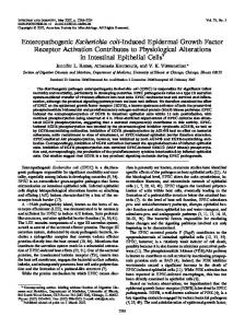
RESULTS Effect of GM-CSF and G-CSF on the E. coli alpha-hemolysin-induced chemiluminescence response from human PMN. Previously, we have shown that E. coli alpha-hemolysin induces a characteristic and dose-dependent chemiluminescence response from human PMN (23). To analyze the effects of GM-CSF and G-CSF on the chemiluminescence response, human PMN (10") were preincubated with buffer, GM-CSF, or G-CSF in various amounts (5, 10, and 50 ng) for 30 min at 37°C; the subsequent incubation proceeded in the presence of hemolysin-positive E. coli bacteria [E. coli 5K(pANN202-312)] at an optimal concentration for chemiluminescence response (hemolytic activity, 50 to 70%; 107 to 108 bacteria) (23) for up to 20 min. GM-CSF pretreatment resulted in an increased basal chemiluminescence response (buffer) in a dose-dependent manner (from 2,000 cpm up to 3,500 cpm with 10 ng of GM-CSF); G-CSF treatment did not modulate the basal chemiluminescence response from human PMN (data not shown). As is apparent from Fig. 1, in which the results of a typical experiment are shown (mean values for one experiment performed in triplicate), pretreatment (30 min) with 5 ng of GM-CSF led to an increase in the E. coli alpha-hemolysininduced chemiluminescence response of up to twofold. GMCSF amounts greater than 5 ng did not further increase the chemiluminescence response from human PMN. A preincubation time of 10 to 60 min was similarly effective with regard to the priming efficacy (data not shown). In the presence of G-CSF, the results are clearly less pronounced; G-CSF pretreatment enhanced the chemiluminescence response maximally by up to 30% ± 10% (10 ng of G-CSF) (data not shown); a preincubation time of 10 to 30 min was most effective with regard to the priming efficacy (data not shown). Effect of GM-CSF and G-CSF on enzyme release (,I-glucuronidase, lysozyme, and LDH) from human neutrophils induced by the E. coli alpha-hemolysin. In the next series of experiments, we analyzed the effects of GM-CSF and G-CSF on enzyme release (3-glucuronidase, lysozyme, and LDH) from human PMN challenged with hemolysin-producing E. coli bacteria. In the first set of experiments, human PMN were preincubated in the absence or presence of GM-CSF or G-CSF (0, 1, 10, or 50 ng) for 30 min at 37°C; the incubation proceeded for a further 30 min at 37°C in the presence of an optimal concentration of the Hly+ bacteria, which lead to 3-glucuronidase and lysozyme release but not to LDH release (hemolytic activity, 55% ± 15%). As is apparent from Table 1, for an incubation time of 30 min the E. coli alpha-hemolysininduced 3-glucuronidase release increased to 24% ±+ 6% of the total cellular enzyme activities. Similar results were obtained when human PMN were challenged with hemolysinpositive E. coli bacteria for a longer time (up to 90 min) or were expressing a hemolytic activity between 10 and 100%. Independently of the GM-CSF and G-CSF concentrations, the release of P-glucuronidase was only slightly enhanced. With regard to lysozyme release, GM-CSF and G-CSF induced a maximal lysozyme release of 11% ± 2% after an incubation time of 90 min; no LDH release was observed. As is apparent from Table 1, with a GM-CSF or G-CSF pretreatment of 30 min and an incubation time of 30 min with Hly+ E. coli
50
2087
-
601 GM-CSF [ng]
-
0
40
-
5
-
E3-
oI
200 10 0
5
10
15
20
Time [min] Effects of GM-CSF on the hemolysin-induced chemilumifrom human PMN. Neutrophils (2 x 10") were preincubated in the absence (buffer control) or presence of GM-CSF at the indicated concentrations (nanograms per 107 cells) for 30 min at 37°C, and then incubation proceeded in the presence of Hly+ E. coli 5K(pANN202-312) (hemolytic activity, 55%l ± 15`1c; 1(7 to 1(8 bacteria) for the indicated times at 37C. Results of one of three typical experiments from three different donors are shown. FIG.
1.
nescence response
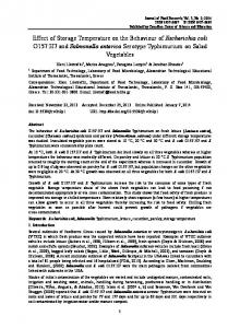
bacteria [SK(pANN202-312); hly+ activity, 55c ±- 15%] a decrease in E. coli alpha-hemolysin-induced lysozyme release was observed. The inhibitory effects were maximal with 10 ng of GM-CSF and were reversed with greater amounts (Table 1). G-CSF pretreatment led to a decrease in lysozyme release in a dose-dependent manner (Table 1). GM-CSF and G-CSF did not affect LDH release (less than 10% of total LDH content) from human PMN challenged with Hly+ E. coli bacteria (data not shown). Hly E. coli bacteria of strain 5K did not induce 3-glucuronidase, lysozyme, or LDH release from human PMN; neither GM-CSF nor G-CSF showed any modulatory effects. Effects of GM-CSF and G-CSF on E. coli alpha-hemolysininduced LTB4 generation. Human PMN were preincubated in the presence of PBS (buffer control) or in the presence of GM-CSF or G-CSF at different concentrations (1, 5, 10, and 50 ng) for 30 min at 37°C; incubation then proceeded for a further 2, 5, 10, or 20 min at 37°C in the presence of buffer or of the Hly+ E. coli bacteria at an optimal concentration for LTB4 generation (Hly+, 55% ± 15%) (22). GM-CSF by itself induced only low amounts of LTB4 (up to 1.7 ± 0.5 ng/107 a total incubation time of 50 min even when the amount was increased to 50 ng; G-CSF failed to do so. Human neutrophils were primed for an enhanced LTB4 generation after challenge with Hly+ E. coli bacteria [SK(pANN202-312)] (Fig. 2). However, depending on the donor, both GM-CSF and
cells) after
2088
KONIG AND KONIG
INFEcr. IMMUN.
TABLE 1. Effects of GM-CSF and G-CSF on 13-glucuronidase and lysozyme release from human PMN challenged by E. coli alpha-hemolysina Release of compound (% of total enzyme activities) with indicated amt (ng) of factor" Factor and stimulus
GM-CSF Buffer only E. coli 5K(pANN202-312) E. coli 5K G-CSF Buffer only E. coli SK(pANN202-312) E. coli 5K
0
1
10
50
,B-Glucoronidase
Lysozome
,B-Glucoronidase
Lysozome
,-Glucoronidase
Lysozome
3-Glucoronidase
Lysozome
7.5 ± 3.2 24.6 ± 6.5 7.8 ± 0.9
10.0 ± 2.3 52.0 ± 4.3 10.3 ± 2.5
9.3 ± 4.2 33.1 ± 7.3 8.4 ± 1.2
8.8 ± 3.1 41.6 ± 5.8 10.4 ± 2.3
10.7 ± 3.1 34.3 ± 6.5 8.0 ± 2.5
7.7 ± 3.1 37.5 ± 3.7 10.2 ± 3.0
10.8 ± 4.5 34.2 ± 5.8 9.3 ± 2.5
11.0 ± 2.8 52.1 ± 5.3 11.8 ± 2.1
7.5 ± 3.2 24.6 ± 6.5 7.8 ± 0.9
10.0 ± 2.3 52.0 ± 4.8 10.3 ± 2.5
4.8 ± 1.5 32.6 ± 7.0 6.8 ± 2.8
10.0 ± 2.1 47.9 ± 2.5 10.4 ± 2
5.9 ± 2.4 31.3 ± 4.8 7.3 ± 1.2
10.0 ± 2.3 43.7 ± 4.0 9.8 ± 1.0
8.8 ± 4.8 31.5 ± 5.2 8.4 ± 1.9
11.0 ± 2.3 41.6 ± 3.8 10.4 ± 0.8
Hemolytic activity was 55% ± 15% (see Materials and Methods). b Data are means ± SDs for three independent experiments.
G-CSF primed human PMN for an enhanced LTB4 generation after challenge with Hly+ E. coli bacteria (Table 2). Nonetheless, the priming efficacy of G-CSF at 10 ng was less than that of GM-CSF. A preincubation time of 10 min for GM-CSF and for G-CSF was necessary to show the priming effects (data not shown). Hly- bacteria of strain E. coli 5K failed to induce significant leukotriene generation from human PMN; neither GM-CSF nor G-CSF showed modulatory effects on leukotriene generation from PMN treated with E. coli 5K. The experiments were performed under noncytotoxic conditions (trypan blue exclusion; LDH release, 5%). Effects of GM-CSF and G-CSF on histamine release from human basophils. The E. coli alpha-hemolysin is a potent inducer for histamine release from human basophils (23). To study the influence of GM-CSF and G-CSF on E. coli alphahemolysin-induced histamine release, an LMB suspension was used. In a first set of experiments, the optimal preincubation time and the optimal concentrations for GM-CSF and G-CSF pretreatment were determined. LMB were preincubated in the presence of GM-CSF (0, 1, 5, 10, or 50 ng) or G-CSF (0, 1, 5, 10, or 50 ng) for 0, 30, 45, or 60 min at 37°C. The incubation then proceeded in the absence (buffer control only) or presence of Hly+ E. coli bacteria expressing hemolysin at an optimal concentration for histamine release (hemolytic activity, 55% ± 15%). GM-CSF pretreatment primed human basophils similarly to human neutrophils over the concentration range tested; maximal priming effects were obtained at a GM-CSF amount of 10 ng (data not shown). The priming effects were quite similar when a preincubation time between 30 and 60 min at 37°C was chosen (data not shown). G-CSF pretreatment did not significantly increase E. coli alphahemolysin-induced histamine release (data not shown). We then studied the priming effects of GM-CSF (10 ng) when human LMB were stimulated with Hly+ E. coli 5K(pANN202312) at different hemolysin concentrations. Human LMB were preincubated in the absence (PBS buffer alone) or in the presence of GM-CSF (10 ng) for 30 min at 37°C; the incubation proceeded in the absence or presence of Hly+ E. coli bacteria (hemolytic activities, 80, 64, and 50%) for a further 60 min at 37°C. As is apparent from Table 3, the priming effect of GM-CSF was observed at all tested hemolysin concentrations. Hly- E. coli bacteria of strain 5K failed to induce histamine release from human LMB; neither GM-CSF nor G-CSF showed modulatory effects on histamine release from LMB treated with E. coli 5K. Effect of GM-CSF and G-CSF on IL-8 release from human PMN and LMB. First, we studied the effects of GM-CSF and
G-CSF by themselves on IL-8 release from human PMN and LMB. As is apparent from Table 4, GM-CSF and G-CSF at concentrations of 10 ng induced a time-dependent increase in IL-8 release from human PMN. The GM-CSF-induced maximal IL-8 release was observed after an incubation time of 100 min; thereafter, IL-8 release declined. A similar activation pattern was obtained for G-CSF. For LMB, identical results were obtained (data not shown). E. coli alpha-hemolysin (hemolytic activity, 55% ± 15%) led to an increase (over values obtained with the buffer control) in IL-8 release from human PMN after an incubation time of 60 min; thereafter, the IL-8 release declined to baseline levels (Table 4). In human LMB, Hly+ E. coli bacteria expressing hemolytic activities from 14.2 to 46% led to a downregulation of IL-8 release compared with values obtained with the buffer control (Fig. 3). To study the effects of GM-CSF and G-CSF on hemolysin-
14
GM-CSF
[ng]
No
12 W
6--
2
10 Time [min]
5
20
FIG. 2. Effects of GM-CSF on hemolysin-induced LTB4 generation from human PMN. Neutrophils (107/500 I.l) were preincubated in the absence (buffer) or presence of GM-CSF (1, 5, 10, or 50 ng/107 cells) for 30 min at 37°C, and incubation then proceeded in the absence or presence of Hly+ E. coli 5K(pANN202-312) (hemolytic activities, 55% ± 15%) bacteria for a further 2, 5, 10, or 20 min at 37°C. Data are means + SDs (bars) for three independent experiments.
EFFECT OF GROWTH FACTORS ON MEDIATOR RELEASE
VOL. 62, 1994
2089
TABLE 2. Effects of GM-CSF and G-CSF on LTB4 generation by the indicated E. coli strains LTB4 generation (ng/1l(7 neutrophils) by the indicated E. coli strain after the indicated time (min) 2) 10
Preincubation with: 5K(pANN202-312)
5K
5K(pANN202-312)
5K
5K(pANN20I2-312)
5K
18.12 ± 4.3 121.70 ± 13.1 41.0 ± 7.3"
18.2 ± 5.8
34.58 ± 7.3 109.79 ± 13.8" 60.40 ± 10.1"
19.4 ± 3.1 17.6 ± 7.9 19.4 ± 5.7
15.84 ± 3.4 71.74 ± 8.5" 18.80 ± 4.5
17.7 ± 2.7 18.9 ± 4.2 16.8 ± 6.4
Buffer only GM-CSF (10 ng) G-CSF (10 ng) " Significant increase (P
0,1 5
-
E. coli*
NDC ND 800 ± 56d
1,400 + 110" 4,800 ± 700" 900 ± 60" ND
incubation time 30 min -
m
r-
0
0) C v-
0,1
-
CD
0,05
" Data are means ± SDs for three independent experiments. The amount of
GM-CSF and of G-CSF was 10 ng. "Hemolytic activity, 55% ± 15%. 'ND, not determined. "Significant increase (P < 0.05) in IL-8 release compared with the buffer control.
cytokines, e.g., interferons, IL-2, and CSFs, were used in the treatment of immunological disorders and cancer. GM-CSF is secreted by a large variety of cells. The principal sources are macrophages, endothelial cells, and activated T cells. GM-CSF was originally described as a factor that stimulated the growth of colonies from immature bone marrow progenitor cells (28). However, it became apparent that, in addition to its activity as a mitogen for granulocyte precursors, GM-CSF has numerous effects on mature PMN (14). To date, enhanced functional activities on mature neutrophils, eosinophils, basophils, and mononuclear phagocytes have been described (14, 28). In this regard, it has been shown that GM-CSF primes neutrophils for enhanced oxidative metabolism upon stimulation with FMLP, C5a, and LTB4 (25). Our results support and extend these findings. GM-CSF pretreatment and, to a lesser degree, GCSF pretreatment increased the E. coli alpha-hemolysin-induced chemiluminescence response from human PMN. The E. coli alpha-hemolysin presents a physiological stimulus released from E. coli bacteria during the logarithmic growth phase. Recent evidence has shown that cytokines can stimulate the production of 5-LO products, e.g., LTB4 (7). LTB4 is a major mediator of leukocyte activation in acute inflammatory reactions that produces chemotaxis, lysosomal enzyme release, and cell aggregation. Other investigators have shown that GM-CSF primes PMN to produce LTB4 when treated with the Ca ionophore A23187, with platelet activating factor, or with the chemotactic peptide FMLP (11, 27). LTB4 induces polymorphonuclear leukocyte migration in vivo and is a potent activator of human leukocytes (24). It thus represents a potent mediator of inflammation. We showed that GM-CSF dramatically increased E. coli alpha-hemolysin-induced generation of LTB4 from human neutrophils, which are the cells of the first line of defense against invading microorganisms. Previously, we have shown that E. coli alpha-hemolysin is also a potent stimulus for the release of histamine, which is responsible for vasodilation (23). GM-CSF pretreatment also enhanced E. coli alpha-hemolysin-induced histamine release. These results indicate that GM-CSF pretreatment primes effector cells for an exaggerated inflammatory event. G-CSF pretreatment primed inflammatory cells for cellular responses to a lesser degree than did GM-CSF. In contrast to inflammatory mediators such as 02- metabolites, leukotrienes, and histamine, the release of cytokines (IL-6, TNF-oa, and IL-1,) from human LMB was downregulated in the presence of the E. coli alpha-hemolysin (22). In addition to LTB4, IL-8 evokes a chemotactic response from
buffer
14
27
40
42
44
46
hemolytic activities of E. coli5K(pANN202-312)
B
incubation time 90 min
1,4
-
1,2
-
-J 1 m -0
0) 0,6
-
_ 0,4
-
0,2
-
r_
buffer
46
44
42
40
27
14
hemolytic activities of
coliK(pANN202-312) FIG. 3. IL-8 release from human LMB challenged with Hly+ E. coli E.
SK(pANN202-312) bacteria. Human LMB (106/ml) were incubated without (buffer control) or with Hly+ SK(pANN202-312) bacteria for 30 min (A) or 90 min (B) at 37°C. The cell supernatants were analyzed for IL-8 by ELISA. The data are means + SDs for three independent experiments. An asterisk indicates a significant difference from the PBS control (P < 0.05). PMN and therefore may be important in recruiting PMN into sites of inflammation (1). IL-8 has also been implicated in a large number of inflammatory diseases in humans including psoriasis, rheumatoid arthritis, and gram-negative sepsis (32). Concomitantly with other investigators, we showed that human PMN and LMB constitutively produced IL-8 mRNA and protein (29). Our data show that up to 12 ng/ml of IL-8 protein is released into the supernatants by human peripheral blood PMN (2 x 10⁷ cells/ml) stimulated with GM-CSF (5 ng/ml) or G-CSF. This event is donor dependent and may be due to individual cell reactivity. In contrast to LTB4 generation from human LMB, E. coli alpha-hemolysin, down to a hemolytic activity of 10% led to a decrease in basal IL-8 level and a decrease in IL-8-specific mRNA. When human PMN were chosen as target cells, E. coli alpha-hemolysin induced a slight increase in IL-8 release in a time-dependent manner; a downregulation was not observed up to an incubation time of 120 min. Evidence that IL-8 interacts with TNF and GM-CSF as well as with G-CSF has been obtained (1, 33). In contrast to the above-described effects of GM-CSF and G-CSF on inflam-
EFFECT OF GROWTH FACTORS ON MEDIATOR RELEASE
VOL. 62, 1994
2091
okadaic acid/GM-CSF -
PBS/GM-CSF okadaic acid/PBS
600
PBS/G-CSF
-
-x-
-x-
E
PBS/GM-CSF
500
PBS/PBS/
400-
E
PBS/PBS
400-
0 0 '-
r-300 -
300-
200
200
100 100
0
10
20
30
40
50
Time [min] FIG. 4. Effects of GM-CSF and G-CSF on NaF-induced chemiluminescence responses from human PMN. Neutrophils (2 x 10⁶) were preincubated in the absence (buffer control) or presence of GM-CSF (10 ng) or G-CSF for 30 min at 37°C; thereafter, incubation proceeded in the presence of buffer or NaF (15 mM) for the indicated times at 37°C. Data are means + SDs for three independent experiments.
0
10
20
30
40
50
Time [min] FIG. 5. Influence of okadaic acid on the GM-CSF priming efficacy for the NaF-induced chemiluminescence response from human PMN. Neutrophils (2 x 10⁶) were preincubated in the absence (buffer control) or presence of okadaic acid (10-8 M) for 10 min at 37°C, and then incubation proceeded in the presence of buffer or GM-CSF (10 ng) for 30 min at 37°C; thereafter, PMN were stimulated with NdF (15 mM) for a further 40 min at 37°C. Data are means + SDs for three independent experiments.
matory mediator release (02- generation, LTB4 generation,
and histamine release) from human PMN and LMB in response to E. coli alpha-hemolysin, the effects of E. coli alpha-hemolysin on IL-8 release were not influenced either by GM-CSF or by G-CSF pretreatment, regardless of the cell type used (PMN or LMB). Thus, we conclude that depending on the mediator, distinct signal transduction pathways which are differently influenced by GM-CSF or G-CSF are involved. Up to now, the specific mechanisms by which GM-CSF and G-CSF activate and prime various neutrophil functions including the generation of LTB4, the release of IL-8 and of superoxide anions, and the degranulation of neutrophils or basophils are not clear. The findings of Bourgoin et al. exclude the involvement of the Ptd1ns(4,5)P2-specific phospholipase C-diacylglycerol pathway in neutrophil priming by GM-CSF (4). Gomez-Cambronero et al. suggested a role of guanine nucleotide-regulatory proteins (G proteins) in GM-CSF priming (17). We observed that GM-CSF pretreatment (with results similar to those of McColl et al.) and to a lesser degree G-CSF pretreatment enhanced chemiluminescence response induced via direct G-protein activation through NaF by sixfold (GMCSF) or fourfold (G-CSF), while PMA-induced chemiluminescence response via direct activation of protein kinase C was only slightly enhanced (25). Similarly, the NaF-induced but not the PMA-induced histamine release was enhanced by GMCSF and G-CSF (by 60% ± 5% and 29% ± 6%, respectively).
These results indicate that signal transduction elements upstream of protein kinase C, e.g., G proteins, are involved in chemiluminescence and histamine priming by GM-CSF and G-CSF. Thus, our new data support our previous findings that G proteins are involved in E. coli alpha-hemolysin-induced cellular signalling (21). Receptors for the hematopoietic growth factors erythropoietin, IL-3, and GM-CSF are members of a structurally related receptor superfamily. Interestingly, while none of these receptors encodes tyrosine kinase activities, tyrosine phosphorylation has been observed in various responsive cells stimulated with each factor, indicating the involvement of tyrosine kinases in the priming effects of GM-CSF and G-CSF (13, 16, 18, 26). Indeed, the tyrosine kinase inhibitor lavendustin inhibited GM-CSF-induced IL-8 release and inhibited GM-CSF-primed IL-8 release induced by FMLP. The FMLP-induced histamine release from GM-CSF-primed basophils was not influenced by lavendustin (data not presented). In contrast, tyrosine kinase inhibitors, e.g., lavendustin and genistein, failed to modulate the priming effects of GM-CSF or of G-CSF in our described test systems. Moreover, as described in Results, the threonine/ serine phosphatase inhibitor okadaic acid diminished the priming efficacy of GM-CSF and G-CSF. Pretreatment of human PMN with okadaic acid abolished GM-CSF and G-CSF priming with regard to the NaF- and E. coli alpha-hemolysin-
2092
KONIG AND KONIG
INFECT. IMMUN.
studies are directed to analysis of the signal transduction pathway involved in the GM-CSF and G-CSF priming effects and its role in microbial pathogenicity. ACKNOWLEDGMENT W.K. was supported in this work by a grant from the Deutsche
Forschungsgemeinschaft (Ko 427/7-5).
1
2
3
4
5
6
7
8
9 10 11 12
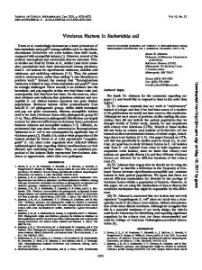
FIG. 6. 5-LO-specific mRNA expression by GM-CSF and G-CSF. PMN (2 x 10⁷/ml) were incubated in the presence of GM-CSF (10 ng) (lanes 1 to 6) or G-CSF (10 ng) (lanes 7 to 12) for 2, 5, 10, 30, 60, or 90 min at 37°C (lanes 1 and 7, 2 and 8, 3 and 9, 4 and 10, 5 and 11, and 6 and 12, respectively). Total RNA was reverse transcribed with was amplified by PCR (35 cycles) as described in Materials and Methods. The amplified product was of the appropriate size (375 bp). A 100-bp ladder (Gibco BRL) was used as a molecular size marker (right lane). The figure represents a typical result out of four experiments.
oligo(dT)12-18, and the product
induced chemiluminescence response. The priming effects of GM-CSF and G-CSF on E. coli alpha-hemolysin-induced histamine release were not affected by okadaic acid or genistein (data not presented). The possible involvement of a threonine/serine kinase, e.g., RAF-1, in GM-CSF priming has also been suggested by other investigators (8). Thus, GM-CSF may act via different signal-transducing elements depending on the inflammatory mediator and/or the cell type. For LTB4 generation, 5-LO and the 5-LO-activating protein play central roles (24, 30). Consistent with results obtained by other investigators, GM-CSF alone did not stimulate the release of detectable amounts of LTB4 or its omega products (12, 27). McColl et al. excluded a direct effect of GM-CSF on 5-LO activity (27). Our results have shown that GM-CSF and G-CSF in a concentration range from 1 to 50 ng/107 cells upregulate 5-LO-specific mRNA expression in human neutrophils within 2 to 5 min, thus offering a higher availability for 5-LO. Recently, a concentration-dependent effect of GM-CSF on 5-LO-activating protein de novo synthesis was reported (31). Nonetheless, maximal effects on mRNA level were observed after 4 h; the total 5-LO-activating protein concentration increased only minimally. Furthermore, neutrophils express the receptor for LTB4, which is downregulated as a consequence of GM-CSF treatment (11). Thus, some of the GM-CSF priming events, e.g., the enhanced LTB4 generation from human neutrophils, may be mediated by direct autocrine stimulation of neutrophils by LTB4. Whether pretreatment predominantly with GM-CSF, leading to an exaggerated response pattern from inflammatory cells, will enhance the host defense against microorganisms and their products or will cause inappropriate inflammation and tissue damage has to be further elucidated. Thus, further
REFERENCES 1. Baggiolini, M., A. Walz, and S. L. Kunkel. 1989. Neutrophilactivating peptide-1/interleukin 8, a novel cytokine that activates neutrophils. J. Clin. Invest. 84:1045-1049. 2. Bazzoni, F., M. A. Cassatella, F. Rossi, M. Ceska, B. Dewald, and M. Baggiolini. 1991. Phagocytosing neutrophils produce and release high amounts of the neutrophil-activating peptide 1/interleukin 8. J. Exp. Med. 173:771-774. 3. Bhakdi, S., M. Muhly, S. Korom, and G. Schmidt. 1990. Effects of Escherichia coli hemolysin on human monocytes-cytocidal action and stimulation of interleukin-1 release. J. Clin. Invest. 85:17461753. 4. Bourgoin, S., E. Plante, M. Gaudry, P. H. Naccache, P. Borgeat, and P. E. Poubelle. 1990. Involvement of a phospholipase D in the mechanism of action of granulocyte-macrophage colony-stimulating factor (GM-CSF): priming of human neutrophils in vitro with GM-CSF is associated with accumulation of phosphatidic acid and diacylglycerol. J. Exp. Med. 172:767-777. 5. Boyum, A. 1976. Isolation of lymphocytes, granulocytes and macrophages. Scand. J. Immunol. 5:9-15. 6. Brom, J., and W. Konig. 1992. Signal transduction and priming of human neutrophils. Int. Arch. Allergy Immunol. 99:387-389. 7. Brom, J., and W. Konig. 1992. Cytokine-induced (interleukin-3, -6, and -8 and tumour necrosis factor-beta) activation and deactivation of human neutrophils. Immunology 75:281-285. 8. Carroll, M. P., L. I. Clark, U. R. Rapp, and W. S. May. 1990. Interleukin 3 and granulocyte-macrophage colony-stimulating factor mediate rapid phosphorylation and activation of cytosolic c-raf. J. Biol. Chem. 265:19812-19817. 9. Chomezynski, P., and N. Sacchi. 1987. Single step method of RNA isolation by acid guanidium thiocyanate-phenol-chloroform-extraction. Anal. Biochem. 162:156-159. 10. Colditz, I. G., R. D. Zwahlen, and M. Baggiolini. 1990. Neutrophil accumulation and plasma leakage induced in vivo by neutrophilactivating peptide-1. J. Leukocyte Biol. 48:129-137. 11. Dahinden, C. A., J. Zingg, and F. E. Maly de Wal. 1988. Leukotriene production in human neutrophils primed by recombinant human granulocyte/macrophage colony-stimulating factor and stimulated with the complement component C5A and FMLP as second signals. J. Exp. Med. 167:1281-1295. 12. DiPersio, J. F., P. Billing, R. Williams, J. C. Gasson, and J. F. DiPersio. 1988. Human granulocyte-macrophage colony-stimulating factor and other cytokines prime human neutrophils for enhanced arachidonic acid release and leukotriene B4 synthesis. J.
Immunol. 140:43315-43318.
13. Evans, J. P., S. A. R. Mire, A. V. Hoffbrand, and R. G. Wickremasinghe. 1990. Binding of G-CSF, GM-CSF, tumor necrosis factor-alpha, and gamma-interferon to cell surface receptors on human myeloid leukemia cells triggers rapid tyrosine and serine phosphorylation of a 75 kd protein. Blood 75:88-92. 14. Fabian, I., Y. Kletter, S. Mor, C. Geller-Bernstein, M. BenYaakov, B. Volovitz, and D. W. Golde. 1992. Activation of human eosinophil and neutrophil functions by haematopoietic growth factors: comparisons of IL-1, IL-3, IL-5 and GM-CSF. Br. J. Haematol. 80:137-143. 15. Goebel, W., and J. Hedgpeth. 1982. Cloning and functional characterization of the plasmid-encoded hemolysin determinant of
Escherichia coli. J. Bacteriol. 151:1290-1298.
16. Gomez-Cambronero, J., C. K. Huang, V. A. Bonak, E. Wang, E. Casnellie, T. Shiraishi, and R. I. Sha'afi. 1989. Tyrosine phosphorylation in human neutrophils. Biochem. Biophys. Res. Commun.
162:1478-1485. 17. Gomez-Cambronero, J., M. Yamazaki, F. Metwally, T. F. P. Melski, V. A. Bonak, C. Huang, E. L. Becker, and R. I. Sha'afi.
VOL. 62, 1994
18. 19.
20.
21.
22.
23.
24.
25.
EFFECIT OF GROWTH FACTORS ON MEDIATOR RELEASE
1989. Granulocyte-macrophage colony-stimulating factor and human neutrophils: role of guanine nucleotide regulatory proteins. Proc. Natl. Acad. Sci. USA 86:3569-3573. Isfort, R. J., and J. N. Ihie. 1990. Multiple hematopoietic growth factors signal trough tyrosine phosphorylation. Growth Factors 2:213-216. Kawakami, M., H. Tsutsumi, T. Kumakawa, H. Abe, M. Hirai, S. Kurosawa, M. Mori, and M. Fukushima. 1990. Levels of serum granulocyte-stimulating factor in patients with infections. Blood 76:1962-1964. Knoller, J., W. Schonfeld, M. Koller, T. Hensler, and W. Konig. 1988. Arachidonic acid metabolites from polymorphonuclear leukocytes of healthy donors, severely burned patients, and children with cystic fibrosis. Routine monitoring by high-performance liquid chromatography. J. Chromatogr. 427:199-208. Konig, B., and W. Konig. 1991. Roles of human peripheral blood leukocyte protein kinase C and G proteins in inflammatory mediator release by isogenic Escherichia coli strains. Infect. Immun. 59:3801-3810. Konig, B., and W. Konig. 1993. Induction and suppression of cytokine release (TNF-a; IL-6; IL-1p) by E. coli pathogenicity factors (adhesins, a-hemolysin). Immunology 78:526-533. Konig, B., W. Konig, J. Scheffer, J. Hacker, and W. Goebel. 1986. Role of Escherichia coli alpha-hemolysin and bacterial adherence in infection: requirement for release of inflammatory mediators from granulocytes and mast cells. Infect. Immun. 54:886-892. Konig, W., W. Schonfeld, M. Raulf, M. Koller, J. Knoller, J. Scheffer, and J. Brom. 1990. The neutrophil and leukotrienesrole in health and disease. Eicosanoids 3:1-22. McColl, S. R., D. Beauzeigle, C. Gilbert, and P. H. Naccache. 1990. Priming of the human neutrophil respiratory burst by granulocytemacrophage colony-stimulating factor and tumor necrosis factoralpha involves regulation and a post-cell surface receptor level. Enhancement of the effect of agents which directly activate G-proteins. J. Immunol. 145:3047-3053.
2093
26. McColl, S. R., F. DiPersio, A. C. Caon, P. Ho, and P. H. Naccache. 1991. Involvement of tyrosine kinases in the activation of human peripheral blood neutrophils by granulocyte-macrophage colonystimulating factor. Blood 78:1842-1852. 27. McColl, S. R, E. Krump, P. H. Naccache, P. E. Poubelle, P. Braquet, M. Braquet, and P. Borgeat. 1991. Granulocyte-macrophage colony-stimulating factor increases the synthesis of leukotriene B4 by human neutrophils in response to platelet-activating factor. Enhancement of both arachidonic acid availability and 5-lipoxygenase activation. J. Immunol. 146:1204-1211. 28. Metcalf, D. 1985. The granulocyte-macrophage colony stimulating factors. Cell 43:5-6. 29. Mielke, V., J. G. J. Baumann, M. Sticherling, T. Ibs, A. G. Zomershoe, K. Seligmann, H.-H. Henneicke, J.-M. Schroder, W. Sterry, and E. Christophers. 1990. Detection of neutrophil-activating peptide NAP/IL-8 and NAP[IL-8 mRNA in human recombinant IL-la and human recombinant tumor necrosis factor-astimulated human dermal fibroblasts. J. Immunol. 144:153-156. 30. Reid, G. K., S. Kargman, P. J. Vickers, J. A. Mancini, C. Lereille, D. Ethier, D. K. Miller, J. W. Gillard, R A. F. Dixon, and J. F. Evans. 1990. Correlation between expression of 5-lipoxygenaseactivating protein, 5-lipoxygenase, and cellular leukotriene synthesis. J. Biol. Chem. 265:19818-19823. 31. Van Zee, K. J., L. E. DeForge, and E. Fischer. 1991. IL-8 in septic shock, endotoxemia, and after IL-1 administration. J. Immunol. 146:3478-3482. 32. Willems, J., M. Joniau, S. Cinque, and J. van Damme. 1991. Human granulocyte chemotactic peptide (IL-8) as a specific neutrophil degranulator; comparison with other cytokines. Immunology 67:540-542. 33. Yoshimura, T. K, S. Matsushima, S. Tanaka, E. A. Robinson, E. Appela, J. Oppenheim, and E. J. Leonard. 1987. Purification of a human monocyte-derived neutrophil chemotactic factor that shares sequence homology with other host defense cytokines. Proc. Natl. Acad. Sci. USA 84:9233-9237.