measured after administration of human recombinant interferon (rIFN)-alpha-2a.
Interferon (IFN) injection caused a dramatic decrease in lymphocyte output from ...
Immunology 1988 64 469-474
Effect of interferon-alpha-2a on the output of recirculating lymphocytes from single lymph nodes W. R. HEIN* & A. SUPERSAXOt *Basel Institute for Immunology and tPharmaceutical ResearchiPreclinical Development, F. Hoffmann-La Roche & Co. Ltd, Basel, Switzerland
Acceptedfor publication 14 March 1988
SUMMARY The output of recirculating lymphocytes from cannulated popliteal lymph nodes in sheep was measured after administration of human recombinant interferon (rIFN)-alpha-2a. Interferon (IFN) injection caused a dramatic decrease in lymphocyte output from lymph nodes. Following a single s.c. or i.d. injection of 2 x 10⁷ U IFN into the drainage area of the popliteal lymph node, lymphocyte output fell to below 1% of the pre-treatment level and remained depressed for up to 35 hr. A substantial decrease in lymphocyte output from cannulated nodes also occurred after IFN was injected either i.v., into the skin of the opposite non-cannulated hind leg or into an afferent lymphatic vessel leading to the popliteal lymph node. After the period of depressed lymphocyte output, a seemingly compensatory surge of cell traffic occurred that lasted 2-3 days. During this phase there was a relative increase in the proportion of CD4+ T cells in lymph. Similar changes occurred after each treatment in animals given multiple doses of IFN. These effects are unlikely to be antigeninduced since there was no blast cell response in any treated animal. The analysis of blood and lymph plasma samples showed that the most severe depression of lymphocyte output was associated with high levels of IFN, while there was no apparent correlation between the reduction in lymphocyte traffic and concentrations of cortisol in plasma. These results suggest that IFN-alpha-2a is involved directly in the regulation of lymphocyte output from lymph nodes.
INTRODUCTION The continual migration of lymphocytes from blood to lymph is an essential prerequisite for a functional immune system. Lymphocyte recirculation through lymph nodes has special significance because it occurs at a tempo many times greater than lymphocyte migration through non-lymphoid tissues and plays a pivotal role in immune surveillance and the dissemination of an immune response (Hay & Cahill, 1982). The rate of lymphocyte recirculation through single lymph nodes changes suddenly after exposure to antigen (Cahill, Frost & Trnka, 1976). However, the mechanisms that control the decrease and increase in the output of lymphocytes from lymph nodes undergoing an immune response are incompletely understood and it is important to identify agents that disrupt this process. Studies done in rodents suggest that IFN may influence lymphocyte recirculation. Gresser et al. (1981) reported that injection of IFN into mice caused a decrease in the number of lymphocytes in the thoracic duct lymph and in the peripheral blood concomitant with an increase in the weight of peripheral lymph nodes. Administration of the potent IFN inducer polyinosinic-polycytidilic polyribonucleotide (poly I: polyC) to Correspondence: Dr W. R. Hein, Basel Institute for Immunology, Grenzacherstrasse 487, Postfach, CH-4005 Basel, Switzerland.
469
mice produced rapid changes in lymphocyte distribution, resulting in lymphopaenia (Schattner, Meshorer & Wallach, 1983) and a decrease in the number of thoracic duct lymphocytes (Korngold, Blank & Murasko, 1983). Recently, Kimber et al. (1987) reported that the incubation of rat thoracic duct lymphocytes with IFN prior to i.v. injection into recipients caused an increased localization into lymph nodes and a reduced output into the thoracic duct. Although these observations are consistent with the notion that IFN inhibits the egress of lymphocytes from lymphoid tissue, it is not feasible to measure this directly in rodents. The lymphocytes that leave the popliteal lymph node (PLN) of sheep can be collected quantitatively under physiological conditions over long periods of time. Nearly all of the lymphocytes that can be collected in the efferent lymph from the PLN are derived by recirculation from the blood (Hall & Morris, 1965; Hall, 1967) and the output of lymphocytes from the node is therefore a direct measure of lymphocyte traffic from blood to lymph. Earlier investigators used partially purified preparations of IFN alpha/beta and in these circumstances it is not clear whether the effects they observed were due to the action of a specific type of IFN. Pure formulations of recombinant IFNs are now available and in this report we describe the effects of IFN-alpha-2a on lymphocyte recirculation in single lymph nodes. Our results suggest that IFN-alpha-2a is a direct mediator of lymphocyte 'shutdown'.
470
W. R. Hein & A. Supersaxo MATERIALS AND METHODS
Animals and surgical procedures White Alpine and Black Jura sheep of both sexes, aged 2-4 years, were obtained from Versuchsbetrieb Sennweid, Olsberg. The cannulation of the efferent and afferent lymphatic vessels of the PLN was done as described elsewhere (Hall & Morris, 1962). Jugular canulae were introduced through the bore of a 14-gauge needle and secured in position with stay sutures. After the operation sheep were maintained in metabolism cages, fed a ration of pellets, and lymph and blood were collected as described elsewhere (Miyasaka & Trnka, 1985). Lymph collection flasks were changed every 10 min for the first hour after injection of IFN and then hourly until 6 hr after injection and then at 8-15 hr intervals. Injection of IFN-alpha-2a Experiments were not started until at least 2-3 days after surgery, by which time the output of lymphocytes in the lymph had stabilized to a baseline level. Two times 107 units of human recombinant IFN-alpha-2a (Roferon®, Roche, Nutley, NJ) were dissolved in 2 ml water and injected s.c. or i.d. into the lower hind leg using a peristaltic pump and special injection device that allowed administration at precise rates and depths (Supersaxo et al., 1988). This dose of IFN was chosen because it is approximately in the middle of the range of doses reported to be used for treatment of human cancer patients (Jones & Itri, 1986). In some experiments IFN was injected i.v. and on one occasion it was infused directly into lymphatic vessels afferent to the PLN.
Table 1. Peak concentrations of IFN-alpha-2a measured in blood and lymph plasma after administration by different routes Peak IFN concentration (U/ml) in
Mode of administration*
Lymph plasma
i.d. into cannulated leg i.d. into non-cannulated leg i.v. into jugular vein
3-1 x 10⁶ 60 14
38 315 8250
* Two millilitres of rIFN-alpha-2a solution injected over 20 min.
>,
z
Blood plasma
(1 x 10⁷ U/ml)
were
5
c
.-
4-
3
0
C
c 0
N,
u
(b)
0
25(
c
)
20(
0-
151
0i
A.
T
aG) -
0
cL
E
-"iflow
"
~~
a),
\ T
.4.....
Lymph
101
(V
Duration o 0 -injection
N
s_I
rate
Lymphocyte
_I
Analysis of IFN-alpha-2a The concentration of IFN-alpha-2a in blood and lymph plasma was measured by an enzyme-linked immunosorbent assay, as described elsewhere (Supersaxo et al., 1988). Analysis of cortisol The concentration of cortisol in blood and lymph plasma was measured by direct radioimmunoassay by Drs U. Hennes and A. Edelmann, Pharmaceutical Research, Hoffmann-La Roche, Basel. The assay was done in antibody-coated tubes using an antiserum prepared in rabbits against a cortisol 21-conjugate with bovine serum albumin. Cortisol standards and '25I-labelled cortisol were purchased from Sorin Biomedica (Saluggia, Italy). Output of lymphocytes in the efferent lymph For each collection period, the concentration of small and large efferent lymph lymphocytes (ELL) was measured with an electronic cell counter (Coulter Electronics, Dunstable, U.K.) and lymphocyte output per hour was calculated as the product of concentration (total cells/ml) and lymph flow rate (ml/hr). In one animal we examined the effect of IFN on the recirculation of lymphocyte subsets. ELL collected at intervals before and after two injections of IFN were analysed for surface expression of differentiation and major histocompatibility antigens using a panel of monoclonal antibodies (mAb). The mAbs used included ST-1 (pan sheep T cell), SBU-T4 (CD4 homologue, provided by Dr M. Brandon, University of Melbourne, Australia), ST-8 (CD8 homologue), 197 (third T-cell subset in sheep) and S064.6 and SW73.2 (MHC class I and class II respectively, provided by Dr J. Hopkins, University of Edinburgh, U.K.).
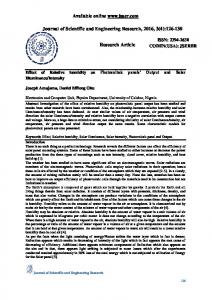
output 0 10 20 30 40 50 60 Time after start of injection (min)
Figure 1. The concentration of IFN-alpha-2a in popliteal efferent lymph (a) and changes in lymph flow rate and lymphocyte output (b) following s.c. or i.d. injection of 2 x I07 units of rIFN-alpha-2a into the lower hind leg over 20 min. Data shown as mean + SE of three experiments.
The properties of these mAbs and their target antigens have been reviewed recently (Miyasaka et al., 1988). Indirect surface immunofluorescent staining and FACS analysis were done as described elsewhere (Ezaki et al., 1987). B lymphocytes were stained directly with FITC-rabbit anti-sheep Ig (H+L) antibody (Cappel Laboratories, Cochranville, PA).
Histology Popliteal lymph nodes removed from sheep 10-20 hr after the s.c. injection of IFN-alpha-2a into the ipsilateral leg were fixed immediately in paraformaldehyde (4% solution in phosphatebuffered saline) at 4°. Popliteal nodes were also removed from non-treated control sheep and handled in a similar way. After routine embedding in methacrylate resin, 5 gim sections were cut and stained with Nocht's azure-eosin (Sigma Chemical Co., St Louis, MO). RESULTS Concentration of IFN in blood and lymph after different modes of administration The concentration of IFN measured in blood and lymph plasma varied greatly, depending on the site and duration of administration. Following either i.d. or s.c. injection, IFN was absorbed
471
IFN-alpha-2a and lymphocyte recirculation Table 2. Changes in lymphocyte output from the popliteal lymph node of sheep after the injection of IFN Lowest recorded
lymphocyte output Time taken to return IFN injection* to pre-injection level Time after % of preExp. Animal no. Route Duration injection level injection (hr) of lymphocyte output (hr) no. 1 2 3 4 7 9 10 11 12 13 14 15 16
3667 3568 3568 3568 3517 3836 3836 3118 3670 3670 3670 3670 3670
s.c. i.d. s.c. i.d. i.ly. s.c.
s.c. s.c. i.v. i.d. i.d. i.d. i.d.
30 min 20 min 20 min 20 min 2 hr 7 min 6 hr 6hr 20 min 20 min 6hr 20 mint 20 min