Reference: BioL BulL 166: 68—77.(February,
1984)
EFFECT OF TEMPERATURE ON INTERACTION BETWEEN EGGS AND SPERMATOZOA OF SEA URCHIN MASATOSHI MITA,I* AKIYA HINO,2 AND IKUO YASUMASU' ‘¿Department ofBiology, School ofEducation. Waseda University, Nishiwaseda, Shinjuku@ku,Tokyo 160. and 2DepartmentofBiology. Faculty ofScience,Nagoya University, Chikusa. Nagoya 464, Japan ABSTRACT
Fertilization
in the sea urchin,
Anthocidaris
crassispina,
showed marked
tem
perature dependence; high temperatures (15°—30°C) were required for fertilization. In contrast, fertilization in Hemicentrotus pukherrimus occurred over a wide range oftemperatures
(O°—30°C). The mechanism
ofthis
temperature
effect in Ant hocidaris
was investigated. The number of sperm bound to the egg surface and the rate of the acrosome reaction were markedly reduced by lower temperatures (O°—1O°C). Fur thermore, an abnormal elongation ofthe sperm head tip occurred with higher frequency at lower temperatures. In contrast, the egg activation with calcium ionophóre A23 187 was not prevented at 10°C.The swimming activity measured by distance traveled was also relatively high at 0°C,although the activity increased as the temperature rose. These results strongly suggest that temperature exerts a direct influence on fertilization in Anthocidaris by acting on the acrosome reaction. tilization rate at higher temperatures in Anthocidaris corresponds perature observed during the breeding season of this species.
The increased fer to the higher tern
INTRODUCTION
It is well known that development of sea urchin eggs and embryos is influenced by the temperature of the sea water. Generally, embryos develop relatively faster at high temperatures. Most sea urchin species have a specific breeding season. It is interesting
that exogastrulation
is induced
by culture
of embryos
at temperatures
lower than environmental condition during the breeding season (Takahashi et al., 1977). Fujisawa and Amemiya (1979, 1980) also reported that adhesion of dissociated cells in sea urchin embryos is related to the environmental temperature in the breeding
season. This is due, to some extent, to energy metabolism which depends on tem perature. Glycogen metabolism during early development in the sea urchin, Hemi centrotus puicherrimus, depends upon temperature. It has been reported that glycogen is metabolized at 15°C(Okabayashi and Nakano, 1980) but not at 20°C(Hino and Yasumasu, 1979). In sea urchin spermatozoa, energy metabolism is influenced by temperature. We already reported (Mita and Yasumasu, 1983) that phospholipid and carbohydrate metabolism in Hemicentrotus sperm are activated at 20°Cand only glycolysis is carried out at 0°C.Yanagisawa (1967) reported that Hemicentrotus sperm rnaintain a certain ATP level but decrease their phospholipid level after dilution at 14°C.Upon fertilization,
a dramatic
change
in metabolism
occurs
in both sperm
(Fujiwara
et a!.,
1983) and eggs (Yasumasu et aL, 1973). However, little is known about the effect of Received 31 May 1983; accepted 7 November * Present
address:
Department
1983.
of Developmental
Biology,
Nishigonaka, Myodaijicho, Okazaki 444, Japan. 68
National
Institute
For
Basic
Biology,
38
TEMPERATURE AND FERTILIZATION
69
temperature on fertilization. In the present study, we compare the effects of temperature on fertilization, using the sea urchins, Anthocidaris crassispina, which breeds in sum mer, and Hemicentrotus pukherrimus, which breeds in winter. MATERIALS AND METHODS
Preparation of gametes Shedding
of gametes of the sea urchins,
Anthocidaris
and Hemicentrotus,
was
induced by injection of 0.5 M KG into the coelomic cavity. Semen was collected as “¿dry sperm―and kept undiluted at 4°C.Eggs were shed directly into sea water. Artificial sea water (ASW) composed of485 mM NaC1, 9.6 mM KC1, 10 mM CaC12, 49 mM MgSO4, and 10 mM Tris-HC1, pH 8.2 was used. The number of eggs and sperm was calculated from the protein concentration as determined by the method of Lowry et a!. (195 1), using bovine serum albumin as standard. An egg and a spermatozoon
contained
100 ng protein and 0.5 pg protein, respectively.
Ferti!ization in artjficia! sea water Eggs were washed 3 times with ASW. The eggs were diluted in ASW (2 X iO@ eggs/ml) at a desired temperature and then 5 @il dry sperm (3 X 10⁸sperm) was added to 1.0 ml egg suspension. Five mm after insemination, 0.04 ml of 1% formaldehyde in ASW was dropped into the suspension. Under a light microscope, the percentage of fertilized eggs was calculated by counting the number of eggs with a fertilization membrane. Measurement of sperm moti!ity Sperm motility was determined by measuring the distance in which sperm traveled through a glass capillary vessel (d = 1 mm, Vitrex, Modulohm uS, Denmark) as described by Turner and Giles (1982) with modifications. Dry sperm were diluted in 0.5 ml ASW and a glass capillary vessel prefilled with ASW was immediately inserted into the suspension. The vessel rested at approximately 10°.At the conclusion of the incubation period, the distance which sperm traveled in the vessel was examined under a light microscope. Oxygen consumption assay Respiration was measured polarographically using a Clark type oxygen electrode (Yellow Spring Co., U.S.A.). Twenty-five @zl of dry sperm (2.3-2.8 X i09 sperm) were incubated in 2.5 ml ASW in a closed vessel equipped with an oxygen electrode. Oxygen consumption was determined as described by Robinson and Cooper (1970). A TP assay Samples were prepared for ATP assay as described by Mita and Yasumasu (1983). ATP concentration was determined enzymatically as described by Lamprecht and Trantsold (1974). Treatment of sperm with jelly water Jelly water was prepared by acidifying the egg suspensions with HC1 to pH 5.5 for 4 to 5 mm. The eggswere removed by a hand-driven centrifuge, and the supernatant
70
M. MITA ET AL.
was adjusted to pH 8.2 with Tris-HC1 buffer. Insoluble matter was removed by cen trifugation at 12,000 X g for 20 mm. The concentration ofjelly water was estimated by a carbohydrate determination using the method of Dische and Shettles (1951) with fucose as standard. Ten @l ofdry sperm (0.9-1.2 X l0@sperm) was treated with 1.0 ml ofjelly water at a concentration of 20 @g fucose equivalent/ml. After 5 mm incubation, the sperm were fixed by adding 1.0 ml of 4% glutaraldehyde in ASW. The percentage of sperm with the acrosome reaction was determined from electron microscopic observation (JEM 100CX, JEOL, and Hitachi HU-l 1-PS). Samples for scanning electron microscopy were fixed with 2% glutaraldehyde, and washed with ASW. A few drops ofconcentrated fixed sperm were placed onto a coverslip previously coated with 1% protamin sulfate (Nakarai Chemical Co., Japan). After 10 mm, one or two drops of 4% OsO4 were added to the droplet on the coverslip. The specimens were critical point dried in carbon dioxide and coated with gold (about 250 A thickness) using an ion coater (IB-2 Eiko Eng'g Co., Japan). Observations were made using a scanning electron microscope
(Aipha-lO,
Akashi Seisaku Co., Japan).
Measurement ofadhesion ofsperm to egg Dejellied eggs were diluted in ASW (2 X i04 eggs/mI) at a desired temperature
and dry sperm (3 X 10⁸)were added. Vacquier (1979) reported that sperm bound to eggs from 0 to 30 s after addition
of sperm
to the egg suspension
and that at 40 s
the sperm began to detach. Therefore, after standing for 40 s the eggs were fixed with 1% glutaraldehyde
in ASW. The fixed eggs were photographed
under a light mm@@pe
and the binding of the sperm to the dejellied eggs was measured by counting the number of sperm bound to the periphery ofthe egg according to the method of Kato and Sugiyama (1978). Artijicia! parthenogenesis by cakium ionophore Eggs were washed three times with ASW and diluted in the ASW (2 X 10― eggs! ml) at a desired temperature. Calcium ionophore A23 187 (Calbiochem Co., U.S.A.) was added
to the egg suspension
at final concentration
of 50
@M.Five
mm
after
treatment, 40 ,ul of 1% formaldehyde in ASW was dropped into the suspension. The number of eggs with fertilization membrane was counted under a light microscope. RESULTS
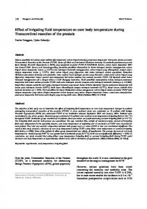
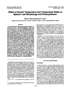
Effect of temperature on frrtihzation Figure 1 shows the effect of temperature on fertilization based on formation of the fertilization membrane in sea urchin eggs. In Anthocidaris which breeds in summer, fertilization did not occur at 5°and 7°C.The fertilization ratio increased between 10°and 15°C,and more than 90% fertilization was obtained at temperatures between 15°and 30°C.On the other hand, fertilization in Hemicentrotus, which breeds in winter, did not depend on temperature and fertilization was observed even at 0°C. These data suggest that fertilization in Anthocidaris but not Hemicentrotus depends on temperature (between 0°—30°C). In order to determine how temperature affects fertilization, therefore, we investigated the effect of temperature on the interaction between eggs and sperm, mainly using Anthocidaris. Effect of temperature on sperm moti!ity When dry sperm of Anthocidaris were diluted in ASW, the ATP level in sperm decreased rapidly within 5 mm (Fig. 2). The constant level of ATP was higher at 0°C
TEMPERATURE AND FERTILIZATION
71
100
C 0
a 50 N 4)'
IL
0
10
20
30
Temperature (°C) FIGURE 1. Effect of temperature
on fertilization
of sea urchin eggs. Eggs were inseminated
by sperm
and the number of eggs that formed a fertilization membrane was calculated. More than 100 eggs were observed. Values represent the mean of three separate experiments. (•):Anthocidaris crassispina, (0): Hemicentrotus pukherrimus.
than at 20°C.ATP in sea urchin sperm is produced by phospholipid metabolism (Mohri, 1957; Mita and Yasumasu, 1983) and is consumed by their movement (Gibbons and Gibbons, 1972). Respiration is indispensable for the phospholipid me
12
10 C
4) 0
I 2
0-
FIGURE 2.
0 5 10 15 20 Time After Dilution (mm)
Change in the level of ATP after dilution of the dry sperm of the sea urchin, Anthocidaris
crassispina. Dry sperm were diluted 100 fold in artificial sea water at 0°C(0) and 20°C(C). Values represent the mean of three separate experiments. Vertical bars show S.E.M.
72
M. MITA ET AL.
tabolism. The respiratory rate in Anthocidaris sperm was correlated with temperature between 5°and 30°C.At low temperature the respiratory rate decreased markedly (Table I). These data suggest that ATP turnover is reduced at low temperatures. Furthermore,
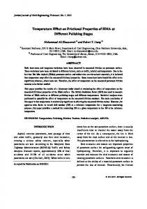
the distance traveled byAnthocidaris
sperm in a glass capillary vessel
was correlated with temperature between 0°and 30°C(Fi& 3). The distance traveled also depends on time after dilution. The distance traveled after 10 mm incubation at 20°and 30°Cwas almost 3 times longer than that at 0°C.However, the Anthocidaris sperm were motile even at 0°C.Thus, swimming activity of sperm remains at low temperatures. Treatment ofsperm with jelly water When sperm of Anthocidaris as well as Hemicentrotus were treated with jelly water, sperm were agglutinated regardlessoftemperature between 0°and 30°C.Below 10°Cthe agglutinated Anthocidaris sperm were not dispersed and sperm became immotile,
whereas the agglutination
in Hemicentrotus
sperm was released regardless
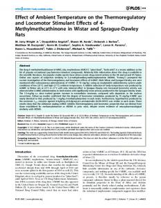
of the temperature between 0°and 30°C.In Anthocidaris, the percentage of sperm with reacted acrosome (judging from filament formation) was very low between 5° and 10°Cbut increased above 15°C(Fig. 4). It is interesting that at low temperatures the acrosome filament is not formed but agglutination is observed. According to scanning electron microscopic observations, Anthocidaris sperm with elongated head tips were more frequently observed at 4°Cbut they did not have the acrosomal process (Figs. Sc and d). The length of head in the elongated sperm was approximately 0.7—1.0 @mlonger than that of the controls (Fig. 5a). The shape of the middle piece of sperm does change during fertilization in several invertebrate species as shown in Figure Sb (Lambert and Epel, 1979; Ikadai and Hoshi, 1981). Change in the shape of the middle piece was also observed among the elongated sperm (Fig. Sc). This may suggest that an incomplete acrosome reaction causes an increase in the number of sperm with elongated head tips at low temperatures. On
the other hand, 40 to 60% of the acrosome reactions in Hemicentrotus sperm were constantly obtained between 0°and 30°C(Fig. 4). Binding of sperm to eggs Figure 6 shows that a number of sperm bound to the periphery of the dejeffied eggs of Anthocidaris
40s after addition
of dry sperm to egg suspension.
The number
of bound sperm depends on temperature in the same way that the fertilization ratio was correlated with temperature (Fig. 1). Below 10°C,no spermatozoa bound to TABLE I
Effect of temperature on oxygen consumption rate of spermatozoa of the sea urchin, Anthocidaris crassispina Temp. protein)S2.60±0.171017.0 (°C)Oxygen 0.522066.9 1.1330117.5
consumption rate (nmoles O2/min/mg ± ± ±0.87
Dry sperm were diluted 100 times in artificial sea water. The value is mean ±S.E.M. obtained in
three separate experiments.
TEMPERATURE AND FERTILIZATION
73
5
E E 4) 4) a
I4) (.) C (5 (I)
0 0
10
20
Time After Dilution(mm) FIGURE 3.
Distance traveled by the sperm of the sea urchin, Anthocidaris crassispina, after dilution
at 0°C(0), 5°C(A), 10°C(ti), 20°C(e), and 30°C(U). Dry sperm were diluted 100 times in artificial sea water. Sperm motility was determined by measurement of the distance which sperm traveled in a glass capillary vessel (d = 1 mm). Each value represents the mean of three separate experiments Vertical bars show S.E.M.
100 S
C 0 U
a
C
50
C
E 0 C 0 U
4
0
10
Temperature FIGURE 4.
20
30
(°C)
Effect of temperature on the acrosome reaction in sea urchin sperm induced by the jelly
water. Dry sperm were treated with jelly water. The percentageof the acrosome reactionjudging from the filament formation
was monitored
by electron microscopy.
More than 100 sperm were observed. Values
represent the mean of three separate experiments. (c) Anthocidaris crassispina, (0): Hemicentrotus pul. cherrimus.
74
M. MITA ET AL.
*.@.@..@ @-_@\
\\,DL@
[email protected]$@
.-—.
FIGURE 5.
The jelly water treatment of the sea urchin sperm, Anthocidaris crassispina at low tem
perature.Dry sperm were treated with the jelly water at 4°C.(a): unreacted sperm, (b): acrosome reacted sperm, and (c) and (d): sperm with elongated head tip. Bar shows 2 aim.
(5 0
N
0
CS
E I-
4) 0. U) ‘¿4-
0 L.
4) .0
E
z
5
1015202530
Temperature
(°C)
FIGURE6. Effect of temperature on adhesion of sperm to egg of Anthocidaris crassispina. Dry sperm were added to the dejellied egg suspension. The binding of the sperm to the egg was measured by counting
the numberof sperm bound to the peripheryof the egg. Each value representsthe mean of five experiments. Vertical bars show S.E.M.
75
TEMPERATURE AND FERTILIZATION
the egg. The number of sperm bound to the egg increased between 10°and 15°C; a constant level of bound sperm was observed above 15°C. Egg activation with cakium ionophore A23187 Calcium ionophore A23187 activates sea urchin eggs accompanied by formation ofthe fertilization membrane (Steinhardt and Epel, 1974). About 40%of Anthocidaris eggs showed the formation
ofthe fertilization
membrane
at 4°C,and more than 90%
of the eggs were activated at or above 10°C(Fig. 7), after 5 mm incubation with the ionophore. The ratio of the fertilization membrane formation did not change at low temperatures,
even when eggs were incubated
with the ionophore
for more than 5
mm (data not shown). On the other hand, the activation of Hemicentrotus eggs with the ionophore A23 187 was not influenced by temperature between 0°and 30°C(Fig. 7) in the same manner that fertilization was not temperature dependent (Fig. 1). DISCUSSION
The ambient sea water temperature during the breeding season is 0°—17°C for Hemicent rotus and 19°—27°C for Anthocidaris (Fujisawa and Amemiya, 1979, 1980). This agrees with the results in the present study that successful fertilization of An thocidaris required a temperature higher than 15°C(Fig. 1). It is interesting that fertilization occurs within the range of the environmental temperature at the breeding season. However, Anthocidaris eggs could be activated with calcium ionophore A23 187 at low temperatures (Fig. 7). This suggests that temperature does not exert a direct influence on activation. Therefore, fertilization of Anthocidaris is apparently regulated by the temperature
dependency
of sperm functions.
Energy metabolism (Fig. 2, Table I) and swimming activity (Fig. 3) in the An thocidaris sperm were correlated with temperature and decreased at low temperatures. The temperature dependency in the swimming activity of sperm may explain the
100
C 0
50
U
4
0 Temperature (°C) FIGURE 7.
Effect of temperature
on sea urchin egg activation
by calcium ionophore
A23 187. Eggs
were activated by the ionophore and the percentage of eggs with fertilization membrane was calculated. More than 100 eggs were observed. Each value represents the mean of three separate experiments. (C): Anthocidaris crassispina, (0): Hemicentrotus puicherrimus.
76
M. MITA ET AL
failure of fertilization at low temperatures. The number of sperm bound to eggs in Anthocidaris was almost zero at temperatures lower than 10°Cbut was remarkably increased above 15°C(Fig. 6). Before adhesion
of egg and spermatozoa,
sea urchin
sperm undergo the acrosome reaction (Dan, 1952; Afzelius and Murray, 1957). The percentage ofthe acrosome-reacted sperm in Anthocidaris also increased above 15°C (Fig. 4). These data suggest that the temperature at which the acrosome reaction occurs is closely related to successful fertilization. On the other hand, the acrosome reaction in Hemicentrotus sperm was not influenced by temperatures between 0°and 30°C(Fig. 4). Thus, fertilization in Hemicentrotus occurred at a wide range of tem peratures from 0°to 30°C(Fig. 1). There may be three reasons why the acrosome reaction in Anthocidaris does not occur frequently at low temperatures. First, activity of an acrosin-like enzyme which contributes
to the acrosome reaction (Levine et a!., 1978) may depend on temperature
in Anthocidaris but may not be influenced by temperature in Hemicentrotus. Second, the cell membrane of Hemicentrotus sperm may be more fluid at low temperatures than that ofAnthocidaris sperm, suggesting that a low temperature condition prevents the acrosome from undergoing exocytosis. Finally, it is also possible that the poly merization ofactin in the acrosomal rod is reduced under a low temperature condition. Unfortunately,
little is known about these phenomena.
In the present study we have
reported that sperm with elongated head tips were observed at low temperature (Figs. Sc and d). The occurrence of the sperm with an elongated head tip may induce the failure of fertilization in Anthocidaris at low temperatures. ACKNOWLEDGMENTS
The authors are indebted to Professor H. Kanatani and Professor Y. Nagahama ofNational Institute For Basic Biology, Okazaki, Japan for their invaluable suggestions regarding this manuscript. Thanks are also due to Dr. S. Nemoto and staff of the Tateyama Marine Laboratory for affording us the opportunities to utilize their facilities. This investigation was supported in part by grants-in-aid from the Ministry of Ed ucation, Science and Culture, of Japan (no. 57740394) to A.H.
LITERATURE AFZELIUS, B. A., AND A. MURRAY. 1957. The acrosome
CITED reaction of spermatozoa
during fertilization
or
treatment with egg water. Exp. Cell Res. 12: 325—337. DAN, J. C. 1952. Studies on the acrosome. I. Reaction to egg-water and other stimuli. Biol. Bull. 103: 54—66. DISCHE, Z., AND L. B. SHETrLES. 1951. A new spectrophotometric test for the detection of methylpentose.
J. Biol. Chem. 192: 579—582. FUJISAWA, H., AND S. AMEMIYA. 1979. Difference
in the temperature-dependency
of reaggregation
and
adhesion of cells isolated from the blastulaeof two kinds of the sea urchinswith differentspawning seasons. Med. BioL 99: 79-83. FUJISAWA, H., AND S. AMEMIYA. 1980. Effect of temperature
on the adhesion
of cells dissociated
from
sea urchin blastulae with different spawning seasons. Med. Biol. 100: 357—359. FUJIWARA,A., M. MrrA, A. HIN0, T. HAMAZAKI,Y. NAITOH,ANDI. YASUMASU.1983. Inhibition of respiration in sea urchin spermatozoa followinginteraction with fixed unfertilized eggs.VII. Decrease in the rateof respirationin the spermatozoaof the sea urchin, Hemicentrotus pulcherrimus,caused by long chain fatty acyl-CoA-induced inhibition of the movement. Dev. Growth Differ.
25: 39—47. GIBBONS,B. H., ANDI. R. GIBBONS.1972. Flagella movement and adenosin triphosphatase activity in sea urchin sperm extracted with Triton X-lOO.J. Cell Biol. 54: 75—97. HIN0, A., ANDYASUMASU. 1979.Change in the glycogencontent of sea urchin eggsduring early development. Dev. Growth Differ. 21: 229—236.
TEMPERATURE AND FERTILIZATION IKADAI, H., AND M. HOSHI. 1981. Biochemical
studies
on the acrosome
77
reaction
ofthe
starfish,
Asterias
amurensis. I. Factors participating in the acrosome reaction. Dev. Growth Differ. 23: 73-80. KATO, K. H., AND M. SUGIYAMA. 1978. Species-specific
adhesion
of spermatozoa
to the surface
of fixed
eggs in sea urchins. Dev. Growth Differ. 20: 337-347. LAMBERT, C., AND D. EPEL. 1979. Calcium-mediated
mitochondrial
movement
in ascidian
sperm during
fertilization. Dev. Biol. 69: 296—304. LAMPRECHT, W., AND I. TRANTSCHOLD.
1974.
Pp. 296-304
in Method
ofEnzymatic
Analysis.
Vol. 4,
H. U. Bergmeyer, ed. Academic Press, New York. LEVINE, A. E., K. A. WALSH, AND E. J. B. F0D0R.
1978. Evidence
of an acrosin-like
enzyme
in sea urchin
sperm. Dev. Biol. 63: 299-306. Loway, 0. H., N. J. ROSEBROUGH, A. L. FARR,ANDR. J. RANDALL1951. Protein measurement with the Folin phenol reagent.I. Biol. Chem. 193: 265—275. MrrA, M., AND I. YASuMASu. 1983. Metabolism
of lipid and carbohydrate
in sea urchin spermatozoa.
GameteRes.7:133-144. MOHRI, H. 1957. Endogenous
substrates
of respiration
in sea urchin spermatozoa.
I. Fac. Sci. Univ. Tokyo
1V8: 51—63. OKABAYASHI,K., AND E. NAKANO. 1980. Glycogen metabolism and changes in the activities of phosphorylase, phosphofructokinase and pyruvate kinase during development of sea urchin eggs. Dev. Growth
Differ. 22: 187—194. ROBINSON, J., AND J. M. COOPER. 1970. Method of determining
oxygen concentrations
in biological media,
suitable for calibration of the oxygen electrode. Anal. Biochem. 33: 390—399. STEINHARDT, R. A., AND D. EPEL 1974. Activation
of sea-urchin eggs by a calcium ionophore.
Proc. Nat.
Acad. Sci. USA 71: 1915—1919. TAKAHASHI, T., M. HOSHI, AND E. ASAHINA.
1977.
Exogastrulation
induced
by chilling
in sea urchin
larvae. Dev. Growth Differ. 19: 131—137. TURNER,
T. T., AND R. D. GILE5.
1982.
The effects
of cyclic
adenine
nucleotides,
phosphodiesterase
inhibitors,and cauda epididymal fluid on the motility of rat epididymal spermatozoa.I. Androl. 3: 134—139. VACQUIER, V. D. 1979. The fertilizing
capacity
of sea urchin
sperm
rapidly
decreases
after induction
of
the acrosome reaction. Dev. Growth Differ. 21: 61-69. YANAGISAWA, T. 1967. Studies
on echinoderm
phosphagens.
Exp. Cell Res. 46: 348-354.
YASUMASU, I., K. ASAMI, R. L SHOGER, AND A. FUJIWARA. 1973. Glycolysis of sea urchin eggs. Exp.
Cell Res. 80: 361—371.